. 2017 Aug 1;102(8):2647-2659.
doi: 10.1210/jc.2016-3806.

β Cells Persist in T1D Pancreata Without Evidence of Ongoing β-Cell Turnover or Neogenesis

Affiliations

β Cells Persist in T1D Pancreata Without Evidence of Ongoing β-Cell Turnover or Neogenesis

Carol J Lam et al. J Clin Endocrinol Metab. .

Abstract

Context: The cellular basis of persistent β-cell function in type 1 diabetes (T1D) remains enigmatic. No extensive quantitative β-cell studies of T1D pancreata have been performed to test for ongoing β-cell regeneration or neogenesis.

Objective: We sought to determine the mechanism of β-cell persistence in T1D pancreata.

Design: We studied T1D (n = 47) and nondiabetic control (n = 59) pancreata over a wide range of ages from the Juvenile Diabetes Research Foundation Network of Pancreatic Organ Donors with Diabetes via high-throughput microscopy.

Intervention and main outcome measures: We quantified β-cell mass, β-cell turnover [via Ki-67 and terminal deoxynucleotide transferase-mediated dUTP nick end labeling (TUNEL)], islet ductal association, and insulin/glucagon coexpression in T1D and control pancreata.

Results: Residual insulin-producing β cells were detected in some (but not all) T1D cases of varying disease duration. Several T1D pancreata had substantial numbers of β cells. Although β-cell proliferation was prominent early in life, it dramatically declined after infancy in both nondiabetic controls and T1D individuals. However, β-cell proliferation was equivalent in control and T1D pancreata. β-cell death (assessed by TUNEL) was extremely rare in control and T1D pancreata. Thus, β-cell turnover was not increased in T1D. Furthermore, we found no evidence of small islet/ductal neogenesis or α-cell to β-cell transdifferentiation in T1D pancreata, regardless of disease duration.

Conclusion: Longstanding β-cell function in patients with T1D appears to be largely a result of β cells that persist, without any evidence of attempted β-cell regeneration, small islet/ductal neogenesis, or transdifferentiation from other islet endocrine cell types.


Figures

Figure 1.
Figure 1.
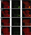
β-cell deficiency in T1D pancreata of longstanding diabetes duration. Pancreatic islet cell and β-cell area images of (a–f) control and (g–l) T1D pancreata stained for (a, d, g, j) synaptophysin-positive (syn; red) and (b, e, h, k) insulin-positive (ins; green) islets with corresponding merged images (c, f, i, l). Insets provide ×4 (left) and ×10 (right) magnification of image. Image scale bar: 5 mm; left inset scale bar: 1 mm; right inset scale bar: 100 μm.
Figure 2.
Figure 2.
β-cell mass is greatly reduced in T1D; substantial β-cells persist in some cases of T1D. (a) β-cell mass (in grams) of adolescent, young, and older adults in control and T1D pancreata. (b–c) β-cell mass (in grams) vs. age (years) in (b) control and (c) T1D pancreata across all ages. β-cell mass increases with age in controls, as seen in (b). β-cell mass does not increase with age in T1Ds, as seen in (c). (d) β-cell mass (in grams) vs. duration of diabetes (years) in T1D pancreata. β-cell mass declines with duration of diabetes. (a) Results are expressed as mean ± standard error of the mean for 28 controls and 33 T1D pancreata. ****P < 0.0001. (b–d) Data points represent the mean value for each of 59 control and 47 T1D pancreata.
Figure 3.
Figure 3.
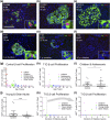
No compensatory β-cell proliferation in T1D. (a–h) β-cell proliferation declines with age in control and T1D pancreata. (a–f) Representative pancreatic islet images for (a–c) control and (d–f) T1D stained for DAPI (blue), insulin (ins; green), and Ki-67 (red). Scale bar: 100μm. (g–h) Ki-67+ β cells (% total β cells) in (g) control and (h) T1D vs. age (years) demonstrate that β-cell proliferation is reduced in T1D pancreata. (i–j) Ki-67+ β cells (% total β cells) were reduced in T1D pancreata from (i) children and adolescents and also in pancreata from (j) young and older adults. (k–l) β-cell proliferation vs. T1D duration (years) reveal that most pancreata from individuals with T1D exhibited no evidence of β-cell proliferation, regardless of duration of diabetes. (l) Panel inset from (k) represents individuals with duration of diabetes <10 years. (i–j) Results are expressed as mean ± standard error of the mean for 28 controls and 33 T1D pancreata. *P < 0.05; ****P < 0.0001. (g–h, k–l) Data points represent the mean value for each of 59 control and 47 T1D pancreata.
Figure 4.
Figure 4.
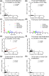
Duration of ICU stay is unassociated with β-cell proliferation in infants to older adults. (a–b) Ki-67+ β cells (% total β cells) vs. duration of ICU stay (days) in (a) control and (b) T1D pancreata. Data points represent the mean value for each of 48 control and 45 T1D pancreata. (c) Control and (d) T1D Ki-67+ acinar cells (% total) vs. age (years). Data points represent the mean value for each of 59 control and 47 T1D pancreata. (e–f) Ki-67+ acinar cells (% total) vs. duration of ICU stay (days) in (e) control and (f) T1D pancreata. Data points represent the mean value for each of 48 control and 45 T1D pancreata. (g–h) Quantification of Ki-67+ β cells vs. Ki-67+ acinar cells (% total) in (g) control and (h) T1D pancreata. Data points represent the mean value for each of 51 control and 47 T1D pancreata.
Figure 5.
Figure 5.
No apparent α-cell to β-cell transdifferentiation. (a, c, e) control and (b, d, f) T1D islets stained for glucagon (gcg; yellow), and ARX (green), insulin (ins; white), and Nkx6.1 (red) demonstrate that α- to β-cell transdifferentiation is not the primary mechanism of β-cell persistence. Insets indicate gcg+ARX+ α cells or insulin-positive Nkx6.1+ β cells.

Similar articles

Cited by

References

    1. Gepts W. Pathologic anatomy of the pancreas in juvenile diabetes mellitus. Diabetes. 1965;14(10):619–633. - PubMed
    1. Gepts W, De Mey J. Islet cell survival determined by morphology. An immunocytochemical study of the islets of Langerhans in juvenile diabetes mellitus. Diabetes. 1978;27(Suppl 1):251–261. - PubMed
    1. Klöppel G, Löhr M, Habich K, Oberholzer M, Heitz PU. Islet pathology and the pathogenesis of type 1 and type 2 diabetes mellitus revisited. Surv Synth Pathol Res. 1985;4(2):110–125. - PubMed
    1. Butler AE, Galasso R, Meier JJ, Basu R, Rizza RA, Butler PC. Modestly increased beta cell apoptosis but no increased beta cell replication in recent-onset type 1 diabetic patients who died of diabetic ketoacidosis. Diabetologia. 2007;50(11):2323–2331. - PubMed
    1. Matveyenko AV, Butler PC. Relationship between beta-cell mass and diabetes onset. Diabetes Obes Metab. 2008;10(Suppl 4):23–31. - PMC - PubMed

Publication types

MeSH terms