. 2017 May 1;158(5):1271-1288.
doi: 10.1210/en.2017-00122.

Loss of Action via Neurotensin-Leptin Receptor Neurons Disrupts Leptin and Ghrelin-Mediated Control of Energy Balance

Affiliations

Loss of Action via Neurotensin-Leptin Receptor Neurons Disrupts Leptin and Ghrelin-Mediated Control of Energy Balance

Juliette A Brown et al. Endocrinology. .

Abstract

The hormones ghrelin and leptin act via the lateral hypothalamic area (LHA) to modify energy balance, but the underlying neural mechanisms remain unclear. We investigated how leptin and ghrelin engage LHA neurons to modify energy balance behaviors and whether there is any crosstalk between leptin and ghrelin-responsive circuits. We demonstrate that ghrelin activates LHA neurons expressing hypocretin/orexin (OX) to increase food intake. Leptin mediates anorectic actions via separate neurons expressing the long form of the leptin receptor (LepRb), many of which coexpress the neuropeptide neurotensin (Nts); we refer to these as NtsLepRb neurons. Because NtsLepRb neurons inhibit OX neurons, we hypothesized that disruption of the NtsLepRb neuronal circuit would impair both NtsLepRb and OX neurons from responding to their respective hormonal cues, thus compromising adaptive energy balance. Indeed, mice with developmental deletion of LepRb specifically from NtsLepRb neurons exhibit blunted adaptive responses to leptin and ghrelin that discoordinate the mesolimbic dopamine system and ingestive and locomotor behaviors, leading to weight gain. Collectively, these data reveal a crucial role for LepRb in the proper formation of LHA circuits, and that NtsLepRb neurons are important neuronal hubs within the LHA for hormone-mediated control of ingestive and locomotor behaviors.


Figures

Figure 1.
Figure 1.
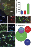
Distribution of neuropeptide-defined and LepRb neurons in the LHA. (A) The distribution of LHA neurons was determined via immunofluorescent labeling of MCH (blue), OX (red), and Nts-EGFP (green) in the brains of NtsEGFP mice (n = 4). The dashed-box area is enlarged in the bottom panels, showing the individual staining for each neuronal population and the merged image. (B) Quantitation of the average number of MCH, OX, and Nts neurons in the LHA ± SEM, n = 4. (C) NtsEGFP mice were treated with leptin (5 mg/kg, IP, 2 hours) and brains were analyzed via immunohistochemistry and immunofluorescence to identify Nts-EGFP neurons (green) and pSTAT3, a marker for LepRb activation (blue). Filled arrows, Nts-EGFP neurons that colocalize with pSTAT3 and are NtsLepRb neurons; unfilled arrows, LepRb neurons that do not express Nts. (D) Schematic depicting the relative size and distribution of the MCH, OX, Nts, NtsLepRb, and LepRb neuronal populations in the LHA. ARC, arcuate nucleus; DMH, dorsomedial hypothalamus; f, fornix; VMH, ventromedial hypothalamus.
Figure 2.
Figure 2.
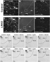
Nts and OX neurons respond to distinct hormonal cues. (A) Male NtsEGFP mice were treated with vehicle or leptin (5 mg/kg, IP, 2 hours) and brains were immunostained for Nts-EGFP (green), OX (red), and pSTAT3, a marker of leptin-activated LepRb neurons (blue). (A, top) Filled arrows identify pSTAT3-labeled nuclei within Nts-EGFP neurons, which are leptin-activated NtsLepRb neurons. (A, bottom) Unfilled arrows identify the same pSTAT3-labeled nuclei, none of which are found within OX neurons. (C) Quantification of the percentage of Nts and OX neurons that contain pSTAT3 (e.g., are activated by leptin) in response to vehicle or leptin treatment (vehicle n = 4, leptin n = 5). (C) Male NtsEGFP mice were treated with ghrelin (100 μg/treatment, IP, 4 hours) and brains were immunostained for Nts-EGFP (green), OX (red), and cFos, a marker of neuronal depolarization (blue). (D, top) Unfilled arrows identify cFos-labeled nuclei that are not found within Nts neurons. (C, bottom) Filled arrows identify the same cFos-labeled nuclei from the top panels, which are found within OX neurons. (D) Quantification of the percentage of Nts and OX neurons that contain cFos (e.g., are activated by ghrelin) in response to vehicle or ghrelin treatment (vehicle n = 4, leptin n = 4). Graphed data represent average values ± SEM. Statistical differences were determined via one-way ANOVA. **P ≤ 0.01, ***P ≤ 0.001, ****P ≤ 0.0001.
Figure 3.
Figure 3.
Loss of leptin action via NtsLepRb neurons blunts the ghrelin-mediated activation of OX neurons. (A) Generation of LRKO mice, which lack functional LepRb only in Nts neurons. (B) Backcrossed control (Nts++;Leprflfl) and LRKO (NtsCre+;Leprflfl) mice were treated with vehicle or leptin (5 mg/kg, IP, 2 hours) and brains were immunostained for pSTAT3 to identify leptin-activated LepRb neurons. LRKO mice have fewer pSTAT3-positive neurons in the LHA compared with controls, confirming loss of functional LepRb from LHA NtsLepRb neurons. (C) Male control and LRKO mice were treated with saline or ghrelin (3 µg, ICV, 4 hours) and brains were analyzed via immunohistochemistry and immunofluorescence for OX (red) and cFos (green). Arrows identify OX neurons that contain cFos-labeled nuclei (e.g., OX:cFos cells), which are activated OX neurons. (D) Quantitation of the percentage of OX neurons that contain cFos (OX:cFos) in treated control and LRKO mice. (Control + saline, n = 6; control + ghrelin, n = 6; LRKO + vehicle, n = 3; LRKO + ghrelin, n = 3.) (E) Quantitation of the total number of LHA OX neurons from three representative LHA sections of control mice (n = 12) and LRKO mice (n = 6). Graphed data represent average values ± SEM. *P ≤ 0.05 by ANOVA.
Figure 4.
Figure 4.
Loss of leptin signaling via NtsLepRb neurons disrupts energy balance. Energy balance was assessed in spontaneously moving adult (8 to 12 weeks of age) male control (n = 13) and LRKO mice (n = 14). (A) Body weight and (B) the percentage of body fat were increased in LRKO mice compared with controls, but (C) chow intake and (D) respiratory quotient were not significantly different. (E) Spontaneous locomotor activity is decreased in LRKO animals relative to controls, and (F) there is a trend for decreased spontaneous VO2. When offered a running wheel, control mice exhibit increased (G) wheel running time and (H) VO2 compared with LRKO mice (n = 10-12 per genotype). Graphed data represent average value ± SEM. *P < 0.05, **P < 0.01 by Student t test.
Figure 5.
Figure 5.
Loss of action via NtsLepRb neurons disrupts adaptive reward preference. (A) Total liquid intake and (B) sucrose preference were similar in control and LRKO mice at baseline (control n = 20, LRKO n = 18). (C) Control mice adaptively decrease chow intake in response to leptin but LRKO mice do not. (D) There is no significant difference in leptin-mediated adaptive sucrose preference between control and LRKO mice (control, n = 14; LRKO, n = 11). (E) Control and LRKO mice adaptively increase chow intake in response to ghrelin treatment (control, n = 11; LRKO, n = 11). (f) Ghrelin treatment adaptively increases sucrose preference in control mice, but not in LRKO mice (control, n = 10; LRKO, n = 11). Graphed data represent average value ± SEM. *P < 0.05 by Student t test.
Figure 6.
Figure 6.
Loss of action via NtsLepRb neurons disrupts adaptive reward wanting. Adult male control and LRKO mice were tested via a PR paradigm for their willingness to work for sucrose rewards (9 to 37 weeks old; control, n = 11; LRKO, n = 15). The PR breakpoint represents how much the sucrose reward is wanted. (A) Control and LRKO mice have similar baseline PR breakpoints at baseline. (B) Leptin treatment adaptively decreases the PR breakpoint in control mice, but not in LRKO mice, which are unable to respond to leptin via NtsLepRb neurons. (C) Ghrelin treatment adaptively increases the PR breakpoint in control mice, but not in LRKO mice. Graphed data represent average value ± SEM. *P < 0.05 by Student t test.
Figure 7.
Figure 7.
Loss of action via NtsLepRb neurons disrupts LHA gene expression. Gene expression was assessed in the brains of adult male (13 to 19 weeks old) control and LRKO mice (n = 7-8 per genotype). Gene expression in the LHA for transcripts specific to LepRb neurons, including (A) Nts, (B) galanin, and (C) LepR. Gene expression in the LHA for transcripts that are specific to OX neurons, including (D) OX, (E) Dlk1, and (F) GHSR. VTA gene expression of transcripts found in mesolimbic dopamine neurons, including (G) TH, (H) DAT, and (I) NtsR1. Graphed data represent average value ± SEM. *P < 0.05 by Student t test.
Figure 8.
Figure 8.
Projections of LHA Nts neurons and activation of mesolimbic dopamine signaling. Immunofluorescent detection of synaptophysin-mCherry in adult (21 to 31 weeks old) male NtsCre mice (control, n = 3) and LRKO mice (n = 4) mice following intra-LHA injection of the cre-inducible anterograde tract tracer, Ad-Syn-mCherry. (A) Representative image of the LHA from a control mouse, showing Nts cell bodies and local projections within the LHA and (B) projections to the VTA. (C) Enlargement of the boxed area from panel B. (D) Representative image of the Nts-containing cell bodies and location projections from a LRKO mouse. (E) Projections to the VTA. (F) Enlargement of the boxed area from panel E. Insets in panels A and D identify the injection site into the LHA. Immunohistochemical detection of cFos in the nucleus accumbens core (NAcC) and nucleus accumbens shell (NAcSh) of adult male control and LRKO mice following treatment with (G, K) amphetamine (control, n = 5; LRKO, n = 5), (H, L) vehicle (control, n = 7; LRKO, n = 6), (I, M) leptin (control, n = 5; LRKO, n = 6), and (J, N) ghrelin (control, n = 4; LRKO, n = 5). Mice were male, 17 to 23 weeks of age.

Similar articles

Cited by

References

    1. Anand BK, Brobeck JR Localization of a “feeding center” in the hypothalamus of the rat. Proc Soc Exp Biol Medi 1951;77(2):323–324. - PubMed
    1. Morrison SD, Barrnett RJ, Mayer J. Localization of lesions in the lateral hypothalamus of rats with induced adipsia and aphagia. Am J Physiol. 1958;193(1):230–234. - PubMed
    1. Brown JA, Woodworth HL, Leinninger GM. To ingest or rest? Specialized roles of lateral hypothalamic area neurons in coordinating energy balance. Front Syst Neurosci. 2015;9:9. - PMC - PubMed
    1. Jennings JH, Ung RL, Resendez SL, Stamatakis AM, Taylor JG, Huang J, Veleta K, Kantak PA, Aita M, Shilling-Scrivo K, Ramakrishnan C, Deisseroth K, Otte S, Stuber GD. Visualizing hypothalamic network dynamics for appetitive and consummatory behaviors. Cell. 2015;160(3):516–527. - PMC - PubMed
    1. Nieh EH, Matthews GA, Allsop SA, Presbrey KN, Leppla CA, Wichmann R, Neve R, Wildes CP, Tye KM. Decoding neural circuits that control compulsive sucrose seeking. Cell. 2015;160(3):528–541. - PMC - PubMed

Publication types

MeSH terms