. 2017 Jun 1;158(6):1701-1714.
doi: 10.1210/en.2017-00027.

Incretin Therapies Do Not Expand β-Cell Mass or Alter Pancreatic Histology in Young Male Mice

Affiliations

Incretin Therapies Do Not Expand β-Cell Mass or Alter Pancreatic Histology in Young Male Mice

Aaron R Cox et al. Endocrinology. .

Abstract

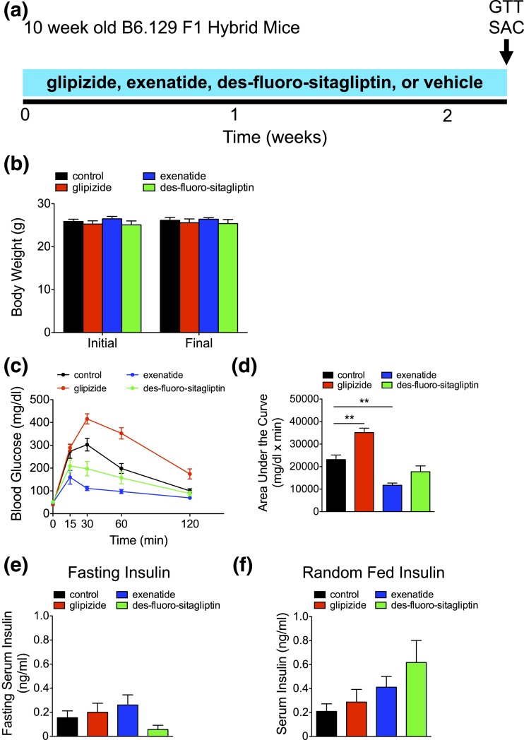
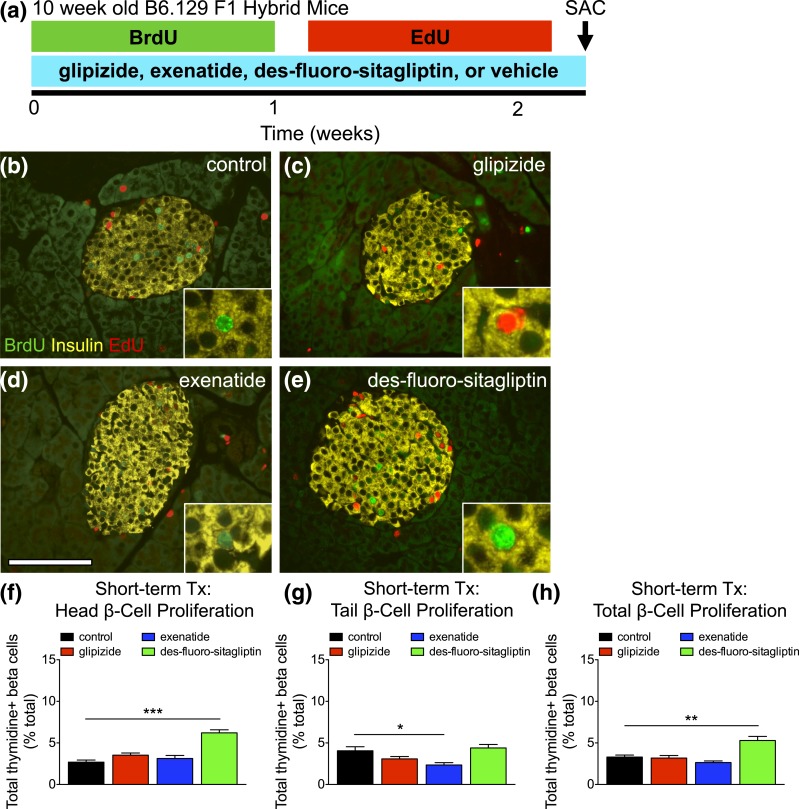
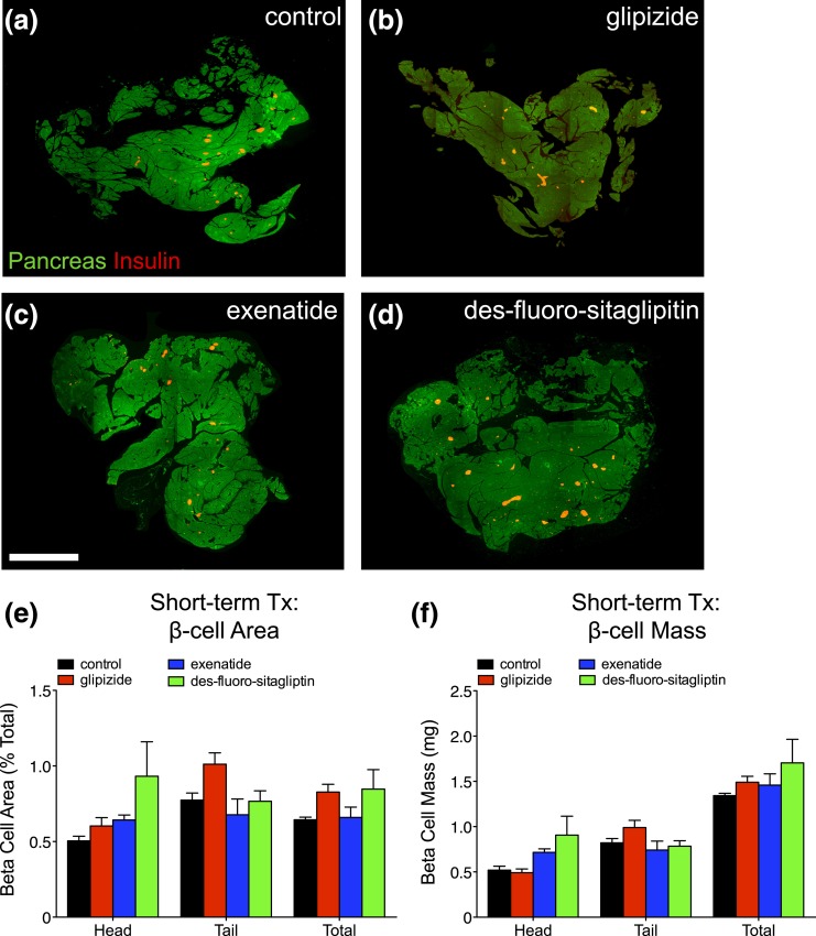
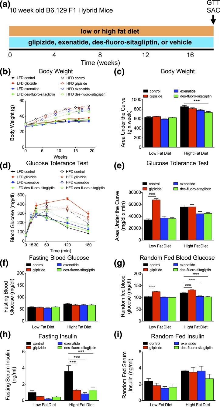
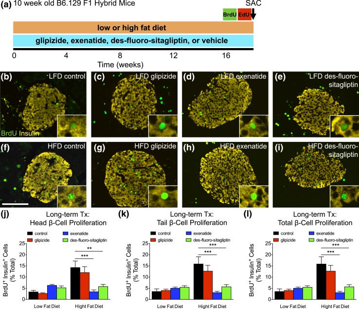
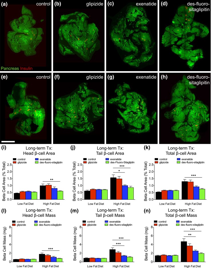
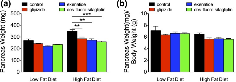
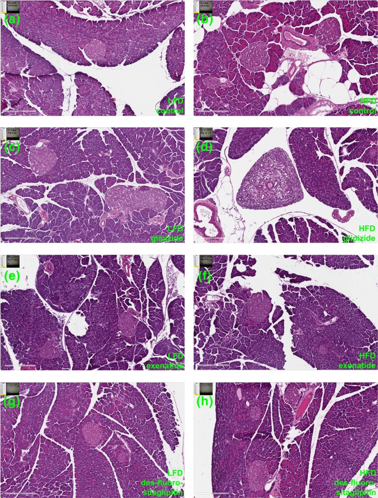
The impact of incretins upon pancreatic β-cell expansion remains extremely controversial. Multiple studies indicate that incretin-based therapies can increase β-cell proliferation, and incretins have been hypothesized to expand β-cell mass. However, disagreement exists on whether incretins increase β-cell mass. Moreover, some reports indicate that incretins may cause metaplastic changes in pancreatic histology. To resolve these questions, we treated a large cohort of mice with incretin-based therapies and carried out a rigorous analysis of β-cell turnover and pancreatic histology using high-throughput imaging. Young mice received exenatide via osmotic pump, des-fluoro-sitagliptin, or glipizide compounded in diet for 2 weeks (short-term) on a low-fat diet (LFD) or 4.5 months (long-term) on a LFD or high-fat diet (HFD). Pancreata were quantified for β-cell turnover and mass. Slides were examined for gross anatomical and microscopic changes to exocrine pancreas. Short-term des-fluoro-sitagliptin increased serum insulin and induced modest β-cell proliferation but no change in β-cell mass. Long-term incretin therapy in HFD-fed mice resulted in reduced weight gain, improved glucose homeostasis, and abrogated β-cell mass expansion. No evidence for rapidly dividing progenitor cells was found in islets or pancreatic parenchyma, indicating that incretins do not induce islet neogenesis or pancreatic metaplasia. Contrasting prior reports, we found no evidence of β-cell mass expansion after acute or chronic incretin therapy. Chronic incretin administration was not associated with histological abnormalities in pancreatic parenchyma; mice did not develop tumors, pancreatitis, or ductal hyperplasia. We conclude that incretin therapies do not generate β-cells or alter pancreatic histology in young mice.


Figures

Figure 1.
Figure 1.
Short-term incretin therapy improves glucose tolerance. (a) Timing of short-term diabetes therapy (glipizide, exenatide, des-fluoro-sitagliptin, or vehicle [control]), GTT, and kill (SAC) in 10-week-old B6.129 F1 hybrid mice. (b) Initial and final body weight (in grams). (c) Intraperitoneal GTT was performed, with the corresponding area under the curve analysis in (d). (e and f) Fasting and random-fed serum insulin (ng/mL). Means ± standard error of the mean (n = 5 animals/group). **P < 0.01.
Figure 2.
Figure 2.
Short-term des-fluoro-sitagliptin treatment (Tx) increases β-cell proliferation. (a) Timing of short-term diabetes therapy (glipizide, exenatide, des-fluoro-sitagliptin, or vehicle [control]), thymidine labeling (BrdU and EdU 1 week each), GTT, and kill (SAC) in 10-week-old B6.129 F1 hybrid mice. (b–e) Islet staining for BrdU (green), insulin (yellow), and EdU (red) in short-term treated mice with (b) vehicle (control), (c) glipizide, (d) exenatide, or (e) des-fluoro-sitagliptin. Scale bar, 100 μm. (f–h) Cumulative β-cell proliferation, measured by thymidine+ (BrdU and EdU) insulin+ cells as percent of total β-cells for the (f) head, (g) tail, and (h) total pancreas. Mean ± standard error of the mean (≥2427 β-cells/pancreas; n = 5 animals/group). *P < 0.05; **P < 0.01; ***P < 0.001.
Figure 3.
Figure 3.
Short-term incretin therapy (Tx) does not expand β-cells. (a–d) Total pancreas (green) and total β-cell area (red) generated by composites of low-power image scans from pancreatic sections of short-term treated mice with (a) vehicle (control), (b) glipizide, (c) exenatide, or (d) des-fluoro-sitagliptin. Scale bar, 2 mm. (e) β-cell area (% total) and (f) β-cell mass (in milligrams) for the head, tail, and total pancreas. Mean ± standard error of the mean (21–28 sections/pancreas; n = 5 animals/group).
Figure 4.
Figure 4.
Long-term incretin therapy improves glucose homeostasis even on a HFD. (a) Timing of long-term diabetes therapy (glipizide, exenatide, des-fluoro-sitagliptin, or vehicle [control]), diet administration (low or high fat), GTT, and kill (SAC) in 10-week-old B6.129 F1 hybrid mice. (b) Body weight (in grams) recorded over time with the corresponding area under the curve analysis in (c). (d) Intraperitoneal GTT was performed, with the corresponding area under the curve analysis in (e). (f) Fasting and (g) average random-fed blood glucose (mg/dL). Means ± standard error of the mean (n = 8–12 animals/group). (h and i) Fasting and random-fed serum insulin (ng/mL). Means ± standard error of the mean (n = 4–12 animals/group). ***P < 0.001 vs control within diet (one-way analysis of variance).
Figure 5.
Figure 5.
HFD-induced β-cell proliferation is abrogated with long-term incretin therapy. (a) Timing of long-term diabetes therapy (glipizide, exenatide, des-fluoro-sitagliptin, or vehicle [control]), thymidine labeling (BrdU and EdU, 1 week each), GTT, and kill (SAC) in 10-week-old B6.129 F1 hybrid mice. (b–i) Islet staining for BrdU (green) or insulin (yellow) in mice placed on a LFD (top row) or a HFD (bottom row). Scale bar, 100 μm. (j–l) Cumulative β-cell proliferation measured by BrdU+ insulin+ cells as percent of total β-cells (unable to quantify EdU staining) for the (j) head, (k) tail, and (l) total pancreas. Means ± standard error of the mean (≥2290 β-cells/pancreas; n = 8–12 animals/group). **P < 0.01; ***P < 0.001 vs control HFD by one-way analysis of variance.
Figure 6.
Figure 6.
HFD-induced β-cell expansion is reduced with long-term incretin therapy. (a–h) Total pancreas (green) and total β-cell area (red) generated by composites of low-power image scans from pancreatic sections of long-term treated mice with (a and e) vehicle (control), (b and f) glipizide, (c and g) exenatide, or (d and h) des-fluoro-sitagliptin. Scale bar, 2 mm. (i–k) β-Cell area (% total) and (l–n) β-cell mass (in milligrams) for the (i and l) head, (j and m) tail, and (k and n) total pancreas. Means ± standard error of the mean (19–37 sections/pancreas; n = 8–12 animals/group). *P < 0.05; **P < 0.01; ***P < 0.001 vs control HFD by one-way analysis of variance.
Figure 7.
Figure 7.
Long-term incretin therapy does not change pancreas weight in HFD-fed mice. (a) Pancreas weight (in milligrams) and (b) pancreas weight (in milligrams) normalized to body weight (in grams) from mice with long-term therapy on a LFD or and HFD. Means ± standard error of the mean (n = 8–12 animals/group). **P < 0.01; ***P < 0.001 vs control by one-way analysis of variance.
Figure 8.
Figure 8.
Long-term incretin therapy does not alter pancreas histology. Hematoxylin and eosin images from Aperio scans of pancreatic sections from mice on a LFD (a–d) or a HFD (e–h) with long-term treatment of (a and e) vehicle (control), (b and f) glipizide, (c and g) exenatide, or (d and h) des-fluoro-sitagliptin. Scale bar, 200 μm.

Comment in

Similar articles

Cited by

References

    1. Ashcroft FM. ATP-sensitive potassium channelopathies: focus on insulin secretion. J Clin Invest. 2005;115(8):2047–2058. - PMC - PubMed
    1. Campbell JE, Drucker DJ. Pharmacology, physiology, and mechanisms of incretin hormone action. Cell Metab. 2013;17(6):819–837. - PubMed
    1. Drucker DJ, Nauck MA. The incretin system: glucagon-like peptide-1 receptor agonists and dipeptidyl peptidase-4 inhibitors in type 2 diabetes. Lancet. 2006;368(9548):1696–1705. - PubMed
    1. Xu G, Stoffers DA, Habener JF, Bonner-Weir S. Exendin-4 stimulates both beta-cell replication and neogenesis, resulting in increased beta-cell mass and improved glucose tolerance in diabetic rats. Diabetes. 1999;48(12):2270–2276. - PubMed
    1. Buteau J, Spatz ML, Accili D. Transcription factor FoxO1 mediates glucagon-like peptide-1 effects on pancreatic beta-cell mass. Diabetes. 2006;55(5):1190–1196. - PubMed

MeSH terms