Review
. 2017 Jun;10(2):182-188.
doi: 10.1007/s12178-017-9413-8.

Interspinous implants to treat spinal stenosis

Affiliations
Review

Interspinous implants to treat spinal stenosis

Raj J Gala et al. Curr Rev Musculoskelet Med. 2017 Jun.

Abstract

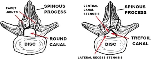
Purpose of review: Lumbar spinal stenosis has historically been treated with open decompressive surgery which is associated with significant morbidity and may give rise to various complications. Interspinous spacers (ISS) have been developed as a less invasive strategy which may serve to avoid many of these risks. The two current spacers that are FDA approved and commercially available are the Coflex and Superion devices. The goal is to review these two implants, their indications, and patient selection.

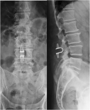
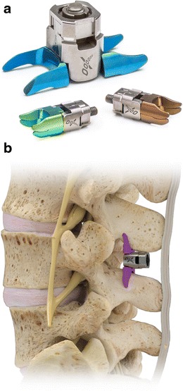
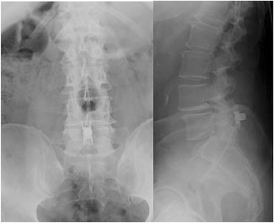
Recent findings: The Coflex device has been shown to be analogous to decompression and fusion when treating moderate spinal stenosis. It provides dynamic stability after a decompression is performed, without the rigidity of pedicle-screw instrumentation. Recent results show improved outcomes in Coflex patients at 3 years of follow-up, as compared to decompression and fusion. The Superion implant is placed percutaneously in the interspinous space with minimal disruption of spinal anatomy. When compared to the X-Stop device (which is no longer available), the Superion implant shows improved outcomes at 3 years of follow-up. ISS are lesser invasive options as compared to formal decompression and fusion for the treatment of lumbar spinal stenosis.

Keywords: Coflex; Interspinous devices; Interspinous spacers; Lumbar spinal stenosis; Neurogenic claudication; Superion.


Conflict of interest statement

Conflict of interest

Peter G. Whang reports personal fees from Paradigm Spine and institutional support from Vertiflex during the conduct of study.

Raj J. Gala and Glenn S. Russo declare that they have no conflict of interest.

Human and animal rights and informed consent

This article does not contain any studies with human or animal subjects performed by any of the authors.

Figures

Fig. 1
Fig. 1
Spinal canals are usually one of the three shapes—round, trefoil, and ovoid (not pictured). Patients with trefoil-shaped canals are predisposed to spinal stenosis symptoms, as these canals have the smallest cross-sectional area
Fig. 2
Fig. 2
a Radiograph of mild anterolisthesis of L4 on L5 in the setting of degenerative discovertebral and facet joints. b. The sagittal view of T2 MRI showing spinal stenosis. Note the disc protrusions and hypertrophic ligamentum flavum.
Fig. 3
Fig. 3
Lateral radiograph taken after implantation of the X-Stop device at two contiguous levels in the lumbar spine. Note the position of the implants
Fig. 4
Fig. 4
Lateral lumbar radiograph of the same patient 5 years later, which shows that the proximal X-Stop implant has migrated in the setting of spinous process fracture
Fig. 5
Fig. 5
© 2017 Paradigm Spine, LLC. All Rights Reserved. Published with permission
Fig. 6
Fig. 6
AP and lateral lumbar radiographs taken after implantation of Coflex device. The device is placed between the spinous processes after decompression is performed
Fig. 7
Fig. 7
Images provided by Vertiflex® and published with permission. a Image of the Superion implant, showing the superior and inferior cam lobes. b The Superion implant is an interspinous device that is delivered percutaneously
Fig. 8
Fig. 8
AP and lateral lumbar radiographs depicting the Superion implant. This device is implanted percutaneously

References

    1. Porter RW. Spinal stenosis and neurogenic claudication. Spine (Phila Pa 1976) 1996;21(17):2046–2052. doi: 10.1097/00007632-199609010-00024. - DOI - PubMed
    1. Katz JN, Harris MB. Clinical practice. Lumbar spinal stenosis. N Engl J Med. 2008;358(8):818–825. doi: 10.1056/NEJMcp0708097. - DOI - PubMed
    1. Kirkaldy-Willis WH, Wedge JH, Yong-Hing K, Reilly J. Pathology and pathogenesis of lumbar spondylosis and stenosis. Spine (Phila Pa 1976) 1978;3(4):319–328. doi: 10.1097/00007632-197812000-00004. - DOI - PubMed
    1. Rosenberg NJ. Degenerative spondylolisthesis. Predisposing factors. J Bone Joint Surg Am. 1975;57(4):467–474. doi: 10.2106/00004623-197557040-00004. - DOI - PubMed
    1. Turner JA, Ersek M, Herron L, Deyo R. Surgery for lumbar spinal stenosis: attempted meta-analysis of the literature. Spine (Phila Pa 1976) 1992;17(1):1–8. doi: 10.1097/00007632-199201000-00001. - DOI - PubMed

LinkOut - more resources