. 2017 Apr 4;25(4):927-934.e3.
doi: 10.1016/j.cmet.2017.02.008. Epub 2017 Mar 16.

The Role of Pancreatic Preproglucagon in Glucose Homeostasis in Mice

Affiliations

The Role of Pancreatic Preproglucagon in Glucose Homeostasis in Mice

Adam P Chambers et al. Cell Metab. .

Abstract

Glucagon-like peptide 1 (GLP-1) is necessary for normal gluco-regulation, and it has been widely presumed that this function reflects the actions of GLP-1 released from enteroendocrine L cells. To test the relative importance of intestinal versus pancreatic sources of GLP-1 for physiological regulation of glucose, we administered a GLP-1R antagonist, exendin-[9-39] (Ex9), to mice with tissue-specific reactivation of the preproglucagon gene (Gcg). Ex9 impaired glucose tolerance in wild-type mice but had no impact on Gcg-null or GLP-1R KO mice, suggesting that Ex9 is a true and specific GLP-1R antagonist. Unexpectedly, Ex-9 had no effect on blood glucose in mice with restoration of intestinal Gcg. In contrast, pancreatic reactivation of Gcg fully restored the effect of Ex9 to impair both oral and i.p. glucose tolerance. These findings suggest an alternative model whereby islet GLP-1 also plays an important role in regulating glucose homeostasis.

Keywords: GLP-1; glucose homeostasis; incretin.


Conflict of interest statement

Competing Interests

DJD has served as an advisor or consultant to Arisaph Pharmaceuticals, Intarcia, Merck Research Laboratories, Novo Nordisk, and Receptors Inc and receives research funding from GSK, Merck, Novo Nordisk, and Sanofi Inc. RJS has received research support from Ethicon Endo-Surgery, Novo Nordisk, Sanofi, and Janssen. RJS has served on scientific advisory boards for, Ethicon Endo-Surgery, Daiichi Sankyo, Janssen, Novartis, Nestle, Takeda, Boehringer-Ingelheim, Sanofi, and Novo Nordisk. RJS is also a paid speaker for Ethicon Endo-Surgery. DAS has received research support from Ethicon Endo-Surgery, Novo Nordisk, and Boehringer Ingelheim.

Figures

Figure 1
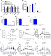
Figure 1. Targeting construct and glucose phenotype of GcgRAΔNull mice
A&B. A Lox P flanked transcriptional blocking cassette consisting of a splice acceptor (SA), an internal ribosomal entry site (IRES), and a green fluorescent protein (GFP) with a poly-A signal (pA) and was inserted into the intron between exons 2 and 3 of the Gcg gene. In the absence of Cre recombinase, expression of the targeted allele is suppressed generating a null allele. C. Gcg gene expression relative to GAPDH was nearly undetectable in GcgRAΔNull vs. WT mice (n=4M-WT and 6M-GcgRAΔNull). D. Glucose response to Ensure® (200μl) was significantly lower at 15 and 30min after the gavage in GcgRAΔNull vs. WT mice (n=4F;6M/genotype). E. Endogenous glucose production (EGP) during baseline and during the final 30min of a hyperinsulinemic euglycemic clamp (n=6M/genotype) was similar in WT and GcgRAΔNull (main effect of time; p<0.05). F. Glucose clearance was similar in WT and GcgRAΔNull at baseline and during the final 30 minutes of the hyperinsulinemic euglycemic clamp (main effect of time; p<0.05). G. Glucose and H. insulin response to an IV glucose load (0.5 g/kg) were similar between WT and GcgRAΔNull (n=8M-WT; 7M- GcgRAΔNull). *p<0.01 vs. GcgRAΔNull. Significance in C was determined using a One-way ANOVA. Signifcance in D–H was determine using a Two-way Anova for genotype and time. Data is represented as mean±SEM.
Figure 2
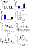
Figure 2. Validation and glucose phenotype of mice with intestinal reactivation of endogenous Gcg
A. Gcg expression was significantly greater in Vil Cre vs. GcgRAΔvilcre in the duodenum but similar in the jejunum, ileum, and colon. Gcg expression was undetectable in the GcgRAΔvilcre in the pancreas and hindbrain and in all tissues in the GcgRAΔNull mice (n=7F;5M/genotype). B. Tissue GLP-1 levels were not significantly different in the duodenum and ileum in Vil Cre vs. GcgRAΔvilcre and were undetectable in the pancreas of GcgRAΔvilcre and in all tissues of the GcgRAΔNull mice (n=9M/genotype). C. Circulating levels of active GLP-1 15 min after a glucose (3 g/kg) gavage was similar between Vil Cre and GcgRAΔvilcre mice and undetectable in GcgRAΔNull (n=7F;5M/genotype). D. Pancreatic protein levels of glucagon were undetectable in GcgRAΔNull and GcgRAΔVilCre mice (n=10M; Vil Cre & GcgRAΔNull; 9M GcgRAΔVilCre). E. Plasma glucagon levels 15min after 1U/kg of insulin were undetectable in GcgRAΔvilcre (n=8 GcgRAΔVilCre and n=10 Vil Cre mice). F. Oral glucose tolerance (2g/kg) was significantly lower at 15, 30 and 45 min in both GcgRAΔNull and GcgRAΔVilCre mice vs. WT mice (n=10M; Vil Cre & GcgRAΔNull; 9M GcgRAΔVilCre). G. IP glucose tolerance (2g/kg) was not significantly different between GcgRAΔNull, GcgRAΔVilCre, and WT mice (n=10M; Vil Cre & GcgRAΔNull; 9M GcgRAΔVilCre). *p<0.01 GcgRAΔNull vs. Vil Cre and GcgRAΔVilCre; **p<0.05 Vil Cre vs. GcgRAΔVilCre; + p<0.01 Vil Cre vs. GcgRAΔNull and GcgRAΔVilCre. H–K. Ex9 (50ug) impaired oral (H–I) and IP (J–K) glucose tolerance over saline (100μl) in WT and Vil Cre vs. GcgRAΔNull and GcgRAΔVilCre, respectively (n=15M; WT and GcgRAΔNull/drug treatment; 15M; Vil Cre and GcgRAΔVilCre/drug treatment). *p<0.05 in WT or Vil Cre vs. all other groups; +p<0.05 Vil Cre Ex9 vs. saline. Significance in A–E was determined using a One-way ANOVA for genotype. Significance in F–G was determine using a Two-way ANOVA for genotype and time. Significance in H–K was determined using a three-way ANOVA for genotype, drug and time. Data is represented as mean±SEM.
Figure 3
Figure 3. Validation and glucose phenotype of mice with pancreatic reactivation of endogenous Gcg
A. Gcg expression was significantly greater in PDXcre control vs. GcgRAΔPDX1Cre mice in the ileum and hindbrain, was not different in the duodenum, and was significantly lower in the pancreas compared to the GcgRAΔPDX1Cre mice. GcgRAΔNull had undetectable Gcg expression across all tissues. n=10M/genotype. B. Plasma glucagon levels 15 min after IP insulin (1U/kg) were similar between control and GcgRAΔPDX1Cre mice and were below detection limits in all GcgRAΔNull mice (GcgRAΔNull and PDX1Cre, n=6M; GcgRAΔPDX1Cre, n=8M). C. Plasma GLP-1 levels 15min after a glucose (3g/kg) gavage (GcgRAΔNull and PDX1Cre, n=10M; GcgRAΔPDX1Cre, n=7M). D. Oral (2g/kg) glucose tolerance in GcgRAΔNull, PDX1Cre, and GcgRAΔPDX1cre mice (n=13M/genotype). E. IP (2g/kg) glucose tolerance in GcgRAΔNull vs. PDX1Cre and GcgRAΔPDX1cre mice (n=10M/genotype). Oral (F.) and IP (G.) glucose tolerance in response to saline or Ex9 (50ug; n=7M/genotype). H. IP glucose tolerance in response to chronic (21d) infusion of saline or Ex9 (50 μg/day; n=11M/genotype). *p<0.05 PDX1Cre vs. GcgRAΔNull and GcgRAΔPDX1Cre mice; +p<0.05 PDX1Cre vs. GcgRAΔNull; **p<0.05 GcgRAΔPDX1Cre vs. GcgRAΔNull; #p<0.05 GcgRAΔPDX1Cre Ex9 vs. saline; @p<0.05 Ex9 vs. Sal in PDX1Cre and GcgRAΔPDX1Cre groups. Significance in A–C was determined using a One-way ANOVA for genotype. Significance in D–E was determine using a Two-way ANOVA for genotype and time. Significance in F-H was determined using a three-way ANOVA for genotype, drug and time. Data is represented as mean±SEM.
Figure 4
Figure 4. Role of extra-pancreatic GLP-1R
A–D. Ex9 (50ug) impaired oral (A&C) and IP (B&D) glucose tolerance over saline (100μl) in GLP-1R f/f vs. GLP-1RKDΔNestin and GLP-1RKDΔNkx2.1, respectively (n=7M; GLP-1R f/f and GLP-1RKDΔNestin/drug treatment; n=10M; GLP-1R f/f and GLP-1RKDΔNkx2.1/drug treatment). *p<0.05 in Ex9 vs. Saline within a both genotypes; +p<0.05 Ex9 vs. saline within GLP1R f/f only. Significance in A–D was determined using a three-way ANOVA for genotype, drug and time. Data is represented as mean±SEM.

Comment in

References

    1. Ali S, Lamont BJ, Charron MJ, Drucker DJ. Dual elimination of the glucagon and GLP-1 receptors in mice reveals plasticity in the incretin axis. J Clin Invest. 2011;121:1917–1929. - PMC - PubMed
    1. Ayala JE, Bracy DP, McGuinness OP, Wasserman DH. Considerations in the design of hyperinsulinemic-euglycemic clamps in the conscious mouse. Diabetes. 2006;55:390–397. - PubMed
    1. Donath MY, Burcelin R. GLP-1 effects on islets: hormonal, neuronal, or paracrine? Diabetes Care. 2013;36(Suppl 2):S145–8. - PMC - PubMed
    1. Drucker DJ, Nauck MA. The incretin system: glucagon-like peptide-1 receptor agonists and dipeptidyl peptidase-4 inhibitors in type 2 diabetes. Lancet. 2006;368:1696–1705. - PubMed
    1. Edwards CM, Todd JF, Mahmoudi M, Wang Z, Wang RM, Ghatei MA, Bloom SR. Glucagon-like peptide 1 has a physiological role in the control of postprandial glucose in humans: studies with the antagonist exendin 9–39. Diabetes. 1999;48:86–93. - PubMed

LinkOut - more resources