. 2017 Mar 21;8(2):e00186-17.
doi: 10.1128/mBio.00186-17.

Pseudomonas aeruginosa Alginate Overproduction Promotes Coexistence with Staphylococcus aureus in a Model of Cystic Fibrosis Respiratory Infection

Affiliations

Pseudomonas aeruginosa Alginate Overproduction Promotes Coexistence with Staphylococcus aureus in a Model of Cystic Fibrosis Respiratory Infection

Dominique H Limoli et al. mBio. .

Abstract

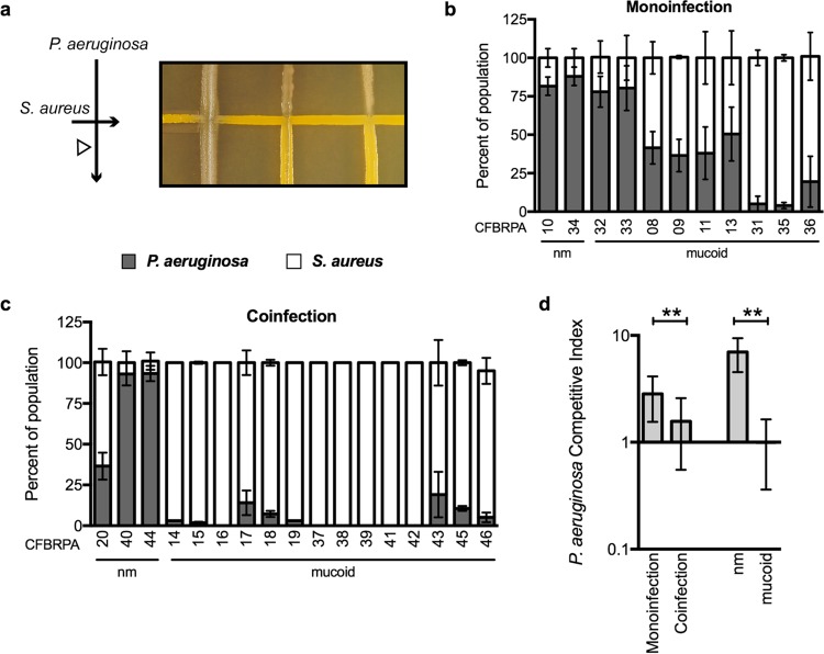
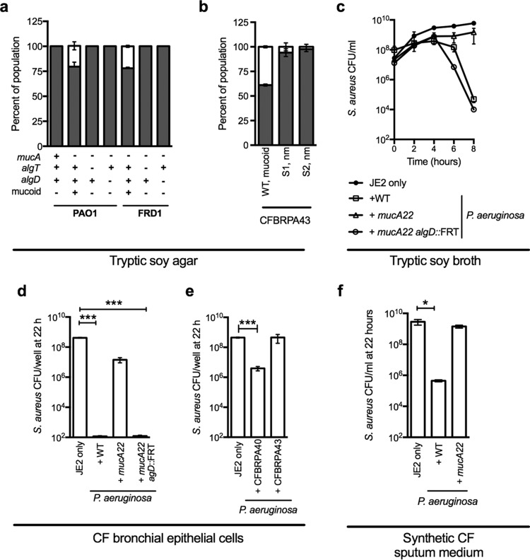
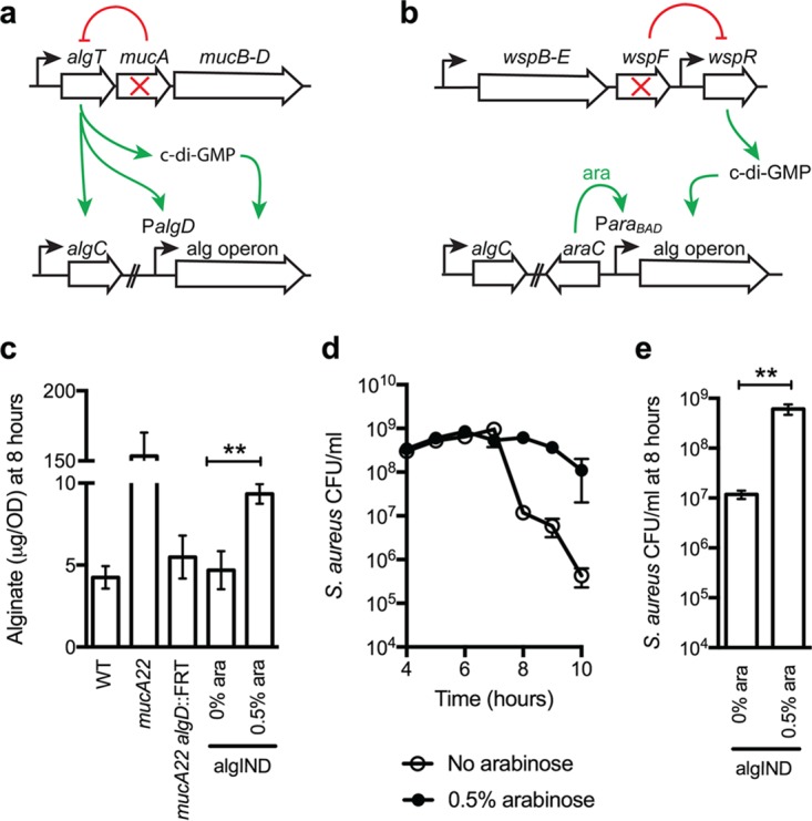
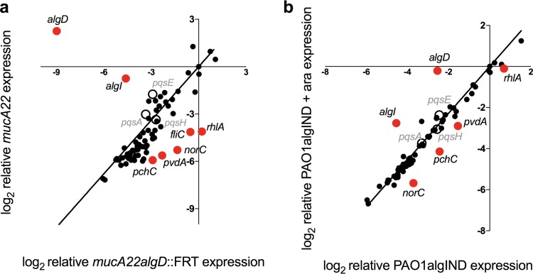
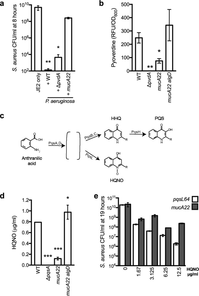
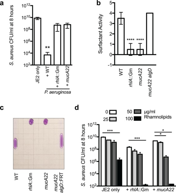
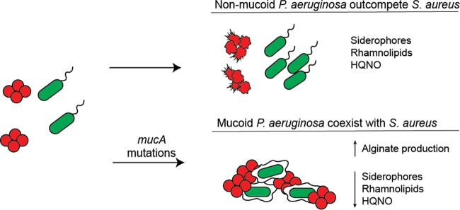
While complex intra- and interspecies microbial community dynamics are apparent during chronic infections and likely alter patient health outcomes, our understanding of these interactions is currently limited. For example, Pseudomonas aeruginosa and Staphylococcus aureus are often found to coinfect the lungs of patients with cystic fibrosis (CF), yet these organisms compete under laboratory conditions. Recent observations that coinfection correlates with decreased health outcomes necessitate we develop a greater understanding of these interbacterial interactions. In this study, we tested the hypothesis that P. aeruginosa and/or S. aureus adopts phenotypes that allow coexistence during infection. We compared competitive interactions of P. aeruginosa and S. aureus isolates from mono- or coinfected CF patients employing in vitro coculture models. P. aeruginosa isolates from monoinfected patients were more competitive toward S. aureus than P. aeruginosa isolates from coinfected patients. We also observed that the least competitive P. aeruginosa isolates possessed a mucoid phenotype. Mucoidy occurs upon constitutive activation of the sigma factor AlgT/U, which regulates synthesis of the polysaccharide alginate and dozens of other secreted factors, including some previously described to kill S. aureus Here, we show that production of alginate in mucoid strains is sufficient to inhibit anti-S. aureus activity independent of activation of the AlgT regulon. Alginate reduces production of siderophores, 2-heptyl-4-hydroxyquinolone-N-oxide (HQNO), and rhamnolipids-each required for efficient killing of S. aureus These studies demonstrate alginate overproduction may be an important factor driving P. aeruginosa coinfection with S. aureusIMPORTANCE Numerous deep-sequencing studies have revealed the microbial communities present during respiratory infections in cystic fibrosis (CF) patients are diverse, complex, and dynamic. We now face the challenge of determining the influence of these community dynamics on patient health outcomes and identifying candidate targets to modulate these interactions. We make progress toward this goal by determining that the polysaccharide alginate produced by mucoid strains of P. aeruginosa is sufficient to inhibit multiple secreted antimicrobial agents produced by this organism. Importantly, these secreted factors are required to outcompete S. aureus, when the microbes are grown in coculture; thus we propose a mechanism whereby mucoid P. aeruginosa can coexist with S. aureus Finally, the approach used here can serve as a platform to investigate the interactions among other CF pathogens.

Keywords: Pseudomonas aeruginosa; Staphylococcus aureus; biofilm; cystic fibrosis; mucoid; polymicrobial.


Figures

FIG 1
FIG 1
P. aeruginosa isolates from coinfected CF patients are less competitive with S. aureus. (a) Schematic of the method for cross-streak assay. S. aureus (USA300 LAC, JE2) was streaked onto tryptic soy agar followed by cross-streaking with P. aeruginosa isolates (CFBRPA) from the CF Biospecimen Registry (CFBR) at Emory+Children's Center for Cystic Fibrosis and Airways Disease Research. Coculture assays were performed by cross-streaking P. aeruginosa CF isolates and S. aureus on an agar surface, and the percentage of the total population of P. aeruginosa (gray bars) and S. aureus (white bars) recovered post-cross-streak (white arrowhead in panel a) were enumerated by plating on selective media and dividing the number of P. aeruginosa or S. aureus CFU by the total CFU (S. aureus plus P. aeruginosa). P. aeruginosa strains from panel b were isolated from CF patients who were infected with only P. aeruginosa (monoinfection), and strains in panel c were isolated from patients who were coinfected with P. aeruginosa and S. aureus. Mucoid and nonmucoid (nm) phenotypes are indicated. In panel d, a summary of the competitive index (CI) of all P. aeruginosa strains is indicated according the patient group from which they were isolated or their mucoid phenotype. CI was calculated by dividing the percentage of P. aeruginosa by the percentage of S. aureus recovered in the post-cross-streak. Error bars indicate standard deviations from three biological replicates performed in triplicate. Statistical significance was determined by performing an unpaired two-tailed t test. **, P < 0.001.
FIG 2
FIG 2
Mucoid conversion prevents P. aeruginosa killing of S. aureus. In vitro P. aeruginosa-S. aureus coculture assays on tryptic soy agar (a and b) and broth (c), in biofilm growth on human CF bronchial epithelial cells (CFBE) (d and e), and in synthetic CF sputum medium (f). In panels a and b, the percentages of the populations of P. aeruginosa (gray) and S. aureus (white) recovered post-cross-streak are indicated. Isogenic P. aeruginosa PAO1 and FRD1 variants, with the mucA, algT, and algD genotypes and the mucoid phenotype indicated below (a), and mucoid P. aeruginosa CFBRPA43 and nonmucoid suppressors (S1, nm and S2, nm) of P. aeruginosa CFRBPA43 (b) were cross-streaked with S. aureus strain JE2. Panel c shows the viable count of S. aureus JE2 over time for the indicated strains. In panels d, e, and f, the viability of S. aureus JE2 after 22 h of competition with the indicated P. aeruginosa strains is indicated. Error bars indicate standard deviations from at least three biological replicates performed in triplicate. Statistical significance was determined by performing an unpaired two-tailed t test. ***, P < 0.0001.
FIG 3
FIG 3
P. aeruginosa alginate production promotes coexistence with S. aureus. (a) Abbreviated schematic of alginate regulation in P. aeruginosa. Disruption of MucA results in release of AlgT, activating transcription of the algC gene, genes encoded in the alginate biosynthetic operon (alg operon, under control of the algD promoter PalgD), and bis-(3′,5′)-cyclic dimeric GMP (c-di-GMP)—all required for alginate synthesis. (b) Schematic of alginate regulation in P. aeruginosa PAO1 ΔwspF PalgD::araC-ParaBAD (PAO1algIND), whereby PalgD was replaced by araC-ParaBAD, to place the alg operon under inducible control of arabinose. Deletion of wspF results in inhibition of WspR and production of c-di-GMP. In panel c, alginate was extracted from nonmucoid wild-type PAO1 (WT) and the PAO1 mucA22, PAO1 mucA22 algD::FRT, and PAO1algIND mutants grown without arabinose (0% ara) and with 0.5% arabinose (0.5% ara) and quantified by a standard carbazole assay. In panels d and e, the log10 CFU/ml for S. aureus JE2 are indicated when grown in the presence of PAO1algIND without (open circles) and with (solid circles) 0.5% arabinose for 10 h (d); S. aureus log10 CFU/ml at 8 h are shown only in panel e. Error bars indicate standard deviations from four biological replicates performed in triplicate. Statistical significance was determined by performing an unpaired two-tailed t test. **, P < 0.01.
FIG 4
FIG 4
Alginate overproduction decreases the expression of P. aeruginosa genes required to reduce S. aureus viability. Relative expression of a subset of P. aeruginosa virulence genes was compared in PAO1 mucA22 (mucoid) and PAO1 mucA22 algD::FRT (nonmucoid) strains (a) and PAO1algIND strains grown with (mucoid) and without 0.5% arabinose (ara) (nonmucoid) (b) by the NanoString nCounter analysis system. The abundance of 75 transcripts was examined with a custom-designed codeset. Transcripts were log2 transformed and normalized to two P. aeruginosa housekeeping genes (rpoD and ppiD). Genes determined to be significantly differentially regulated when alginate is produced by an unpaired t test followed by the two-stage linear step-up procedure of Benjamini, Krieger, and Yekutieli (with q = 1% for false discovery) are indicated in red. Genes involved in the PQS pathway are indicated with open circles.
FIG 5
FIG 5
Alginate overproduction inhibits antistaphylococcal exoproducts. (a) In vitro P. aeruginosa-S. aureus coculture assays in planktonic culture with the indicated strains. Log10 CFU/ml for S. aureus JE2 are indicated after 8 h of incubation. (b) Pyoverdine was quantified as relative fluorescence units (RFU)/OD600 produced by planktonic P. aeruginosa strains grown for 8 h. (c) Schematic of 2-heptyl-4-hydroxyquinoline-N-oxide (HQNO) synthesis. The PqsA to -E, -L, and -H enzymes catalyze the synthesis of a series of 4-hydroxy-2-alkylquinolones (HAQs). The conversion of anthranilic acid to uncharacterized intermediates (indicated by the brackets) is catalyzed by PqsA and -D, followed by conversion to either 4-hydroxy-2-heptylquinoline (HHQ) by PqsB and -C (which can be converted to 3,4-dihydroxy-2-heptylquinoline [PQS] by PqsH) or converted to HQNO by PqsL. (d) HQNO quantification by LC-MS from supernatants derived from the indicated strains following 8 h of incubation in planktonic culture and (e) in vitro P. aeruginosa-S. aureus coculture assays in planktonic culture with P. aeruginosa PAO1 pqsL64 (white) and PAO1 mucA22 (gray) in the presence of the indicated concentrations of HQNO. Log10 CFU/ml for S. aureus JE2 are indicated after 19 h of incubation. Error bars indicate standard deviations from three biological replicates performed in triplicate. In panels a and b, statistical significance was determined by performing an analysis of variance (ANOVA) followed by a Dunnett’s multiple comparison test comparing each condition to the WT in panels a and d (*, P < 0.05; **, P < 0.01; ***, P < 0.0001) and to JE2 only in panel b. In panel e, statistical significance was determined by performing independent ANOVA analyses for PAO1 pqsL64 and PAO1 mucA22 followed by a Dunnett’s multiple comparison test comparing the viability of S. aureus JE2 in the presence of each HQNO concentration to the condition without HQNO, and the viability was significantly decreased at each concentration: for PAO1 pqsL64; P ≤ 0.0001, and for PAO1 mucA22, P ≤ 0.01.
FIG 6
FIG 6
Alginate inhibits the production of rhamnolipids required to kill S. aureus. In vitro P. aeruginosa-S. aureus coculture assays in planktonic culture. In panels a and d, the log10 CFU/ml for S. aureus JE2 are indicated after 8 h of incubation, and in panels b and c, the drop collapse assay was used to measure surfactant activity. Clarified supernatants were serially diluted (1:1) with water plus 0.005% crystal violet for visualization. Twenty microliters of each dilution was spotted onto the underside of the lid of a petri plate and tilted at a 90° angle. As surfactant quantities are reduced by dilution, surface tension increases, resulting in the beading of the droplet. Surfactant scores are equal to the reciprocal of the greatest dilution at which there was surfactant activity (a collapsed drop that migrates down the plate). Quantification is indicated in panel b, and a representative image of all strains at the 1/8 dilution is shown in panel c. In panel d, competitions were performed in the presence of the indicated concentrations of rhamnolipids. Error bars indicate the standard deviation of three biological replicates performed in triplicate (two biological replicates for panel d). In panels a and b, statistical significance was determined by performing an ANOVA followed by a Dunnett’s multiple comparison test comparing each condition to JE2 in panel a and to the WT only in panel b. In panel d, statistical significance was determined by performing an ANOVA followed by a Tukey’s multiple comparison test to compare the mean survival of S. aureus in the presence of each rhamnolipid concentration within strains. *, P ≤ 0.05; **, P < 0.01; ***, P < 0.0001.
FIG 7
FIG 7
Proposed model of P. aeruginosa coinfection with S. aureus. Nonmucoid isolates produce a range of antimicrobial agents that can kill S. aureus, including siderophores, rhamnolipids, and HQNO, which allows P. aeruginosa to outcompete S. aureus. If P. aeruginosa acquires mucA mutations during infection leading to overproduction of the polysaccharide alginate, the expression of genes required for siderophore, HQNO, and rhamnolipid synthesis are decreased. These modifications reduce the capacity of P. aeruginosa to outcompete S. aureus, and the two species coexist in the CF lung.

Comment in

  • Evolution of Bacterial "Frenemies".
    Darch SE, Ibberson CB, Whiteley M. Darch SE, et al. mBio. 2017 May 23;8(3):e00675-17. doi: 10.1128/mBio.00675-17. mBio. 2017. PMID: 28536291 Free PMC article.

Similar articles

Cited by

References

    1. Taylor-Robinson D, Whitehead M, Diderichsen F, Olesen HV, Pressler T, Smyth RL, Diggle P. 2012. Understanding the natural progression in %FEV1 decline in patients with cystic fibrosis: a longitudinal study. Thorax 67:860–866. doi:10.1136/thoraxjnl-2011-200953. - DOI - PMC - PubMed
    1. Filkins LM, O’Toole GA. 2015. Cystic fibrosis lung infections: polymicrobial, complex, and hard to treat. PLoS Pathog 11:e1005258. doi:10.1371/journal.ppat.1005258. - DOI - PMC - PubMed
    1. Lister PD, Wolter DJ, Hanson ND. 2009. Antibacterial-resistant Pseudomonas aeruginosa: clinical impact and complex regulation of chromosomally encoded resistance mechanisms. Clin Microbiol Rev 22:582–610. doi:10.1128/CMR.00040-09. - DOI - PMC - PubMed
    1. McCallum N, Berger-Bächi B, Senn MM. 2010. Regulation of antibiotic resistance in Staphylococcus aureus. Int J Med Microbiol 300:118–129. doi:10.1016/j.ijmm.2009.08.015. - DOI - PubMed
    1. Talwalkar JS, Murray TS. 2016. The approach to Pseudomonas aeruginosa in cystic fibrosis. Clin Chest Med 37:69–81. doi:10.1016/j.ccm.2015.10.004. - DOI - PubMed

MeSH terms