. 2017 Mar 21;10(471):eaah5756.
doi: 10.1126/scisignal.aah5756.

Structural basis for chemokine recognition by a G protein-coupled receptor and implications for receptor activation

Affiliations

Structural basis for chemokine recognition by a G protein-coupled receptor and implications for receptor activation

Joshua J Ziarek et al. Sci Signal. .

Abstract

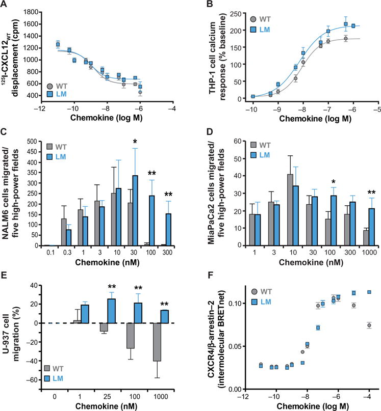
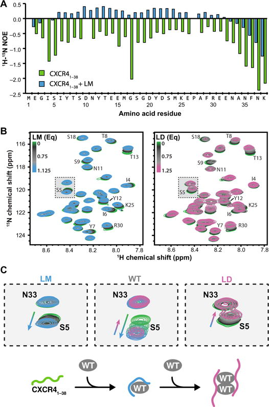
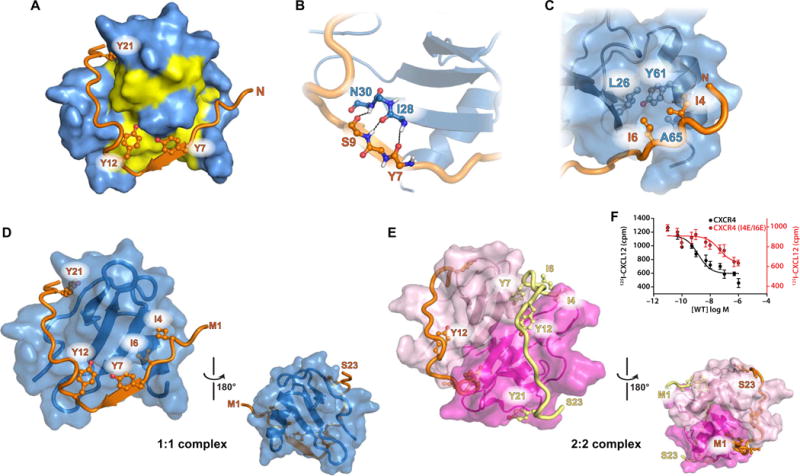
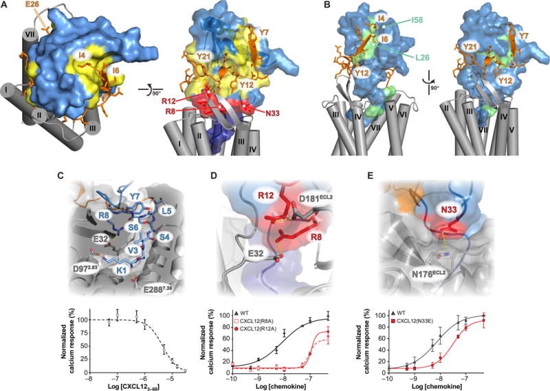
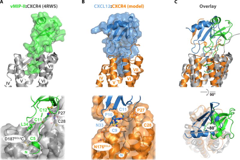
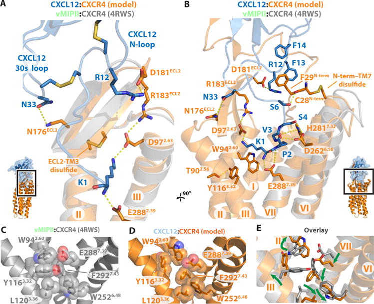
Chemokines orchestrate cell migration for development, immune surveillance, and disease by binding to cell surface heterotrimeric guanine nucleotide-binding protein (G protein)-coupled receptors (GPCRs). The array of interactions between the nearly 50 chemokines and their 20 GPCR targets generates an extensive signaling network to which promiscuity and biased agonism add further complexity. The receptor CXCR4 recognizes both monomeric and dimeric forms of the chemokine CXCL12, which is a distinct example of ligand bias in the chemokine family. We demonstrated that a constitutively monomeric CXCL12 variant reproduced the G protein-dependent and β-arrestin-dependent responses that are associated with normal CXCR4 signaling and lead to cell migration. In addition, monomeric CXCL12 made specific contacts with CXCR4 that are not present in the structure of the receptor in complex with a dimeric form of CXCL12, a biased agonist that stimulates only G protein-dependent signaling. We produced an experimentally validated model of an agonist-bound chemokine receptor that merged a nuclear magnetic resonance-based structure of monomeric CXCL12 bound to the amino terminus of CXCR4 with a crystal structure of the transmembrane domains of CXCR4. The large CXCL12:CXCR4 protein-protein interface revealed by this structure identified previously uncharacterized functional interactions that fall outside of the classical "two-site model" for chemokine-receptor recognition. Our model suggests a mechanistic hypothesis for how interactions on the extracellular face of the receptor may stimulate the conformational changes required for chemokine receptor-mediated signal transduction.


Conflict of interest statement

Competing interests: B.F.V., F.C.P., M.B.D., and C.A.K. each has a substantial financial interest in Protein Foundry, LLC (a manufacturer of recombinant proteins for research).

Figures

Fig. 1
Fig. 1. LM enhances CXCR4-mediated Ca2+ flux, cell migration, and β-arrestin-2 recruitment
(A) Binding of the indicated CXCL12 proteins was measured by radioligand displacement of 125I-CXCL12 from CXCR4-containing membrane fragments made from human embryonic kidney (HEK) 293E cells. Kd values for CXCR4 binding of WT and LM CXCL12 were calculated as 1.45 ± 1.5 nM and 0.98 ± 1.5 nM (SD), respectively, from their corresponding log EC50 (median effective concentration) values of −8.84 ± 0.17 and −9.01 ± 0.17 (SD), respectively. Data are in duplicate from three experiments. cpm, counts per minute. (B) Dose-dependent treatment of THP-1 cells with either LM or WT CXCL12 induced CXCR4-dependent intracellular Ca2+ responses, with EC50 values of 7.1 ± 1.3 nM and 8.7 ± 1.7 nM (SD), respectively. Data are in triplicate from two experiments. (C) NALM6 cell migration in response to the indicated concentrations of WT or LM CXCL12 was quantified after 90 min of stimulation. Chemotaxis was determined by counting the number of migrated cells in five high-power magnification fields. Data are means ± SD of at least nine experiments per concentration. (D) Migration of MiaPaCa2 cells was monitored after 6 hours of stimulation with the indicated concentrations of WT and LM CXCL12 using Transwell migration chambers. Chemotaxis was determined by counting the number of migrated cells in five high-power magnification fields. Data are means ± SD of five fields from four experiments. (E) U-937 cells were confined to 1-μl agarose droplets, and migration was observed after 18 to 24 hours of incubation with test medium containing WT or LM CXCL12. The percentage of migration inhibition is presented as means ± SD; the chemokine-free control was normalized to zero. Data are means ± SD of four experiments. (F) HEK 293E cells transiently cotransfected with plasmids encoding GFP10-β-arrestin-2 as a BRET donor and CXCR4-RLuc3 as a BRET acceptor were stimulated with increasing concentrations of WT and LM CXCL12, resulting in EC50 values of 17.6 ± 1.1 nM for WT and 30.6 ± 1.1 nM (SD) for LM. Data are means ± SD of three experiments. *P < 0.01; **P < 0.001.
Fig. 2
Fig. 2. LM CXCL12 and LD CXCL12 have distinct interactions with the CXCR4 N terminus
(A){1H}-15N heteronuclear NOE experiment of 250 mM[U-15N]-CXCR41–38 in the absence (green) and presence (blue) of 500 μM LM CXCL12. CXCR41–38 residues 4 to 7 exhibited a more stable interaction with LM CXCL12 than with LD CXCL12 (10). Data are from a single experiment. (B) Two-dimensional (2D) 1H/15N HSQC spectra of [U-15N]-CXCR41–38 titrated with increasing concentrations of LM CXCL12 (left) or LD CXCL12 (right). [U-15N]-CXCR41–38 (750 μM) was titrated with LM CXCL12 (0, 187.5, 375, 562.5, 750, and 843.75 μM). [U-15N]-CXCR41–38 (200 μM) was titrated with LD CXCL12 (0, 50, 100, 150, 200, and 250 μM). Data are from a single experiment. ppm, parts per million. (C) The disparate directions of chemical shift perturbations underscore that LM and LD form distinct interfaces with CXCR41–38. In some instances, as illustrated with Ser5, their trajectories can be visually concatenated to reproduce the progression of WT from a 1:1 to 2:2 complex.
Fig. 3
Fig. 3. NMR structure of LM CXCL12 in complex with CXCR41–38
(A) Surface representation of LM (blue)in complex with CXCR41–38 (orange). To simplify the visualization, only CXCR41–38 residues 1 to 23 are visible, and tyrosine residues are shown in ball-and-stick representation. Previously published changes in LM 1H/15N chemical shift upon CXCR41–38 addition are mapped onto the chemokine surface (yellow) (16). (B) CXCR4 residues 7 to 9 add an intermolecular strand parallel to β1 of the three-stranded antiparallel β sheet of the chemokine; the hydrogen bond network is represented by dashed lines. (C) CXCR41–38 residues Ile4 and Ile6 pack into a cleft between the β sheet and helix contacting LM residues Leu26 and Tyr61.(D and E) Comparison of the NMR structures of the LM:CXCR41–38 (PDB 2N55) (D) and LD:CXCR41–38 (PDB 2K04) (E) complexes. (F)Binding of WT CXCL12 was measured by radioligand displacement of 125I-CXCL12 from CXCR4WT or CXCR4 (I4E/I6E) in membranes prepared from transiently transfected HEK 293E cells. Kd values for the binding of CXCR4WT and CXCR4 (I4E/I6E) to CXCL12 were calculated as 1.45 ± 1.5 nM and 45.7 ± 1.9 nM (SD), respectively, from their corresponding log EC50 values of −8.84 ± 0.17 and −7.34 ± 0.29 (SD), respectively. Data are means ± SD of three experiments.
Fig. 4
Fig. 4. Hybrid model of the full-length CXCL12:CXCR4 complex and experimental validation
(A) Combining the LM:CXCR41–38 NMR structure and the CXCR4 crystal structure enabled modeling of the intact 1:1 signaling complex. Model generation and coordinates are located in data file S1. CXCL12 is colored light blue, with site 1, 1.5, and 2 contacts shown in yellow, red, and dark blue, respectively. CXCR4 residues 4 to 28 are colored orange, and the TM region is shaded in gray. (B) CXCL12 methyl groups that exhibited NMR intensity reductions of at least 10% from CXCR4-mediated TCS (16) are highlighted in green. (C) The N-terminal residues of CXCL12 occupy the orthosteric pocket, where salt bridges from the Lys1 α-amine and ε-amino groups to Glu288 and Asp97 of CXCR4 contribute substantially to the binding energy. N-terminal truncation of the first two residues abolishes the Ca2+ flux agonist activity of CXCL12 (fig. S8B), and the CXCL123–68 protein competes only weakly with 10 nM WT CXCL12 [IC50 = 4.5 ± 0.9 μM (SD)]. Data are means ± SD of four replicates from two experiments. (D) Arg8 and Arg12 of CXCL12 form salt bridges with Glu32 and Asp181 of CXCR4, respectively. As predicted, mutagenesis reduced the Ca2+ flux response from 7.3 ± 2.2 nM for WT to 110 ± 11 nM for CXCL12(R8A) or 95 ± 10 nM for CXCL12(R12A). Data are means ± SD of four replicates from two experiments. (E) Our model suggests that CXCL12 Asn33 contributes to binding and signaling but is not predicted to be a component of either site 1 or site 2. A fourfold change in the magnitude of Ca2+ flux [Kd = 21 ±5 nM versus 5.2 ± 2nM (SD)] confirms the contributions of Asn33 to receptor activation. Data are means ±SD of four experiments.
Fig. 5
Fig. 5. Comparison of site 1 structures
The CCL11:CCR3 NMR structure (PDB 2MPM; left), a portion of the vMIP-II:CXCR4 x-ray structure (PDB 4RWS; middle), and a portion of the CX3CL1 :US28 x-ray structure (PDB 4XT1 ; right) were aligned pairwise to the LM:CXCR4 NMR structure (PDB 2N55). The chemokines were aligned from the first cysteine to the last cysteine in each globular domain, yielding root mean square deviations (RMSDs) of 2.7 Å (CCL11), 2.2 Å (vMIP-II), and 1.9 Å (CX3CL1). For reference, the conserved cysteine in the receptor N terminus is at position 24 in CCR3, position 28 in CXCR4 (mutated to alanine in the CXCR41–38 peptide), and position 23 in US28.
Fig. 6
Fig. 6. Comparison of the CXCL12:CXCR4 hybrid model to the vMIP-II:CXCR4 crystal structure
(A) vMIP-II:CXCR4 x-ray structure (PDB 4RWS). vMIP-II forms an intermolecular β strand between the CC-motif and CXCR4 residues Pro27 and Cys28, which positions the globular domain near TM1 and TM2. (B) CXCL12:CXCR4 hybrid model derived from docking the LM:CXCR41–38 NMR structure (PDB 2N55) to the CXCR4 x-ray structure (PDB 3ODU). The globular domain of the chemokine makes contacts with TM4, TM5, and TM6, making distinct site 1, 1.5, and 2 contacts relative to the vMIP-II:CXCR4 structure. (C) Pairwise alignment between CXCR4 residues 28 to 300 (1972 atoms) of the vMIP-II:CXCR4 structure and the CXCL12:CXCR4 model, yielding an RMSD of 2.2 Å.
Fig. 7
Fig. 7. Receptor ECL2 and N terminus may translate CXCL12 binding into conformational changes of the TM bundle
(A) CXCL12:CXCR4 model derived from docking the LM CXCL12:CXCR41–38 NMR structure (PDB 2N55) to the CXCR4 x-ray structure (PDB 3ODU). Inset: Magnified view of ECL2, TM2, and TM3 with the interaction network labeled. Dashed yellow lines indicate likely hydrogen bond or electrostatic interactions. The structure of vMIPII:CXCR4 (PDB 4RWS) is shown in gray. (B) The CXCL12:CXCR4 model demonstrates extensive hydrogen bond and electrostatic interactions spanning sites 1, 1.5, and 2. Apolar and polar interactions at sites 1.5 and 2, formed between CXCR4 Cys28N-term-CXCL12 Ser6 and CXCR4 Phe29N-term-CXCL12 Arg12, may work in tandem with an extensive site 1 network to pull the extracellular portion of TM6 and TM7 toward the bundle during receptor activation. (C and D) Comparison of key amino acid positions in the vMIPII:CXCR4 structure (PDB 4RWS) (C) and the CXCL12:CXCR4 model (D). (E) Overlay of the structures from (C) and (D). Green arrows indicate differences between the vMIPII:CXCR4 structure and the CXCL12:CXCR4 model.

Similar articles

Cited by

References

    1. Rajagopal S, Rajagopal K, Lefkowitz RJ. Teaching old receptors new tricks: Biasing seven-transmembrane receptors. Nat Rev Drug Discov. 2010;9:373–386. - PMC - PubMed
    1. Dell’Agnola C, Biragyn A. Clinical utilization of chemokines to combat cancer: The double-edged sword. Expert Rev Vaccines. 2007;6:267–283. - PMC - PubMed
    1. Veldkamp CT, Peterson FC, Pelzek AJ, Volkman BF. The monomer-dimer equilibrium of stromal cell-derived factor-1 (CXCL 12) is altered by pH, phosphate,sulfate, and heparin. Protein Sci. 2005;14:1071–1081. - PMC - PubMed
    1. Baryshnikova OK, Sykes BD. Backbone dynamics of SDF-1α determined by NMR: Interpretation in the presence of monomer-dimer equilibrium. Protein Sci. 2006;15:2568–2578. - PMC - PubMed
    1. Ohnishi Y, Senda T, Nandhagopal N, Sugimoto K, Shioda T, Nagal Y, Mitsui Y. Crystal structure of recombinant native SDF-1α with additional mutagenesis studies: An attempt at a more comprehensive interpretation of accumulated structure-activity relationship data. J Interferon Cytokine Res. 2000;20:691–700. - PubMed