. 2017 May 12;292(19):8103-8121.
doi: 10.1074/jbc.M116.766816. Epub 2017 Mar 21.

Structural model of dodecameric heat-shock protein Hsp21: Flexible N-terminal arms interact with client proteins while C-terminal tails maintain the dodecamer and chaperone activity

Affiliations

Structural model of dodecameric heat-shock protein Hsp21: Flexible N-terminal arms interact with client proteins while C-terminal tails maintain the dodecamer and chaperone activity

Gudrun Rutsdottir et al. J Biol Chem. .

Abstract

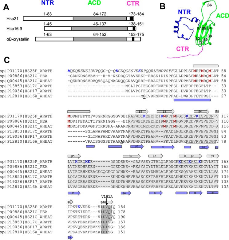
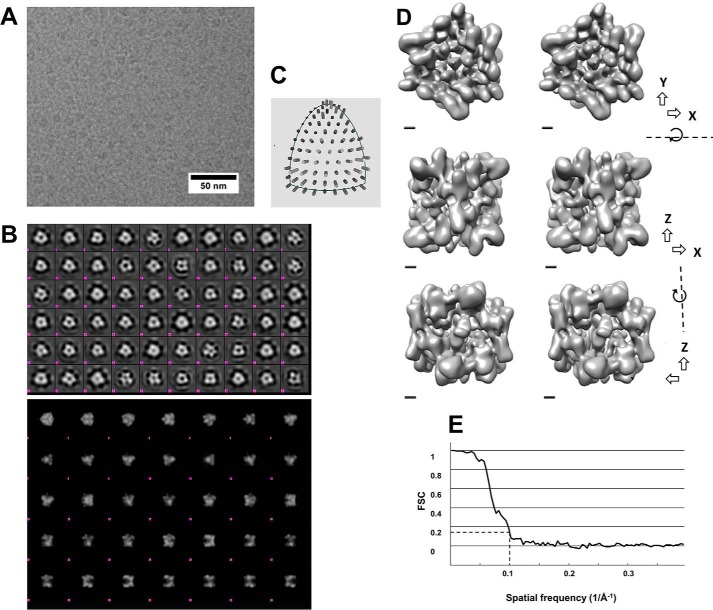
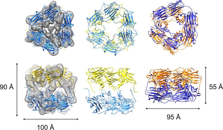
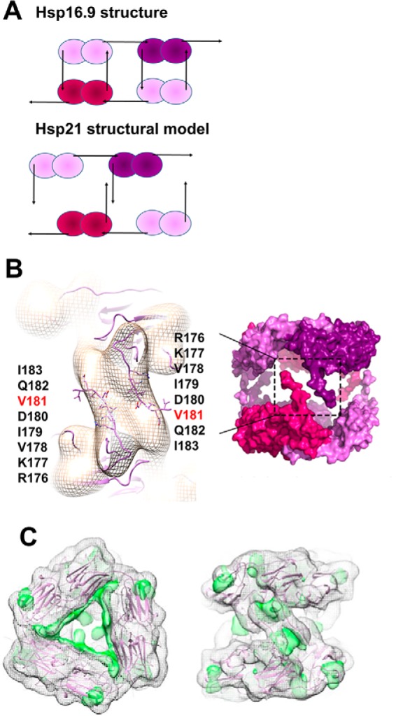
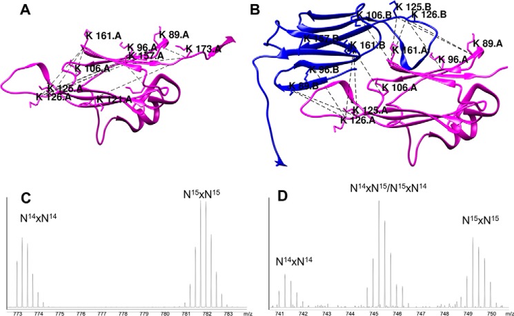
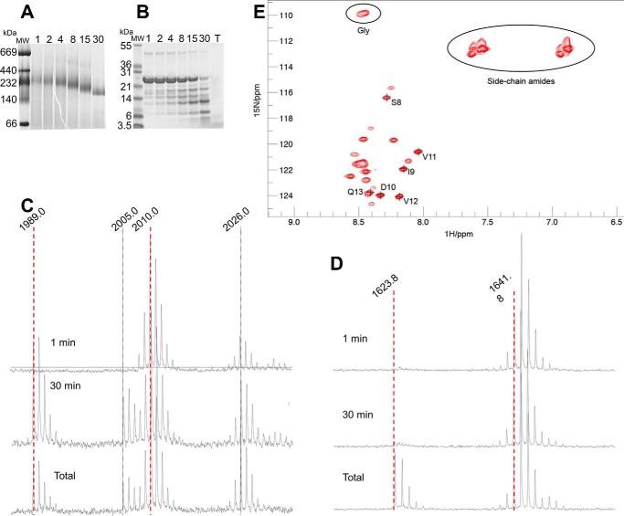
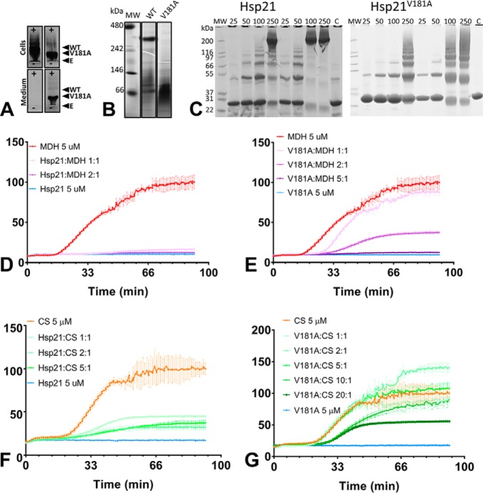
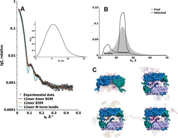
Small heat-shock proteins (sHsps) prevent aggregation of thermosensitive client proteins in a first line of defense against cellular stress. The mechanisms by which they perform this function have been hard to define due to limited structural information; currently, there is only one high-resolution structure of a plant sHsp published, that of the cytosolic Hsp16.9. We took interest in Hsp21, a chloroplast-localized sHsp crucial for plant stress resistance, which has even longer N-terminal arms than Hsp16.9, with a functionally important and conserved methionine-rich motif. To provide a framework for investigating structure-function relationships of Hsp21 and understanding these sequence variations, we developed a structural model of Hsp21 based on homology modeling, cryo-EM, cross-linking mass spectrometry, NMR, and small-angle X-ray scattering. Our data suggest a dodecameric arrangement of two trimer-of-dimer discs stabilized by the C-terminal tails, possibly through tail-to-tail interactions between the discs, mediated through extended IXVXI motifs. Our model further suggests that six N-terminal arms are located on the outside of the dodecamer, accessible for interaction with client proteins, and distinct from previous undefined or inwardly facing arms. To test the importance of the IXVXI motif, we created the point mutant V181A, which, as expected, disrupts the Hsp21 dodecamer and decreases chaperone activity. Finally, our data emphasize that sHsp chaperone efficiency depends on oligomerization and that client interactions can occur both with and without oligomer dissociation. These results provide a generalizable workflow to explore sHsps, expand our understanding of sHsp structural motifs, and provide a testable Hsp21 structure model to inform future investigations.

Keywords: Arabidopsis thaliana; aggregation; chloroplast; cryo-electron microscopy; homology modeling; molecular chaperone; oligomerization; protein cross-linking; small-angle X-ray scattering (SAXS); structural model.


Conflict of interest statement

The authors declare that they have no conflicts of interest with the contents of this article

Figures

Figure 1.
Figure 1.
Hsp21 has long N-terminal arms and short C-terminal tails with an extended IXVXI motif. A, the sHsps share a folded ACD, a CTR, and an NTR with varying length and composition between sHsps. The NTR is longer in Hsp21 as compared with the structurally characterized homologues wheat Hsp16.9 and human αB-crystallin. The (I/V)X(I/V) motif, located in the CTR and shared by all sHsps, is extended to IXVXI in Hsp21. B, one subunit of the Hsp16.9 from T. aestivum (wheat); the dodecamer structure has been determined to atomic resolution (PDB code 1GME) (25) and was used as template to generate the structural model of Hsp21. C, sequence alignment of three chloroplast-localized (Uniprot codes P1170, P09886, and Q00445) and three cytosolic Cl I (Uniprot codes P13853, P19036, and P12810) plant sHsps. The (I/V)X(I/V) motif, which is conserved within all sHsps, and the extended IXVXI motif in Hsp21 are shaded in dark gray, and the ACD is shaded in light gray. In the Hsp21 sequence, the N-terminal part not included in the structural model, because the NTR is longer in Hsp21 than in Hsp16.9, is indicated in italic type; the six unique and conserved methionine residues are shown in red; the residues involved in cross-linking are shown in blue; and the V181A point mutation is indicated with a black arrow. In the Hsp16.9 sequence, the most N-terminal amino acids in the PDB file in chain A (Ser2) and chain B (Asn43) are indicated (boldface type and underlined), and the hydrophobic groove identified in Hsp16.9 (PDB code 1GME) (25) is underlined in both sequences. The secondary structure elements (α-helix (cylinder) and β-strand (arrow)), are indicated above and below the aligned sequences, with data for Hsp21 taken from secondary structure prediction (HH_pred, J-pred, NetSurfP, SCRATCH 3 class, Porter, and PsiPred) and for Hsp16.9 from the structure file (PDB code 1GME). The sequence names are the names of the FASTA files in the UniProt database, where 25 in the Hsp21 sequence from A. thaliana refers to the full mass (25 Da) of a protein subunit before the chloroplast presequence is cleaved off, resulting in a subunit mass of 21 kDa. It may be confusing that UniProt is using “25” in the sequence name for the Arabidopsis sequence and “21” for the orthologues in pea and wheat. There is only one known chloroplast sHsp homologue in known species so far.
Figure 2.
Figure 2.
Cryo-EM of Hsp21 to generate a density map at 10 Å resolution. A, cryo-electron microscopy image of Hsp21 oligomers. The sample was plunge-frozen in liquid ethane and imaged in a JEOL JEM2100F electron microscope. Frame sets covering 2-s exposures were recorded on a DE-20 direct electron detector. The images were drift-corrected by aligning the acquired frames. B, top, class averages from a 2D classification of boxed out regions containing Hsp21 particles. There are several averages with approximate 3-fold and 2-fold symmetry (e.g. 3-fold: row 5 column 9, row 2 column 3; 2-fold: row 2 column 5). The classification was performed using Relion. The size of each box is ∼24 nm. Bottom, reprojections of the final 3D reconstruction with D3 symmetry low-pass filtered to 30 Å resolution. Several projections are similar to class averages in the top panel; for example, the side views in row 5 can be recognized in class averages with approximate mirror symmetry in the top panel (e.g. row 6 column 7). C, distribution of angles according to EMAN2. D, 3D map of the Hsp21 oligomer. Surface-rendered views at contour level 4.5σ are illustrated in a cross-eye stereo representation using Chimera. The views are along the 3-fold axis (top), 2-fold axis from one side (middle), and 2-fold axis from the other side (bottom). Scale bar, 10 Å. E, Fourier shell correlation curve between reconstructions produced by splitting the data set into two halves. Both halves were reconstructed separately. The resolution of the final 3D map was calculated to 10.0 Å from the curve at FSC = 0.143.
Figure 3.
Figure 3.
To fit the Hsp21 structural model into the Hsp21 cryo-EM density map, the discs are rotated 30° and further separated by 35 Å. Left, Hsp21 cryo-EM density map is shown with partially transparent mesh surface representation at contour level 4.5σ. The Hsp21 structural model is illustrated with the hexamer discs in yellow and blue. The views are along the 3-fold axis (top) and the 2-fold axis (bottom). The two discs were fitted separately to the map using the Fit in Map feature in Chimera. Middle, structural model of Hsp21. The Hsp21 structural model is illustrated with the discs in yellow and blue. The views are along the 3-fold axis (top) and the 2-fold axis (bottom). The top view (top) shows a relative rotation of the discs around the 3-fold axis of ∼30°, and the side view (bottom) demonstrates that the distance between the discs is extended by 35 Å in Hsp21, compared with the Hsp16.9 structure (PDB code 1GME) (25) that was used as a template for Hsp21 modeling. These differences in the Hsp21 model can also be described as an imaginary screw movement along the 3-fold axis (see supplemental Movie 1). Right, the structure of Hsp16.9 used as a template for Hsp21 modeling is illustrated with the discs in yellow and blue. The views are along the 3-fold axis (top) and the 2-fold axis (bottom).
Figure 4.
Figure 4.
Structural model of Hsp21; CTR interactions within and between the discs and NTR visible as positive difference density. A, schematic outline of the difference between the Hsp16.9 dodecamer (25) and the Hsp21 dodecamer where the upper disc is rotated 30° and the distance between discs is expanded (see Fig. 3), such that the CTR tails from dimers in the upper and lower disc are close enough for tail-to-tail interactions and too distant for tail-to-groove interactions. B, left, magnification of the Hsp21 density map at 10.0 Å resolution with approximate fitting in Chimera of the CTR tails of the Hsp21 structure model and the amino acids in the extended IXVXI motif (179IDVQI183) and in the preceding sequence RKV presented as sticks. The extended IXVXI motif, as shown in Fig. 1, has a high β-strand propensity. Pairs of C-terminal tails may interact through hydrophobic interactions in β-sheets. Right, the rigid body used for simulation of the SAXS data (i.e. after removing all 82 amino acids in the NTR). C, positive difference density at 7σ depicted by a green surface rendering, following fitting of the atomic model Hsp16.9 (PDB code 1GME) (25), as seen from the top (left) and from the side approximately along a 2-fold axis (right), suggesting 3 + 3 major positive densities inside the dodecamer and 3 + 3 minor positive densities on the dodecamer outside. The fitted Hsp16.9 map is shown with a mesh representation at 2σ.
Figure 5.
Figure 5.
Detected cross-links validate the structural model of Hsp21. The detected cross-linked peptides are listed in Table 1. A, intrasubunit cross-links presented in one subunit of the Hsp21 structural model. B, intersubunit cross-links at the dimer interface presented in a dimeric subunit of the Hsp21 structural model with reciprocal swapping of the β6-strands into the β-sandwich of the neighboring subunit. C, MS spectrum after mixed isotope labeling, with the signature typical for when only 14N-14N or 15N-15N but no 14N-15N or 15N-14N peaks are detected as hybrid cross-linked peptides. This is evidence for intrasubunit cross-linking, in this example for Lys121–Lys126. D, MS spectrum after mixed isotope labeling, with the signature typical for when not only 14N-14N or 15N-15N but also 14N-15N and 15N-14N peaks are detected as hybrid cross-linked peptides. This is evidence for intersubunit cross-linking, in this example for Lys126–Lys161.
Figure 6.
Figure 6.
Limited proteolysis and NMR provide further evidence for N-terminal arms on the outside of the Hsp21 dodecamer. A, native PAGE; Hsp21 samples withdrawn during 1–30 min of limited proteolysis (trypsin/protein molar ratio 1:2,000), showing Hsp21 dodecamers with a gradual shift in mobility. B, SDS-PAGE; same samples as in A plus a total proteolysis sample (T), showing that there is only a small amount of uncleaved Hsp21 remaining after 30 min of limited proteolysis. C, NTR peptide; MALDI-MS spectra after 1 min, 30 min, and total proteolysis, showing a peptide from the NTR (amino acids 33–50, LTMDVSPFGLLPDPLSPMR, m/z = 1,989.9), diluted with a 15N-labeled reference (m/z = 2,010.0). Peptide variants with oxidized methionine (Δ16 Da) are also visible at m/z = 2,005.0 and 2,026.0, respectively. Mass spectra for this peptide indicate that the NTR is completely cleaved after 30 min. D, ACD peptide; MALDI-MS spectra after 1 min, 30 min, and total proteolysis, showing a peptide from ACD (amino acids 84–96, APWDIKEEEHEIK, m/z = 1623.8), diluted with a 15N-labeled reference (m/z = 1641.8). Mass spectra for this peptide indicate that the ACD is hardly cleaved after 30 min. E, 1H-15N HSQC spectra of the Hsp21 dodecamers with signals detected that are assigned to 8SIDVV12 in the NTR. The relative intensities of other signals were evaluated by integrating the cross-peaks using PINT (76) and comparing with the intensity of the well-resolved peak from Ser8.
Figure 7.
Figure 7.
The V181A mutational variant of Hsp21 is non-dodecameric and has decreased chaperone activity. A, non-denaturing agarose gel electrophoresis. Panels show aliquots withdrawn from cells and medium before harvesting the cells; in each panel pair there is Hsp21 (left) and Hsp21V181A (right). Labels indicate the positions of Hsp21 (WT) and Hsp21V181A (V181A) and the entry point for loading (E). The image shows the overexpressed proteins as they migrate toward the anode (+) and that, already before purification, the mutational variant Hsp21V181A is of smaller size compared with Hsp21 and that Hsp21V181A leaks out into the medium. C, denaturing SDS-PAGE of Hsp21 WT and Hsp21V181A mutational variant cross-linked with lysine-specific cross-linker at various protein concentrations (25–250 μm) at protein/cross-linker ratios of 1:10 and 1:60. C, control samples without cross-linker. A protein concentration of 250 μm Hsp21 corresponds to ∼5 mg/ml (calculated based on monomeric mass 21 kDa). D, chaperone activity of Hsp21, determined as its suppression of heat-induced aggregation of the model substrate protein MDH at molar ratios of 1:1 and 2:1. E, chaperone activity of Hsp21V181A mutational variant, determined as its suppression of heat-induced aggregation of the model substrate protein MDH at molar ratios of 1:1, 2:1, and 5:1. F, chaperone activity of Hsp21, determined as its suppression of heat-induced aggregation of the model substrate protein CS at molar ratios of 1:1, 2:1, and 5:1. G, chaperone activity of Hsp21V181A mutational variant, determined as its suppression of heat-induced aggregation of the model substrate protein CS at molar ratios of 1:1, 2:1, 5:1, 10:1, and 20:1.
Figure 8.
Figure 8.
Fit to SAXS data further validates the Hsp21 structure model based on cryo-EM and suggests that there are N-terminal arms that are flexible on the outside of the dodecamer. A, Hsp21 wild-type SAXS data (circles) and the corresponding fits using a compact dodecamer with the N-terminal arms on the inside (blue dashed line, χ2 = 31.6 using CRYSOL), flexible extended dodecamers (yellow dotted line, modeled using EOM, χ2 = 5.52), and flexible extended hexamers and dodecamers (red line, modeled using EOM, χ2 = 1.88). The inset shows the Hsp21 wild-type distance distribution function calculated from the experimental SAXS data using GNOM. B, Rg distribution for the random pool of 10,000 hexamers and 5,000 dodecamers that was used to fit the SAXS data in A is shown in the gray area. The black line shows the Rg distribution of the optimized ensemble fitting our Hsp21 wild-type SAXS data. Whereas the selected hexamer distribution was overall compact, the selected dodecamer distribution was found at the center of the random pool. C, representative models from the selected pools of the hexamer and dodecamer, created by EOM. The rigid core is shown in a surface representation, where the three dimers of each disc are colored in blue, light blue, and teal, respectively, and the modeled flexible parts are shown as spheres. Two hexamer models are shown in one image because they were very similar, whereas the dodecamer models are shown as single objects to better see the flexible parts.
Figure 9.
Figure 9.
Online SEC-SAXS shows that Hsp21V181A mutational variant is a heterogeneous mixture of mainly dimers and some hexamers. A, experimental SAXS data of Hsp21V181A mutational variant after size-exclusion chromatography. The number below each curve represents the respective retention time in minutes on the column, and the red line is the EOM fit of the data using a pool of dimers and hexamers. The inset shows the Hsp21V181A distance distribution function calculated from the experimental SAXS data using GNOM. B, Rg distribution for the random pool of 10,000 dimers and 10,000 hexamers, which was used to fit the SAXS data in A, is shown in the gray area. The colored lines show the Rg distribution of the optimized ensemble fitting our Hsp21V181A SAXS data. The selected distributions consist of dimers with between 13% (V181_80) and 8% (V181_84) hexamers. C, representative models from the selected pools of the hexamer and dimers, created by EOM. The rigid core is shown in a surface representation where the dimers are colored in light blue, blue, and teal, and the modeled flexible parts are shown as spheres. The numbers are as in A, including the following number of conformers: 80, four conformers; 81, four conformers; 82, four conformers; 83, eight conformers; 80–84, five hexameric conformers (due to space limitations, we left out the dimer representations from retention time 84 min).

References

    1. Hilton G. R., Lioe H., Stengel F., Baldwin A. J., and Benesch J. L. P. (2013) Small heat-shock proteins: paramedics of the cell. Top. Curr. Chem. 328, 69–98 - PubMed
    1. Basha E., O'Neill H., and Vierling E. (2012) Small heat shock proteins and α-crystallins: dynamic proteins with flexible functions. Trends Biochem. Sci. 37, 106–117 - PMC - PubMed
    1. Haslbeck M., and Vierling E. (2015) A first line of stress defense: small heat shock proteins and their function in protein homeostasis. J. Mol. Biol. 427, 1537–1548 - PMC - PubMed
    1. Kampinga H. H., and Garrido C. (2012) HSPBs: small proteins with big implications in human disease. Int. J. Biochem. Cell Biol. 44, 1706–1710 - PubMed
    1. Beck M., Malmström J. A., Lange V., Schmidt A., Deutsch E. W., and Aebersold R. (2009) Visual proteomics of the human pathogen Leptospira interrogans. Nat. Methods 6, 817–823 - PMC - PubMed

Publication types

MeSH terms