. 2017 Jun 1;131(11):1123-1136.
doi: 10.1042/CS20170074. Epub 2017 Mar 21.

Pretreatment with the ALDH2 agonist Alda-1 reduces intestinal injury induced by ischaemia and reperfusion in mice

Affiliations

Pretreatment with the ALDH2 agonist Alda-1 reduces intestinal injury induced by ischaemia and reperfusion in mice

Qiankun Zhu et al. Clin Sci (Lond). .

Abstract

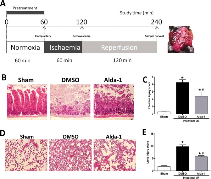
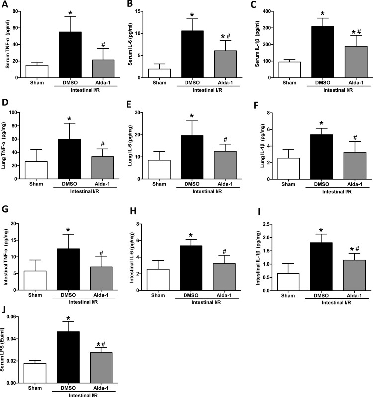
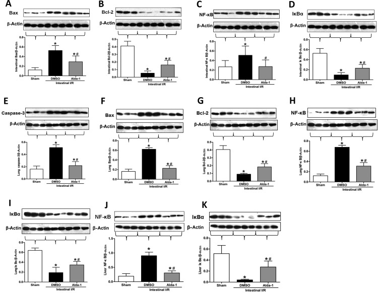
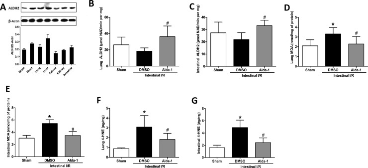
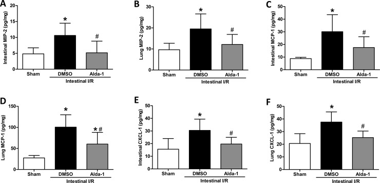
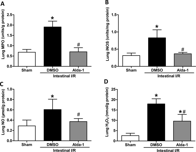
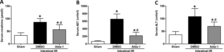
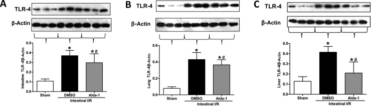
Many studies demonstrate that activation of aldehyde dehydrogenase 2 (ALDH2) protects against oxidative stress via detoxification of cytotoxic aldehydes, and could attenuate cardiac, cerebral, lung and renal ischaemia-reperfusion (I/R) injuries. However, the effect of ALDH2 in intestinal I/R is unknown. The present study was set up to determine whether an ALDH2 agonist, Alda-1, could alleviate intestinal injury after gut I/R. In a mouse model of intestinal I/R injury, histological grading, proinflammatory cytokines, oxidative stress, cellular apoptosis, chemokine contents, ALDH2 activity, 4-hydroxy-trans-2-nonenal (4-HNE) and malondialdehyde (MDA) were evaluated. The results indicated that I/R treatment conferred elevation in pathological scores, proinflammatory cytokines, oxidative stress, cellular apoptosis and chemokine levels, accompanied by accumulated 4-HNE and MDA. No significant changes in ALDH2 activity were observed after I/R. However, Alda-1 pretreatment significantly decreased these injurious indicators, concomitant with up-regulated ALDH2 activity, and lessened 4-HNE and MDA accumulation. Taken together, our results implicate activation of ALDH2 by Alda-1 in the significant abatement intestinal I/R injury.

Keywords: 4-hydroxy-trans-2-nonenal; ALDH2; Alda-1; intestine; ischaemia–reperfusion injury; multiple organ dysfunction syndrome.


Conflict of interest statement

The Authors declare that there are no competing interests associated with the manuscript.

Figures

Figure 1
Figure 1. Alteration of gut and lung morphology by Alda-1 after intestinal I/R injury
(A) Schematic panel, (B) representative H&E section of intestinal tissue, (C) histological grading of intestine injury, (D) morphological lung H&E section and (E) histological grading of lung. Original magnification ×200. Data were given as means±S.D.s (n=5–6/group). Pathological scores were compared using the Kruskal–Wallis test and the Mann–Whitney U test. #P<0.05 vs DMSO and *P<0.05 vs sham.
Figure 2
Figure 2. Changes of proinflammatory cytokines in serum, lung and intestine by Alda-1 treatment after intestinal I/R
The superior mesenteric artery was clamped for 60 min, followed by 120 min of reperfusion. Serum and tissue cytokines were measured using ELISA. Data are presented as means±S.D.s (n=5–6/group) and compared using one-way ANOVA and LSD tests. #P<0.05 vs DMSO and *P<0.05 vs sham. Abbreviation: Eu, endotoxin unit.
Figure 3
Figure 3. Effect of Alda-1 treatment on apoptotic marker expression
Tissue protein expression was measured using Western blotting: (A) intestinal Bax, (B) intestinal Bcl-2, (C) intestinal NF-κB, (D) intestinal IκBα, (E) lung caspase-3, (F) lung Bax, (G) lung Bcl-2, (H) lung NF-κB, (I) lung IκBα, (J) liver NF-κB and (K) liver IκBα. Representative blots are presented and β-actin is used as a loading control. Each bar represents means±S.D.s (n=5–6/group). Data were compared using one-way ANOVA followed by the LSD tests. #P<0.05 vs DMSO and *P<0.05 vs sham.
Figure 4
Figure 4. Alteration of ALDH2 activity and toxic aldehyde by Alda-1 pretreatment after intestinal I/R
(A) Expression of ALDH2 in various tissues was measured by Western blotting. Representative blots against ALDH2 and β-actin are shown. ALDH2 activity was observed in (B) lung and (C) intestine at 2 h after intestinal I/R or sham operation. Aldehyde accumulation was evaluated by determining the MDA and 4-HNE levels in the lung (D and F) and intestine (E and G). Data were expressed as means±S.D.s (n=5–6/group) and compared using one-way ANOVA and LSD tests. #P<0.05 vs DMSO and *P<0.05 vs sham.
Figure 5
Figure 5. Effect of Alda-1 on chemokine production in gut and lung after I/R
The superior mesenteric artery was occluded for 60 min, followed by 120 min of reperfusion, to achieve intestinal I/R. (A) Intestinal MIP-2, (B) lung MIP-2, (C) intestinal MCP-1, (D) lung MCP-1, (E) intestinal CXCL-1 and (F) lung CXCL-1 were measured using ELISA in the sham, DMSO (vehicle) and Alda-1-treated groups. Bar graphs show the means±S.D.s. #P<0.05 vs DMSO and *P<0.05 vs sham by one-way ANOVA and LSD tests.
Figure 6
Figure 6. Suppressive effects of Alda-1 on oxidative stress after intestinal I/R
Mice were subjected to 60 min of ischaemia by clamping the superior mesenteric artery and then 120 min of reperfusion. Sham surgery involved the same surgical procedure except for clamping the artery. (A) MPO, (B) iNOS, (C) NO and (D) H2O2 levels were assessed in the lung tissues. Data are given as means±S.D.s (n=5–6 per group). #P<0.05 vs DMSO and *P<0.05 vs sham by one-way ANOVA and LSD tests.
Figure 7
Figure 7. Alda-1 reduces kidney and liver injury variables after intestinal I/R
Serum (A) creatinine, (B) AST and (C) ALT levels were measured in mice exposed to Alda-1, vehicle or sham treatment (n=5–6/group). Data are presented as means±S.D.s and compared using one-way ANOVA and LSD tests. #P<0.05 vs DMSO and *P<0.05 vs sham.
Figure 8
Figure 8. Effects of Alda-1 on TLR-4 expression in the intestine, lung and liver after gut I/R
TLR-4 expression in (A) the intestine, (B) the lung and (C) liver tissues was determined using Western blotting. Representative blots were presented and β-actin was used as a loading control. Data are given as means±S.D.s (n=5–6/group) and compared using one-way ANOVA and LSD tests. #P<0.05 vs DMSO and *P<0.05 vs sham.

Comment in

References

    1. Wu M.C., Brennan F.H., Lynch J.P., Mantovani S., Phipps S., Wetsel R.A.. et al. (2013) The receptor for complement component C3a mediates protection from intestinal ischemia–reperfusion injuries by inhibiting neutrophil mobilization. Proc. Natl. Acad. Sci. U.S.A. 110, 9439–9444 10.1073/pnas.1218815110 - DOI - PMC - PubMed
    1. Chen L.W., Egan L., Li Z.W., Greten F.R., Kagnoff M.F. and Karin M. (2003) The two faces of IKK and NF-kappaB inhibition: prevention of systemic inflammation but increased local injury following intestinal ischemia-reperfusion. Nat. Med. 9, 575–581 10.1038/nm849 - DOI - PubMed
    1. Rubenfeld G.D., Caldwell E., Peabody E., Weaver J., Martin D.P., Neff M.. et al. (2005) Incidence and outcomes of acute lung injury. N. Engl. J. Med. 353, 1685–1693 10.1056/NEJMoa050333 - DOI - PubMed
    1. Wang G., Yao J., Li Z., Zu G., Feng D., Shan W.. et al. (2016) miR-34a-5p inhibition alleviates intestinal ischemia/reperfusion-induced reactive oxygen species accumulation and apoptosis via activation of SIRT1 signaling. Antioxid. Redox. Signal. 24, 961–973 10.1089/ars.2015.6492 - DOI - PubMed
    1. Cerqueira N.F., Hussni C.A. and Yoshida W.B. (2005) Pathophysiology of mesenteric ischemia/reperfusion: a review. Acta Cir. Bras. 20, 336–343 - PubMed

MeSH terms