. 2017 Mar 7:11:100.
doi: 10.3389/fnins.2017.00100. eCollection 2017.

Disease-Specific Regions Outperform Whole-Brain Approaches in Identifying Progressive Supranuclear Palsy: A Multicentric MRI Study

Affiliations

Disease-Specific Regions Outperform Whole-Brain Approaches in Identifying Progressive Supranuclear Palsy: A Multicentric MRI Study

Karsten Mueller et al. Front Neurosci. .

Abstract

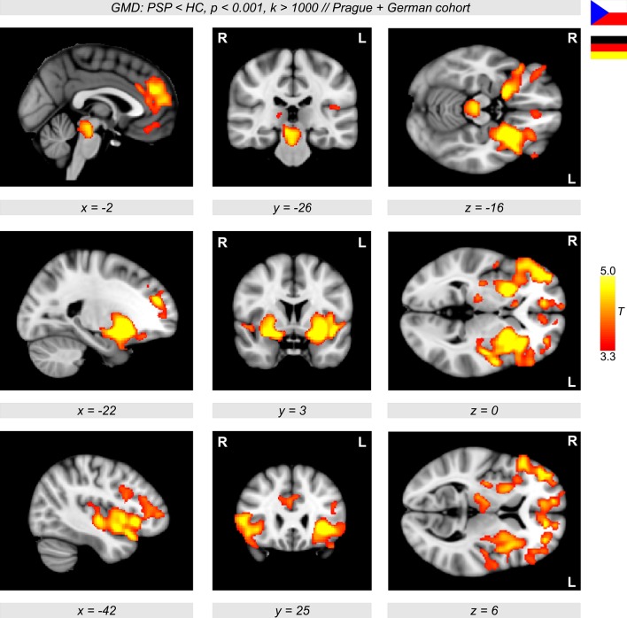
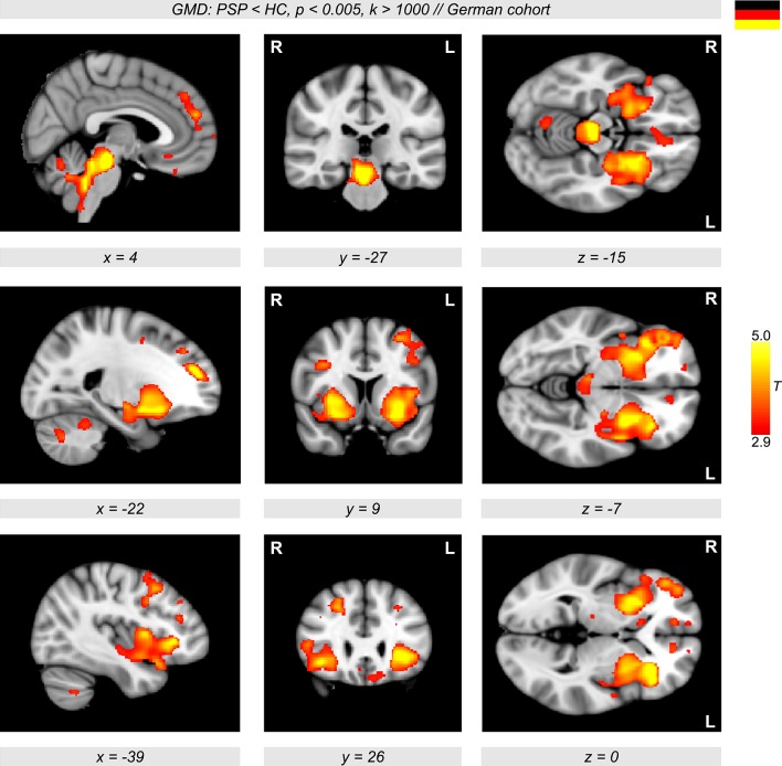
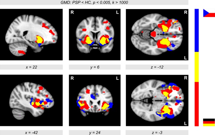
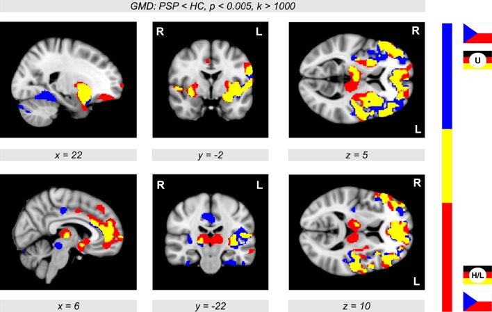
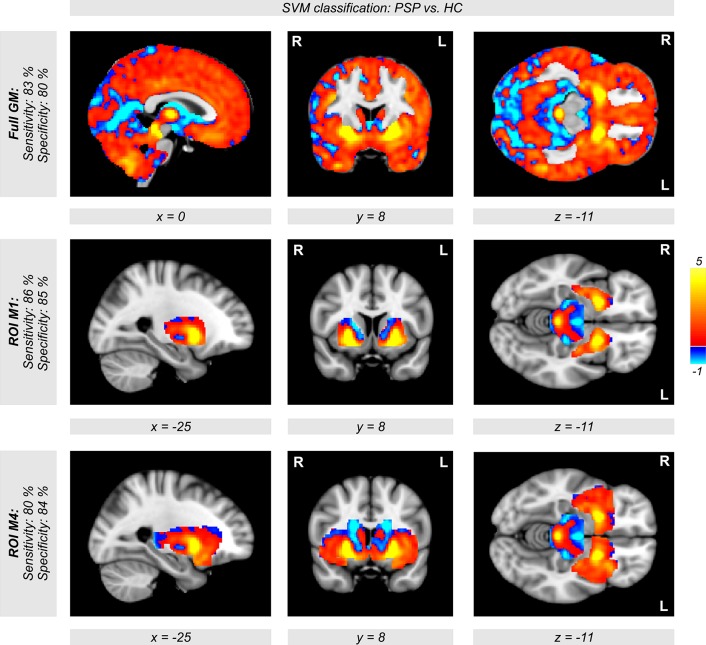
To identify progressive supranuclear palsy (PSP), we combined voxel-based morphometry (VBM) and support vector machine (SVM) classification using disease-specific features in multicentric magnetic resonance imaging (MRI) data. Structural brain differences were investigated at four centers between 20 patients with PSP and 20 age-matched healthy controls with T1-weighted MRI at 3T. To pave the way for future application in personalized medicine, we applied SVM classification to identify PSP on an individual level besides group analyses based on VBM. We found a major decline in gray matter density in the brainstem, insula, and striatum, and also in frontomedian regions, which is in line with current literature. Moreover, SVM classification yielded high accuracy rates above 80% for disease identification in imaging data. Focusing analyses on disease-specific regions-of-interest (ROI) led to higher accuracy rates compared to a whole-brain approach. Using a polynomial kernel (instead of a linear kernel) led to an increased sensitivity and a higher specificity of disease detection. Our study supports the application of MRI for individual diagnosis of PSP, if combined with SVM approaches. We demonstrate that SVM classification provides high accuracy rates in multicentric data-a prerequisite for potential application in diagnostic routine.

Keywords: atypical parkinsonism; magnetic resonance imaging; progressive supranuclear palsy; support vector machine classification; voxel-based morphometry.


Figures

Figure 1
Figure 1
Sagittal, coronal, and axial brain slices showing significant gray matter density (GMD) differences between 20 patients with progressive supranuclear palsy (PSP) and 20 age- and sex-matched healthy controls (HC) (color-coded in red/yellow, p < 0.001, k > 1,000, controlled for multiple comparisons using family-wise error-correction with p < 0.05 on the cluster level). In PSP patients, a diminished GMD was observed in the brain stem, insula, frontal cortex, and also in wide regions of the putamen extending to the pallidum.
Figure 2
Figure 2
Orthogonal brain sections showing significant gray matter density (GMD) differences between patients with progressive supranuclear palsy (PSP) and healthy controls (HC) (color-coded in red/yellow, p < 0.005, k > 1,000) in the German cohort. Significant clusters are shown using family-wise error-correction with p < 0.05 on the cluster level.
Figure 3
Figure 3
Conjunction analysis showing both the unicentric Prague cohort (color-coded in blue) and the multicentric German sample (Ulm, Homburg, and Leipzig, color-coded in red, see also Figure 2) showing significant gray matter density (GMD) differences between patients with progressive supranuclear palsy (PSP) and healthy controls (HC) (p < 0.005, k > 1,000). The overlap (color-coded in yellow) shows a reduced GMD in patients in the putamen extending to the pallidum, and also in the insular cortex. The German cohort showed prominent GMD reductions in the brainstem, while the Prague cohort also showed diminished GMD in frontomedian regions.
Figure 4
Figure 4
Conjunction analysis showing significant gray matter density (GMD) differences (p < 0.005, k > 1,000) in two groups of participants merging the participants from Prague and Ulm (U), and the participants from Prague, Homburg, and Leipzig (H/L). The overlap (color-coded in yellow) shows major reductions of GMD in patients with progressive supranuclear palsy (PSP) in the thalamus, putamen, insula, and also in frontomedian and frontolateral brain regions in comparison with healthy controls (HC).
Figure 5
Figure 5
Weights of voxels most relevant for support vector machine (SVM) classification between both groups of patients with progressive supranuclear palsy (PSP) and healthy controls (HC) after SVM training. Note that these weights are relative and have no applicable units. Relevant voxels are located in the brain stem, putamen, and pallidum. Classification accuracy was obtained with a polynomial kernel using cross-validation generating a set of 400 models leaving a patient and a control subject out when building the classifier. SVM classification was performed on all voxels within the SPM's gray matter mask (tissue probability >0.4, top row). The middle and the bottom row show the same analysis using regions of interest instead of all gray matter voxels (middle row: M1 = putamen+pallidum+midbrain; bottom row: M4 = M1+caudate+thalamus+insulae).

References

    1. Ashburner J., Friston K. J. (2005). Unified segmentation. Neuroimage 26, 839–851. 10.1016/j.neuroimage.2005.02.018 - DOI - PubMed
    1. Belmokhtar N., Benamrane N. (2012). Classification of Alzheimer's Disease from 3 D structural MRI data. Int. J. Compt. Appl. 47, 40–44. 10.5120/7171-9798 - DOI
    1. Boxer A. L., Geschwind M. D., Belfor N., Gorno-Tempini M. L., Schauer G. F., Miller B. L., et al. . (2006). Patterns of brain atrophy that differentiate corticobasal degeneration syndrome from progressive supranuclear palsy. Arch. Neurol. 63, 81–86. 10.1001/archneur.63.1.81 - DOI - PubMed
    1. Brenneis C., Seppi K., Schocke M., Benke T., Wenning G. K., Poewe W. (2004). Voxel based morphometry reveals a distinct pattern of frontal atrophy in progressive supranuclear palsy. J. Neurol. Neurosurg. Psychiatr. 75, 246–249. 10.1136/jnnp.2003.015297 - DOI - PMC - PubMed
    1. Chang C.-C., Lin C.-J. (2011). LIBSVM: a library for support vector machines. ACM Trans. Intell. Syst. Technol. 2:27 10.1145/1961189.1961199 - DOI

LinkOut - more resources