A Dynamic Circuit Hypothesis for the Pathogenesis of Blepharospasm
- PMID: 28326032
- PMCID: PMC5340098
- DOI: 10.3389/fncom.2017.00011
A Dynamic Circuit Hypothesis for the Pathogenesis of Blepharospasm
Abstract
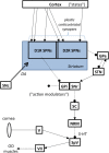
Blepharospasm (sometimes called "benign essential blepharospasm," BEB) is one of the most common focal dystonias. It involves involuntary eyelid spasms, eye closure, and increased blinking. Despite the success of botulinum toxin injections and, in some cases, pharmacologic or surgical interventions, BEB treatments are not completely efficacious and only symptomatic. We could develop principled strategies for preventing and reversing the disease if we knew the pathogenesis of primary BEB. The objective of this study was to develop a conceptual framework and dynamic circuit hypothesis for the pathogenesis of BEB. The framework extends our overarching theory for the multifactorial pathogenesis of focal dystonias (Peterson et al., 2010) to incorporate a two-hit rodent model specifically of BEB (Schicatano et al., 1997). We incorporate in the framework three features critical to cranial motor control: (1) the joint influence of motor cortical regions and direct descending projections from one of the basal ganglia output nuclei, the substantia nigra pars reticulata, on brainstem motor nuclei, (2) nested loops composed of the trigeminal blink reflex arc and the long sensorimotor loop from trigeminal nucleus through thalamus to somatosensory cortex back through basal ganglia to the same brainstem nuclei modulating the reflex arc, and (3) abnormalities in the basal ganglia dopamine system that provide a sensorimotor learning substrate which, when combined with patterns of increased blinking, leads to abnormal sensorimotor mappings manifest as BEB. The framework explains experimental data on the trigeminal reflex blink excitability (TRBE) from Schicatano et al. and makes predictions that can be tested in new experimental animal models based on emerging genetics in dystonia, including the recently characterized striatal-specific D1R dopamine transduction alterations caused by the GNAL mutation. More broadly, the model will provide a guide for future efforts to mechanistically link multiple factors in the pathogenesis of BEB and facilitate simulations of how exogenous manipulations of the pathogenic factors could ultimately be used to prevent and reverse the disorder.
Keywords: blepharospasm; dopamine; dystonia; rodent models; striatum.
Figures


Similar articles
-
Animal models for investigating benign essential blepharospasm.Curr Neuropharmacol. 2013 Jan;11(1):53-8. doi: 10.2174/157015913804999441. Curr Neuropharmacol. 2013. PMID: 23814538 Free PMC article.
-
Enhanced long-term potentiation-like plasticity of the trigeminal blink reflex circuit in blepharospasm.J Neurosci. 2006 Jan 11;26(2):716-21. doi: 10.1523/JNEUROSCI.3948-05.2006. J Neurosci. 2006. PMID: 16407569 Free PMC article.
-
Animal model explains the origins of the cranial dystonia benign essential blepharospasm.J Neurophysiol. 1997 May;77(5):2842-6. doi: 10.1152/jn.1997.77.5.2842. J Neurophysiol. 1997. PMID: 9163399
-
Chapter 33: the history of movement disorders.Handb Clin Neurol. 2010;95:501-46. doi: 10.1016/S0072-9752(08)02133-7. Handb Clin Neurol. 2010. PMID: 19892136 Review.
-
Facial dystonias and rosacea: is there an association?Orbit. 2014 Aug;33(4):276-9. doi: 10.3109/01676830.2014.904379. Epub 2014 May 15. Orbit. 2014. PMID: 24831933 Review.
Cited by
-
Blepharospasm in Japan: A Clinical Observational Study From a Large Referral Hospital in Tokyo.Neuroophthalmology. 2018 Jan 9;42(5):275-283. doi: 10.1080/01658107.2017.1409770. eCollection 2018 Oct. Neuroophthalmology. 2018. PMID: 30258472 Free PMC article.
-
Blink reflex recovery cycle distinguishes patients with idiopathic normal pressure hydrocephalus from elderly subjects.J Neurol. 2022 Feb;269(2):1007-1012. doi: 10.1007/s00415-021-10687-3. Epub 2021 Jul 2. J Neurol. 2022. PMID: 34213613
-
Blepharospasm as a tardive manifestation of COVID-19 disease: A case report.Indian J Ophthalmol. 2023 Feb;71(2):669-670. doi: 10.4103/ijo.IJO_1658_22. Indian J Ophthalmol. 2023. PMID: 36727386 Free PMC article.
-
Transcranial alternating current stimulation can modulate the blink reflex excitability. Effects of a 10- and 20-Hz tACS session on the blink reflex recovery cycle in healthy subjects.Neurol Sci. 2025 Jan;46(1):401-409. doi: 10.1007/s10072-024-07719-x. Epub 2024 Aug 3. Neurol Sci. 2025. PMID: 39096396 Free PMC article.
-
Writing, reading, and speaking in blepharospasm.J Neurol. 2019 May;266(5):1136-1140. doi: 10.1007/s00415-019-09243-x. Epub 2019 Feb 19. J Neurol. 2019. PMID: 30783748
References
-
- Augood S. J., Hollingsworth Z., Albers D. S., Yang L., Leung J., Breakefield X. O., et al. . (2004). Dopamine transmission in DYT1 dystonia. Adv. Neurol. 94, 53–60. - PubMed
LinkOut - more resources
Full Text Sources
Other Literature Sources
Miscellaneous

