Malignant renal tumors in children
- PMID: 28326263
- PMCID: PMC5345535
- DOI: 10.15586/jkcvhl.2015.29
Malignant renal tumors in children
Abstract
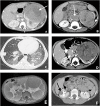
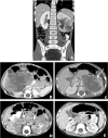
Renal malignancies are common in children. While the majority of malignant renal masses are secondary to Wilms tumor, it can be challenging to distinguish from more aggressive renal masses. For suspicious renal lesions, it is crucial to ensure prompt diagnosis in order to select the appropriate surgical procedure and treatment. This review article will discuss the common differential diagnosis that can be encountered when evaluating a suspicious renal mass in the pediatric population. This includes clear cell sarcoma of the kidney, malignant rhabdoid tumor, renal medullary carcinoma and lymphoma.
Figures


References
-
- Dome JS, et al. COG Renal Tumors Committee 2013, Children’s Oncology Group’s 2013 blueprint for research: Renal tumors. Pediatr Blood Cancer. 2013;60(6):994–1000. Doi: http://dx.doi.org/ 10.1002/pbc.24419. - DOI - PMC - PubMed
-
- Julian JC, Merguerian PA, Shortliffe LM. Pediatric genitourinary tumors. Curr Opin Oncol. 1995;7(3):265–74. Doi: http://dx.doi.org/10.1097/00001622-199505000-00012. - DOI - PubMed
-
- Ritchey ML, Azizkhan RG, Beckwith JB, Hrabovsky EE, Haase GM. Neonatal wilms tumor. J Pediatr Surg. 1995;30(6):856–9. Doi: http://dx.doi.org/10.1016/0022-3468(95)90764–5. - DOI - PubMed
-
- Grovas A, Fremgen A, Rauck A, Ruymann FB, Hutchinson CL, Winchester DP, Menck HR. The National Cancer Data Base report on patterns of childhood cancers in the United States. Cancer. 1997;80(12):2321–32. Doi: http://dx.doi.org/10.1002/(SICI)1097-0142(19971215)80:12<2321::AID-CNCR1.... - DOI - PubMed
-
- Charles AK, Vujanić GM, Berry PJ. Renal tumours of childhood. Histopathology. 1998;32(4):293–309. Doi: http://dx.doi.org/10.1046/j.1365-2559.1998.00344.x. - DOI - PubMed
LinkOut - more resources
Full Text Sources
Other Literature Sources
