

Three-Dimensional Human Cardiac Tissue Engineered by Centrifugation of Stacked Cell Sheets and Cross-Sectional Observation of Its Synchronous Beatings by Optical Coherence Tomography
- PMID: 28326324
- PMCID: PMC5343287
- DOI: 10.1155/2017/5341702
Three-Dimensional Human Cardiac Tissue Engineered by Centrifugation of Stacked Cell Sheets and Cross-Sectional Observation of Its Synchronous Beatings by Optical Coherence Tomography
Abstract
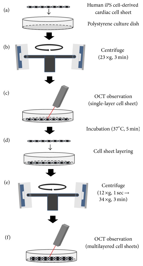
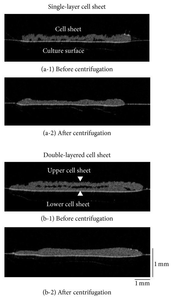
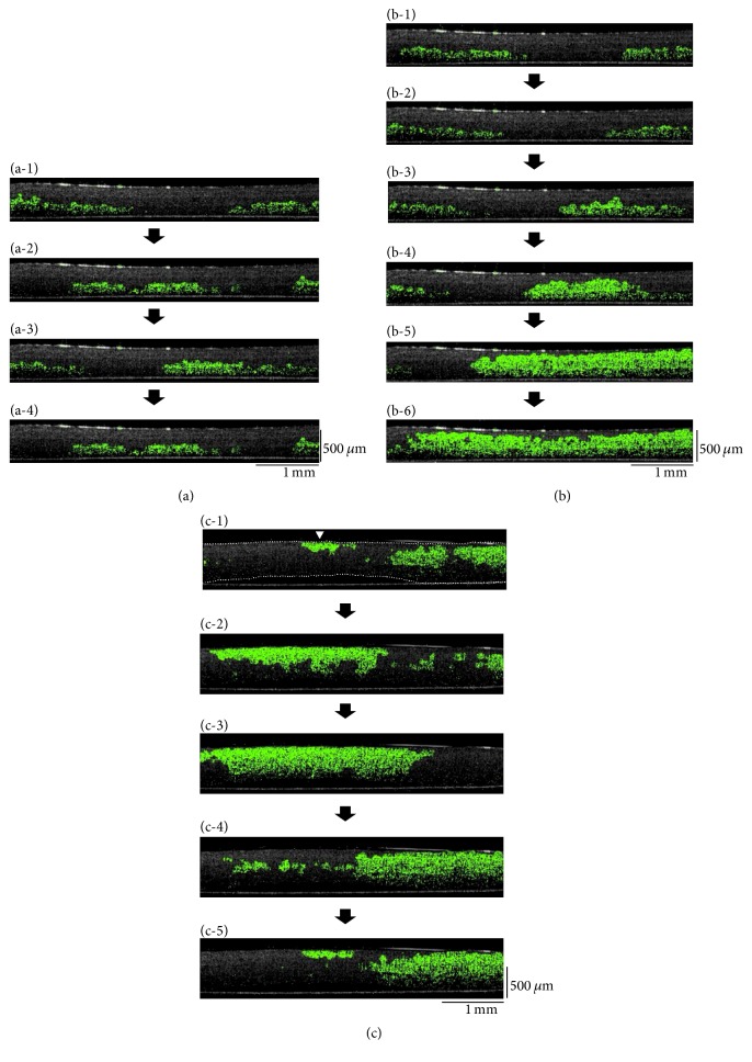
Three-dimensional (3D) tissues are engineered by stacking cell sheets, and these tissues have been applied in clinical regenerative therapies. The optimal fabrication technique of 3D human tissues and the real-time observation system for these tissues are important in tissue engineering, regenerative medicine, cardiac physiology, and the safety testing of candidate chemicals. In this study, for aiming the clinical application, 3D human cardiac tissues were rapidly fabricated by human induced pluripotent stem (iPS) cell-derived cardiac cell sheets with centrifugation, and the structures and beatings in the cardiac tissues were observed cross-sectionally and noninvasively by two optical coherence tomography (OCT) systems. The fabrication time was reduced to approximately one-quarter by centrifugation. The cross-sectional observation showed that multilayered cardiac cell sheets adhered tightly just after centrifugation. Additionally, the cross-sectional transmissions of beatings within multilayered human cardiac tissues were clearly detected by OCT. The observation showed the synchronous beatings of the thicker 3D human cardiac tissues, which were fabricated rapidly by cell sheet technology and centrifugation. The rapid tissue-fabrication technique and OCT technology will show a powerful potential in cardiac tissue engineering, regenerative medicine, and drug discovery research.
Conflict of interest statement
Tatsuya Shimizu is the stakeholder of CellSeed Inc. Katsuhisa Matsuura and Tatsuya Shimizu are inventors of a bioreactor system for culturing human iPS cells and differentiating human iPS cells into cardiac cells. Mari Kobayashi and Shin-ichi Iwana are employees of Panasonic Healthcare Co., Ltd. Yasuhiro Kabetani is an employee of Panasonic Corporation. Tokyo Women's Medical University was receiving research funds from CellSeed Inc., Panasonic Healthcare Co., Ltd., and Panasonic Corporation.
Figures
Similar articles
-
Rapid fabrication system for three-dimensional tissues using cell sheet engineering and centrifugation.J Biomed Mater Res A. 2015 Dec;103(12):3825-33. doi: 10.1002/jbm.a.35526. J Biomed Mater Res A. 2015. PMID: 26097136
-
Noninvasive cross-sectional observation of three-dimensional cell sheet-tissue-fabrication by optical coherence tomography.Biochem Biophys Rep. 2015 May 12;2:57-62. doi: 10.1016/j.bbrep.2015.05.001. eCollection 2015 Jul. Biochem Biophys Rep. 2015. PMID: 29124144 Free PMC article.
-
Rapid creation system of morphologically and functionally communicative three-dimensional cell-dense tissue by centrifugation.Biotechnol Prog. 2018 Nov;34(6):1447-1453. doi: 10.1002/btpr.2691. Epub 2018 Oct 2. Biotechnol Prog. 2018. PMID: 30009512
-
Construction of three-dimensional vascularized cardiac tissue with cell sheet engineering.J Control Release. 2015 May 10;205:83-8. doi: 10.1016/j.jconrel.2014.12.016. Epub 2014 Dec 16. J Control Release. 2015. PMID: 25523520 Review.
-
Three-dimensional cardiac tissue fabrication based on cell sheet technology.Adv Drug Deliv Rev. 2016 Jan 15;96:103-9. doi: 10.1016/j.addr.2015.05.002. Epub 2015 May 14. Adv Drug Deliv Rev. 2016. PMID: 25980939 Review.
Cited by
-
Human Induced Pluripotent Stem Cells as a Disease Model System for Heart Failure.Curr Heart Fail Rep. 2021 Feb;18(1):1-11. doi: 10.1007/s11897-020-00497-5. Epub 2020 Nov 19. Curr Heart Fail Rep. 2021. PMID: 33215357 Free PMC article. Review.
-
Development of High-Cell-Density Tissue Method for Compressed Modular Bioactuator.Micromachines (Basel). 2022 Oct 12;13(10):1725. doi: 10.3390/mi13101725. Micromachines (Basel). 2022. PMID: 36296079 Free PMC article.
-
Three-dimensional tissue fabrication system by co-culture of microalgae and animal cells for production of thicker and healthy cultured food.Biotechnol Lett. 2021 Jun;43(6):1117-1129. doi: 10.1007/s10529-021-03106-0. Epub 2021 Mar 10. Biotechnol Lett. 2021. PMID: 33689062
-
The preparation methods and types of cell sheets engineering.Stem Cell Res Ther. 2024 Sep 27;15(1):326. doi: 10.1186/s13287-024-03937-4. Stem Cell Res Ther. 2024. PMID: 39334404 Free PMC article. Review.
-
A First in Human Trial Implanting Microalgae Shows Safety of Photosynthetic Therapy for the Effective Treatment of Full Thickness Skin Wounds.Front Med (Lausanne). 2021 Nov 30;8:772324. doi: 10.3389/fmed.2021.772324. eCollection 2021. Front Med (Lausanne). 2021. PMID: 34917636 Free PMC article.
References
MeSH terms
LinkOut - more resources
Full Text Sources
Other Literature Sources
Research Materials
