

Morphological and phenotypical features of ovarian metastases in breast cancer patients
- PMID: 28327103
- PMCID: PMC5361796
- DOI: 10.1186/s12885-017-3191-y
Morphological and phenotypical features of ovarian metastases in breast cancer patients
Abstract
Background: Autotransplantation of frozen-thawed ovarian tissue is a method to preserve ovarian function and fertility in patients undergoing gonadotoxic therapy. In oncology patients, the safety cannot yet be guaranteed, since current tumor detection methods can only exclude the presence of malignant cells in ovarian fragments that are not transplanted. We determined the need for a novel detection method by studying the distribution of tumor cells in ovaries from patients with breast cancer. Furthermore, we examined which cell-surface proteins are suitable as a target for non-invasive tumor-specific imaging of ovarian metastases from invasive breast cancer.
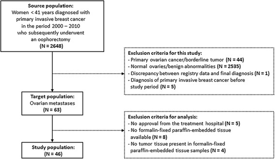
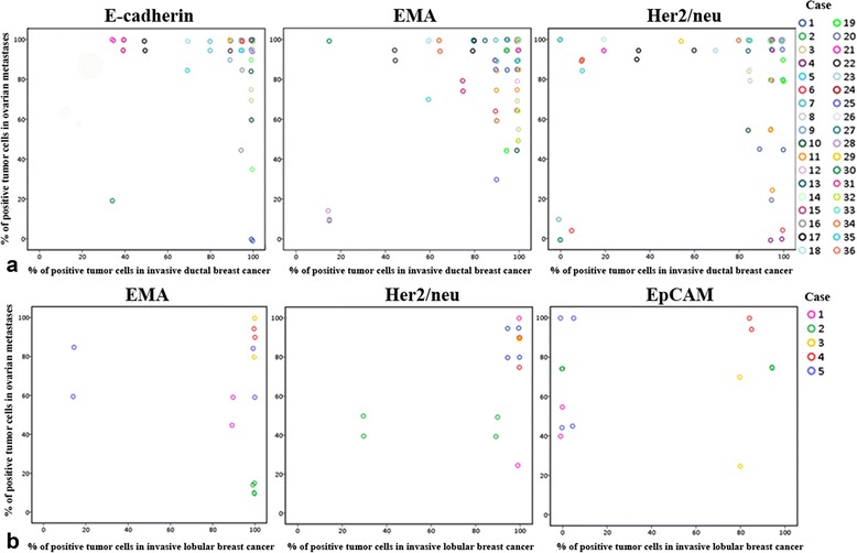
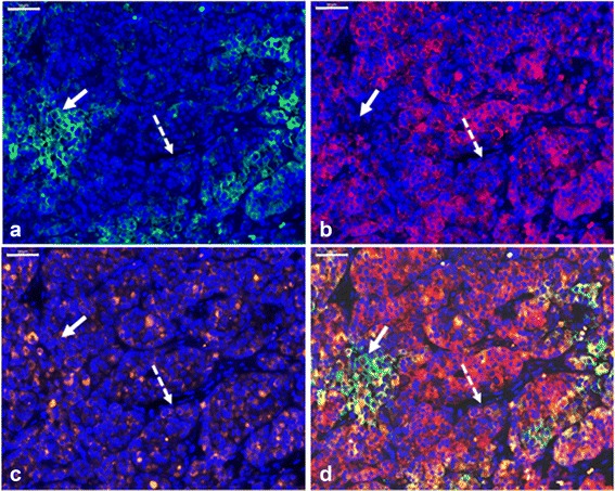
Methods: Using the nationwide database of the Dutch Pathology Registry (PALGA), we identified a cohort of 46 women with primary invasive breast cancer and ovarian metastases. The localization and morphology of ovarian metastases were determined on hematoxylin-and-eosin-stained sections. The following cell-surface markers were immunohistochemically analyzed: E-cadherin, epithelial membrane antigen (EMA), human epidermal growth receptor type 2 (Her2/neu), carcinoembryonic antigen (CEA), αvβ6 integrin and epithelial cell adhesion molecule (EpCAM).
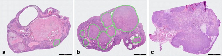
Results: The majority of ovarian metastases (71%) consisted of a solitary metastasis or multiple distinct nodules separated by uninvolved ovarian tissue, suggesting that ovarian metastases might be overlooked by the current detection approach. Combining the targets E-cadherin, EMA and Her2/neu resulted in nearly 100% detection of ductal ovarian metastases, whereas the combination of EMA, Her2/neu and EpCAM was most suitable to detect lobular ovarian metastases.
Conclusions: Examination of the actual ovarian transplants is recommended. A combination of targets is most appropriate to detect ovarian metastases by tumor-specific imaging.
Keywords: breast cancer; fertility preservation; ovarian metastases; ovarian tissue autotransplantation; tumor markers; tumor-specific imaging.
Figures
Similar articles
-
Identification of cell-surface markers for detecting breast cancer cells in ovarian tissue.Arch Gynecol Obstet. 2016 Aug;294(2):385-93. doi: 10.1007/s00404-016-4036-7. Epub 2016 Mar 5. Arch Gynecol Obstet. 2016. PMID: 26946151 Free PMC article.
-
Prevalence and Risk Factors of Ovarian Metastases in Breast Cancer Patients < 41 Years of Age in the Netherlands: A Nationwide Retrospective Cohort Study.PLoS One. 2017 Jan 26;12(1):e0168277. doi: 10.1371/journal.pone.0168277. eCollection 2017. PLoS One. 2017. PMID: 28125710 Free PMC article.
-
New methods to improve the safety assessment of cryopreserved ovarian tissue for fertility preservation in breast cancer patients.Fertil Steril. 2015 Dec;104(6):1493-502.e1-2. doi: 10.1016/j.fertnstert.2015.08.009. Epub 2015 Oct 1. Fertil Steril. 2015. PMID: 26364839
-
[Genital metastases from breast cancer: study of 3 cases and literature review].Pan Afr Med J. 2018 May 4;30:7. doi: 10.11604/pamj.2018.30.7.14839. eCollection 2018. Pan Afr Med J. 2018. PMID: 30123410 Free PMC article. Review. French.
-
Immunohistochemistry applied to the differential diagnosis between ductal and lobular carcinoma of the breast.Appl Immunohistochem Mol Morphol. 2013 Jan;21(1):1-12. doi: 10.1097/PAI.0b013e318255bafa. Appl Immunohistochem Mol Morphol. 2013. PMID: 22595945 Review.
Cited by
-
Long disease-free survival (48 months) in a breast cancer patient with ovarian and pelvic metastasis-a case report.Transl Cancer Res. 2020 Dec;9(12):7669-7675. doi: 10.21037/tcr-20-2031. Transl Cancer Res. 2020. PMID: 35117367 Free PMC article.
-
Ovarian metastasis from breast cancer in three Chinese females: Three case reports.Medicine (Baltimore). 2019 Apr;98(17):e15395. doi: 10.1097/MD.0000000000015395. Medicine (Baltimore). 2019. PMID: 31027137 Free PMC article.
-
Diagnostic Accuracy of Whole-Body Computed Tomography for Incidental Ovarian Tumors in Patients with Prior Breast Cancer.Diagnostics (Basel). 2022 Jan 29;12(2):347. doi: 10.3390/diagnostics12020347. Diagnostics (Basel). 2022. PMID: 35204438 Free PMC article.
-
Ovarian metastasis from breast cancer: a comprehensive review.Clin Transl Oncol. 2019 Jul;21(7):819-827. doi: 10.1007/s12094-018-02007-5. Epub 2018 Dec 17. Clin Transl Oncol. 2019. PMID: 30560347 Review.
-
Heterogeneity of CEACAM5 in breast cancer.Oncotarget. 2020 Oct 27;11(43):3886-3899. doi: 10.18632/oncotarget.27778. eCollection 2020 Oct 27. Oncotarget. 2020. PMID: 33196697 Free PMC article.
References
-
- Bastings L, Beerendonk CCM, Westphal JR, Braat DDM, Peek R. Cryopreservation and Autotransplantation of Ovarian Tissue in Cancer Patients: Is It Safe? J Adolesc Young Adult Oncol. 2013;2(1):31–34. doi: 10.1089/jayao.2012.0017. - DOI
Publication types
MeSH terms
Substances
LinkOut - more resources
Full Text Sources
Other Literature Sources
Medical
Research Materials
Miscellaneous
