. 2017 Mar 21;16(1):67.
doi: 10.1186/s12943-017-0634-7.

TRIM8 restores p53 tumour suppressor function by blunting N-MYC activity in chemo-resistant tumours

Affiliations

TRIM8 restores p53 tumour suppressor function by blunting N-MYC activity in chemo-resistant tumours

Francesca Mastropasqua et al. Mol Cancer. .

Abstract

Background: TRIM8 plays a key role in controlling the p53 molecular switch that sustains the transcriptional activation of cell cycle arrest genes and response to chemotherapeutic drugs. The mechanisms that regulate TRIM8, especially in cancers like clear cell Renal Cell Carcinoma (ccRCC) and colorectal cancer (CRC) where it is low expressed, are still unknown. However, recent studies suggest the potential involvement of some microRNAs belonging to miR-17-92 and its paralogous clusters, which could include TRIM8 in a more complex pathway.

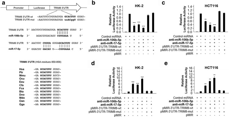
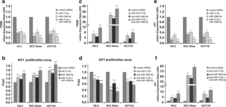
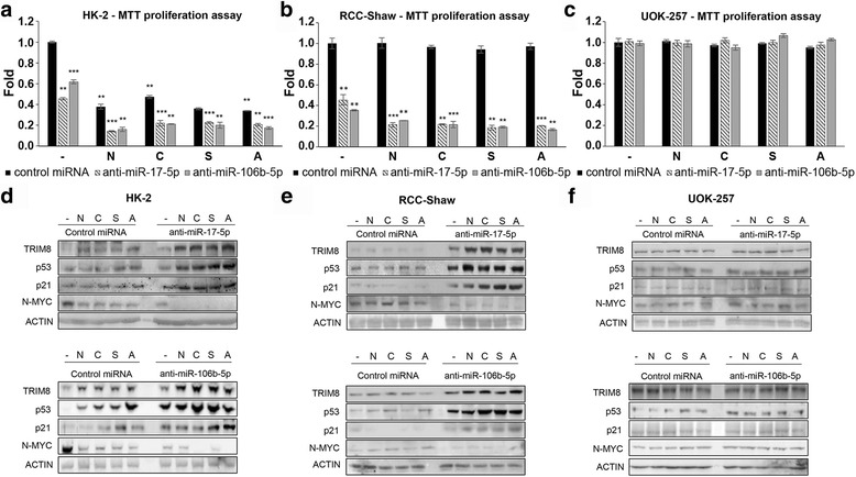
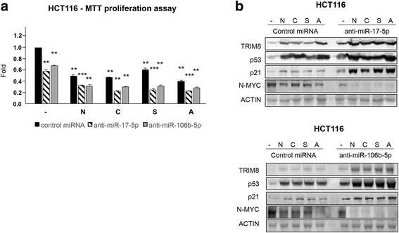
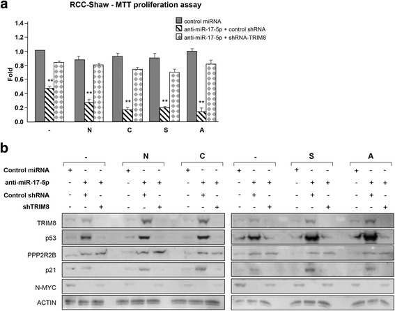
Methods: We used RCC and CRC cell models for in-vitro experiments, and ccRCC patients and xenograft transplanted mice for in vivo assessments. To measure microRNAs levels we performed RT-qPCR, while steady-states of TRIM8, p53, p21 and N-MYC were quantified at protein level by Western Blotting as well as at transcript level by RT-qPCR. Luciferase reporter assays were performed to assess the interaction between TRIM8 and specific miRNAs, and the potential effects of this interaction on TRIM8 expression. Moreover, we treated our cell models with conventional chemotherapeutic drugs or tyrosine kinase inhibitors, and measured their response in terms of cell proliferation by MTT and colony suppression assays.

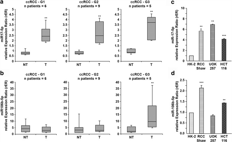
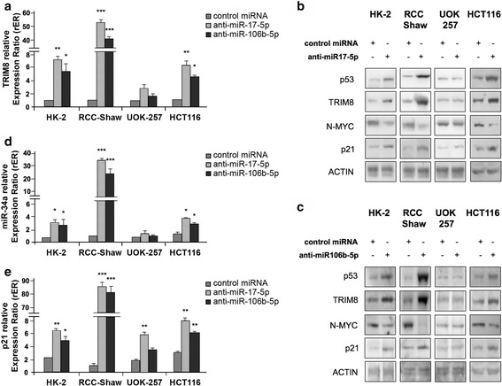
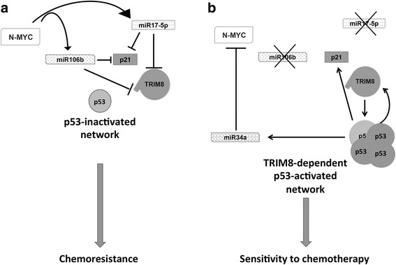
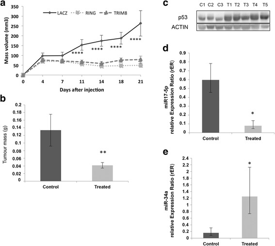
Results: We showed that TRIM8 is a target of miR-17-5p and miR-106b-5p, whose expression is promoted by N-MYC, and that alterations of their levels affect cell proliferation, acting on the TRIM8 transcripts stability, as confirmed in ccRCC patients and cell lines. In addition, reducing the levels of miR-17-5p/miR-106b-5p, we increased the chemo-sensitivity of RCC/CRC-derived cells to anti-tumour drugs used in the clinic. Intriguingly, this occurs, on one hand, by recovering the p53 tumour suppressor activity in a TRIM8-dependent fashion and, on the other hand, by promoting the transcription of miR-34a that turns off the oncogenic action of N-MYC. This ultimately leads to cell proliferation reduction or block, observed also in colon cancer xenografts overexpressing TRIM8.

Conclusions: In this paper we provided evidence that TRIM8 and its regulators miR-17-5p and miR-106b-5 participate to a feedback loop controlling cell proliferation through the reciprocal modulation of p53, miR-34a and N-MYC. Our experiments pointed out that this axis is pivotal in defining drug responsiveness of cancers such ccRCC and CRC.

Keywords: Drug resistance; N-MYC; TRIM8; miR-17 family; p53.


Figures

Fig. 1
Fig. 1
miR-17-5p and miR-106b-5p expression in ccRCC samples. a, b miR-17-5p and miR-106b-5p expression in ccRCC samples and their paired non-tumour tissues. The analysis was performed considering the Fuhrman grading of the tumour samples. Data are represented in box-and-whisker plots showing median and 10th, 25th, 75th and 90th percentiles for each category of sample. Expression data were measured respect to one normal sample chosen arbitrarily as calibrator and then normalized by the expression levels of U6 snRNA. **p-value < 0.005. c, d miR-17-5p and miR-106b-5p expression in the colon cancer HCT116 (p53wt) cell line and in three different renal cell lines: the human proximal tubular epithelial cells HK-2, the human renal cell carcinoma RCC-Shaw (p53wt) and the human renal carcinoma of BHD (Birt-Hogg-Dubè) origin UOK-257 cells (mutated-p53). Expression data were measured respect to HK-2 sample chosen as calibrator, and normalized by the expression levels of U6 snRNA. **p-value < 0.005; *** p-value < 0.001
Fig. 2
Fig. 2
Structure and functional characterization of the putative miR-17-5p/miR-106b-5p target identified in the TRIM8 3’UTR sequence. a Schematic representation of the pMIR luciferase reporter construct containing the TRIM8 3’UTR sequence (wild-type or mutated) cloned downstream the Luciferase gene. Below it is shown the sequence alignment between the miR-17-5p/miR-106b-5p “seed” sequence and the TRIM8 3’UTR, as well as the evolutionary conservation across species. b, c, d, e Luciferase assays. The HK-2 and HCT116 cells were transfected with Negative Control miRNA Mimic, miR-17-5p or miR-106b-5p (alone or together), anti-miR-17-5p or anti-miR-106b-5p (alone or together), along with pMIR luciferase reporter construct containing TRIM8 3’UTR (wt or mut). Cells were lysed and luciferase activity was determined as described in the Material and Methods section. Transfection efficacy was normalized by Renilla Luciferase activity. Data represent the averages of at least three independent experiments with their standard deviations. ** p-value < 0.005; *** p-value < 0.001
Fig. 3
Fig. 3
Effects of overexpression/silencing of miR-17-5p and miR-106b-5p on TRIM8 and p21 expression and cell proliferation. a, c, e, f Expression levels of TRIM8 and p21 were measured by RT-qPCR in HK-2, RCC-Shaw and HCT116 cells transfected with Negative Control miRNA Mimic, miR-17-5p or miR-106b-5p (alone or together) anti-miR-17-5p or anti-miR-106b-5p (alone or together). Relative expression ratios were measured respect to the sample transfected with the Negative Control miRNA Mimic and normalized by the expression levels of RPL13 (* p-value < 0.01; ** p-value < 0.005). b, d Cell proliferation was measured by MTT reduction assay in HK-2, RCC-Shaw and HCT116 transfected with Negative Control miRNA Mimic, miR-17-5p or miR-106b-5p (alone or together), anti-miR-17-5p or anti-miR-106b-5p (alone or together). Measurements were normalized respect to the sample transfected with the Negative Control miRNA Mimic. Data are shown as the average with standard deviation of at least 3 independent experiments (** p-value < 0.005; *** p-value < 0.001)
Fig. 4
Fig. 4
miR-17-5p and miR-106b-5p link p53 to the N-MYC pathway. a, d, e Expression levels of TRIM8, p21 and miR-34a were measured by RT-qPCR in HK-2, RCC-Shaw, UOK-257 and HCT116 transfected with Negative Control miRNA Mimic, anti-miR-17-5p or anti-miR-106b-5p. Relative expression ratios were measured respect to the sample transfected with the Negative Control miRNA Mimic and normalized by the expression levels of RPL13 for TRIM8 and p21, and by the expression level of U6 snRNA for miR-34a (** p-value < 0.005; *** p-value < 0.001). b, c Western blotting analysis of p53, TRIM8, N-MYC and p21 proteins were measured in the indicated cell lines transfected with Negative Control miRNA Mimic, anti-miR-17-5p or anti-miR-106b-5p. Western blot of Actin was conducted as control. Data are shown as the average with standard deviation of at least 3 independent experiments (** p-value < 0.005; *** p-value < 0.001)
Fig. 5
Fig. 5
Schematic representation of the opposite p53-TRIM8 and N-MYC-miR17-5p/miR106b-5p networks in cellular response to treatments. a, b Schematic representation of the molecular pathway that shows how TRIM8 recovery in TRIM8-deficient cells reactivates the p53 tumour suppressor pathway and blunts the N-MYC oncogenic activity
Fig. 6
Fig. 6
Anti-miR-17-5p and anti-miR-106b-5p render chemotherapy treatments effective in ccRCC. a, b, c Cell proliferation by MTT reduction assay in HK-2 (p53 wt), RCC-Shaw (p53 wt), and UOK-257 (mutated p53) transfected with Negative Control miRNA Mimic, anti-miR-17-5p or anti-miR-106b-5p, and treated with Nutlin-3 (N)(10 μM), Cisplatin (C)(7.5 μM), Sorafenib (S)(10 μM), Axitinib (A)(10 μM) or drug-untreated cells(-). For each cell line, drug-untreated sample transfected with control miRNA has been used as calibrator (fold 1.0). Cells transfected with anti-miR-17-5p or anti-miR-106b-5p, or with control miRNA and treated with the different drugs have been normalized with respect to this calibrator. Data are shown as the average with standard deviation of at least 3 independent experiments (** p-value < 0.005; *** p-value < 0.001). d, e, f Western Blotting of the indicated proteins in HK-2 (control), RCC-Shaw and UOK-257 after transfection with Negative Control miRNA Mimic, anti-miR-17-5p, anti-miR-106b-5p without drug (-) or after chemotherapeutic drug treatment with Nutlin-3 (N)(10 μM), Cisplatin (C)(7.5 μM), Sorafenib (S)(10 μM) or Axitinib (A)(10 μM). Western blot of Actin was conducted as control
Fig. 7
Fig. 7
Anti-miR-17-5p and anti-miR-106b-5p render chemotherapy treatments effective in CRC. a Cell proliferation by MTT reduction assay in HCT116 (p53 wt) cells transfected with Negative Control miRNA Mimic, anti-miR-17-5p or anti-miR-106b-5p, and treated with Nutlin-3 (N)(10 μM), Cisplatin (C)(7.5 μM), Sorafenib (S)(10 μM), Axitinib (A)(10 μM) or drug-untreated cells(-). For each cell line, drug-untreated sample transfected with control miRNA has been used as calibrator (fold 1.0). Cells transfected with anti-miR-17-5p or anti-miR-106b-5p, or with control miRNA and treated with the different drugs have been normalized with respect to this calibrator. Data are shown as the average with standard deviation of at least 3 independent experiments (** p-value < 0.005; *** p-value < 0.001). b Western Blotting of the indicated proteins in HCT116 cells after transfection with Negative Control miRNA Mimic, anti-miR-17-5p, anti-miR-106b-5p without drug (-) or after chemotherapeutic drug treatment with Nutlin-3 (N)(10 μM), Cisplatin (C)(7.5 μM), Sorafenib (S)(10 μM) or Axitinib (A)(10 μM). Western blot of Actin was conducted as control
Fig. 8
Fig. 8
TRIM8 is pivotal in controlling chemotherapy cell sensitivity. a Cell proliferation by MTT reduction assay in RCC-Shaw transfected with Negative Control miRNA Mimic or anti-miR-17-5p plus control short hairpin-RNA (control shRNA) or specific short hairpin against TRIM8 (shRNA-TRIM8) (** p-value < 0.005). After transfection the cells were treated with Nutlin-3 (N) (10 μM), Cisplatin (C) (7.5 μM), Sorafenib (S) (10 μM), Axitinib (A) (10 μM) or drug-untreated cells(-). For each cell line, drug-untreated sample transfected with control miRNA has been used as calibrator (fold 1.0). Cells transfected with anti-miR-17-5p plus control shRNA or shRNA-TRIM8, or with control miRNA and treated with the different drugs have been normalized with respect to this calibrator. b Western blotting analysis of the indicated proteins in RCC-Shaw transfected with Negative Control miRNA Mimic or anti-miR-17-5p plus control short hairpin-RNA or specific short hairpin against TRIM8 (shRNA-TRIM8). After transfection the cells were treated with Nutlin-3 (N) (10 μM), Cisplatin (C) (7.5 μM), Sorafenib (S) (10 μM), Axitinib (A) (10 μM) or drug-untreated cells (-). Western blot of Actin was conducted as control
Fig. 9
Fig. 9
Treatment of xenograft tumours in vivo with recombinant adenovirus expressing HA-TRIM8, HA-RING or LacZ (Control). a Growth curves of xenograft tumours in nude mice treated for 3 weeks. The volume of the tumours was measured two times weekly (**** p- value <0.0005). b Tumour masses weight at the moment of the tumour excision in treated (HA-TRIM8, HA-RING) vs. control (LacZ) samples (** p-value < 0.005). c, d, e The stabilization of p53, miR-34a and down-regulation of miR-17-5p in xenografts treated with HA-TRIM8, HA-RING (T1-T5) or LacZ (C1-C3) was demonstrated by western blotting analysis of p53 (Actin was used as loading control) and by RT-qPCR on miR-17-5p (* p-value < 0.05) and miR-34a expression (* p-value < 0.05). The bars represent the Standard Error of the Mean

References

    1. Sato Y, Yoshizato T, Shiraishi Y, Maekawa S, Okuno Y, Kamura T, Shimamura T, Sato-Otsubo A, Nagae G, Suzuki H, et al. Integrated molecular analysis of clear-cell renal cell carcinoma. Nat Genet. 2013;45:860–867. doi: 10.1038/ng.2699. - DOI - PubMed
    1. Caratozzolo MF, Micale L, Turturo MG, Cornacchia S, Fusco C, Marzano F, Augello B, D’Erchia AM, Guerrini L, Pesole G, et al. TRIM8 modulates p53 activity to dictate cell cycle arrest. Cell Cycle. 2012;11:511–523. doi: 10.4161/cc.11.3.19008. - DOI - PubMed
    1. Cambiaghi V, Giuliani V, Lombardi S, Marinelli C, Toffalorio F, Pelicci PG. TRIM proteins in cancer. Adv Exp Med Biol. 2012;770:77–91. doi: 10.1007/978-1-4614-5398-7_6. - DOI - PubMed
    1. Napolitano LM, Meroni G. TRIM family: pleiotropy and diversification through homomultimer and heteromultimer formation. IUBMB Life. 2012;64:64–71. doi: 10.1002/iub.580. - DOI - PubMed
    1. Li Q, Yan J, Mao AP, Li C, Ran Y, Shu HB, Wang YY. Tripartite motif 8 (TRIM8) modulates TNFalpha- and IL-1beta-triggered NF-kappaB activation by targeting TAK1 for K63-linked polyubiquitination. Proc Natl Acad Sci U S A. 2011;108:19341–19346. doi: 10.1073/pnas.1110946108. - DOI - PMC - PubMed

Publication types

MeSH terms