Review
. 2017 Mar 21;15(1):7.
doi: 10.1186/s12947-017-0099-2.

The clinical use of stress echocardiography in ischemic heart disease

Affiliations
Review

The clinical use of stress echocardiography in ischemic heart disease

Rosa Sicari et al. Cardiovasc Ultrasound. .

Abstract

Stress echocardiography is an established technique for the assessment of extent and severity of coronary artery disease. The combination of echocardiography with a physical, pharmacological or electrical stress allows to detect myocardial ischemia with an excellent accuracy. A transient worsening of regional function during stress is the hallmark of inducible ischemia. Stress echocardiography provides similar diagnostic and prognostic accuracy as radionuclide stress perfusion imaging or magnetic resonance, but at a substantially lower cost, without environmental impact, and with no biohazards for the patient and the physician.The evidence on its clinical impact has been collected over 35 years, based on solid experimental, pathophysiological, technological and clinical foundations. There is the need to implement the combination of wall motion and coronary flow reserve, assessed in the left anterior descending artery, into a single test. The improvement of technology and in imaging quality will make this approach more and more feasible. The future issues in stress echo will be the possibility of obtaining quantitative information translating the current qualitative assessment of regional wall motion into a number. The next challenge for stress echocardiography is to overcome its main weaknesses: dependance on operator expertise, the lack of outcome data (a widesperad problem in clinical imaging) to document the improvement of patient outcomes. This paper summarizes the main indications for the clinical applications of stress echocardiography to ischemic heart disease.


Figures
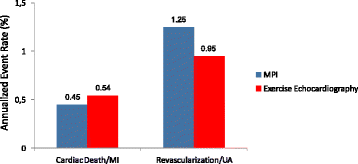
Fig. 1
Fig. 1
Annual cardiac events associated to a normal stress echocardiogram and a normal stress myocardial perfusion scan. From [43]
Fig. 2
Fig. 2
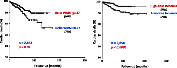
Kaplan-Meier survival curves (considering cardiac death as end point) in patients with pharmacologic stress echocardiography positive for ischemia stratified on the basis of the extent of ischemia, as expressed by delta wall motion score index (WMSI) set at 0.37 (left panel), and the dose to induce ischemia (left panel). From [47]
Fig. 3
Fig. 3
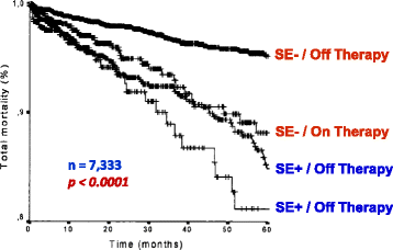
Kaplan-Meier survival curves (considering total mortality as end point) in patients stratified according to presence (DET +) or absence (DET -) of myocardial ischemia at pharmacological stress echocardiography on and off antianginal medical therapy. From [59]
Fig. 4
Fig. 4
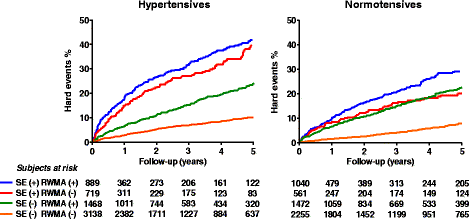
Hard event rates for hypertensive and normotensive patients separated on the basis of presence (+) or absence (−) of ischemia at stress echocardiography, and presence (+) or absence (−) of resting wall motion abnormality (RWMA). From [72]
Fig. 5
Fig. 5
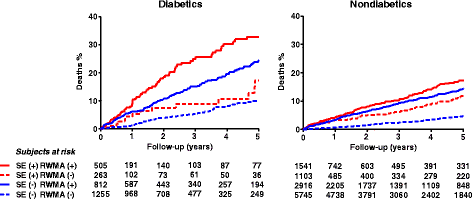
Mortality for diabetic and nondiabetic patients separated on the basis of presence (+) or absence (−) of ischemia at stress echocardiography, and presence (+) or absence (−) of resting wall motion abnormality (RWMA). From [85]
Fig. 6
Fig. 6
Incremental prognostic value of pharmacological stress echocardiography to clinical and exercise electrocardiography data, as determined by the comparison of the global chi-square at each step. From [93]
Fig. 7
Fig. 7
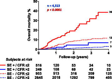
Mortality rates in patients with known or suspected coronary artery disease separated on the basis of presence (+) or absence (−) of ischemia at stress echocardiography (SE) and coronary flow reserve (CFR) of left anterior descending artery >2 or ≤2. From [145]
Fig. 8
Fig. 8
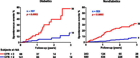
Hard event rates in diabetic and nondiabetic patients with stress echo negative for ischemia separated on the basis of coronary flow reserve (CFR) of left anterior descending artery >2 or ≤2. From [146]
Fig. 9
Fig. 9
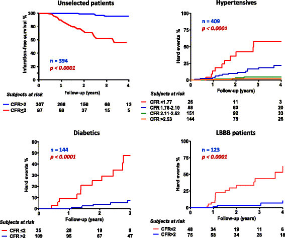
Hard event rates in unselected patients (left upper panel), hypertensives patients (right upper panel), diabetic patients (left lower panel), and patients with left bundle branch block (LBBB) (right lower panel) with normal or near normal coronary arteries separated on the basis of coronary flow reserve (CFR) values. From [, –151]
Fig. 10
Fig. 10
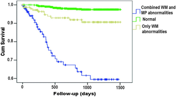
Kaplan-Meier curves based on the combination of presence or absence of wall motion (WM) abnormalities and myocardial perfusion (MP) abnormalities. From [152]
Fig. 11
Fig. 11
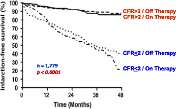
Kaplan-Meier survival curves (considering hard events as end point) in patients stratified on the basis of coronary flow reserve (CFR) of left anterior descending artery >2 or ≤2 on and off antianginal medical therapy. From [155]

References

    1. Picano E. Stress echocardiography. 6. Heidelberg: Springer Verlag; 2015.
    1. Picano E. Dipyridamole-echocardiography test: historical background and physiologic basis. Eur Heart J. 1989;10:365–376. doi: 10.1093/oxfordjournals.eurheartj.a059494. - DOI - PubMed
    1. Ross J., Jr Mechanisms of regional ischaemia and antianginal drug action during exercise. Prog Cardiovasc Dis. 1989;31:455–466. doi: 10.1016/0033-0620(89)90020-0. - DOI - PubMed
    1. Gallagher KP, Matsuzaki M, Koziol JA, Kemper WS, Ross J., Jr Regional myocardial perfusion and wall thickening during ischaemia in conscious dogs. Am J Physiol. 1984;247:H727–738. - PubMed
    1. Picano E, Lattanzi F, Masini M, Distante A, L’Abbate A. Usefulness of a high-dose dipyridamole-echocardiography test for diagnosis of syndrome X. Am J Cardiol. 1987;60:508–512. doi: 10.1016/0002-9149(87)90295-5. - DOI - PubMed

MeSH terms

LinkOut - more resources