

Integrated local binary pattern texture features for classification of breast tissue imaged by optical coherence microscopy
- PMID: 28327449
- PMCID: PMC5479412
- DOI: 10.1016/j.media.2017.03.002
Integrated local binary pattern texture features for classification of breast tissue imaged by optical coherence microscopy
Abstract
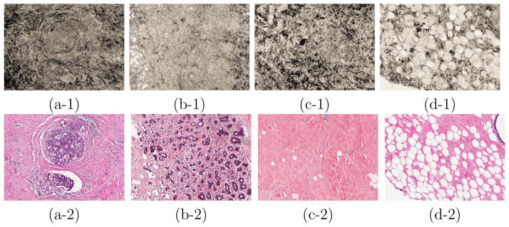
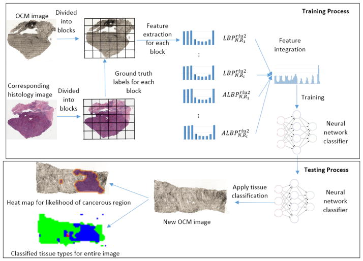
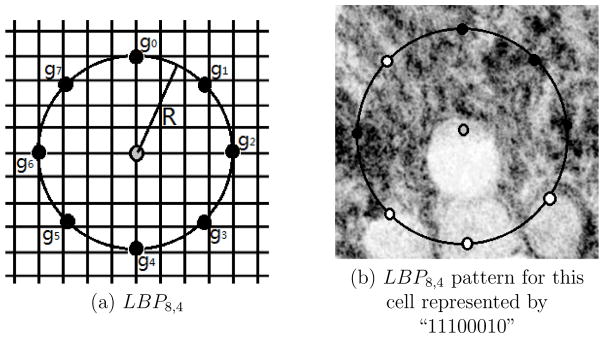
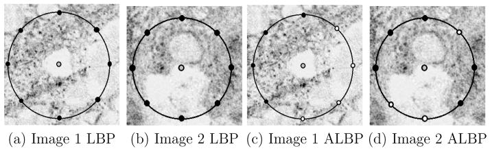
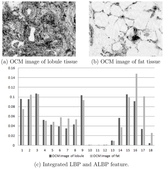
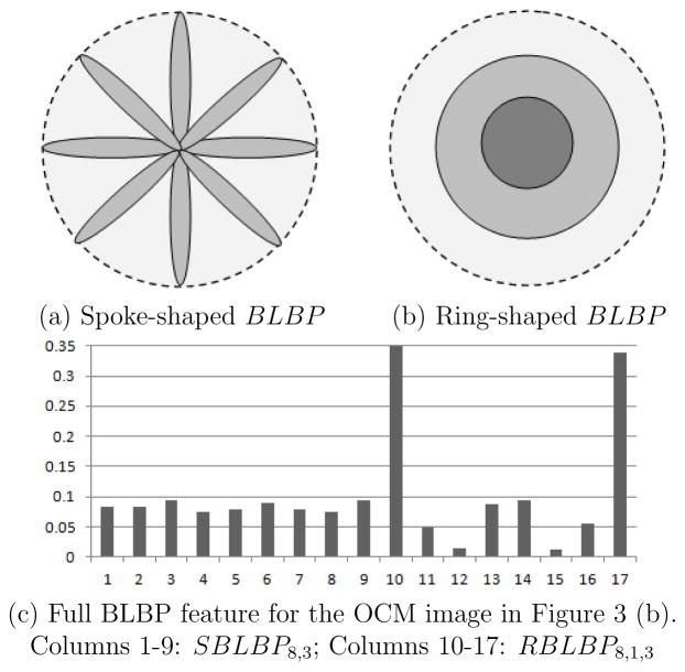
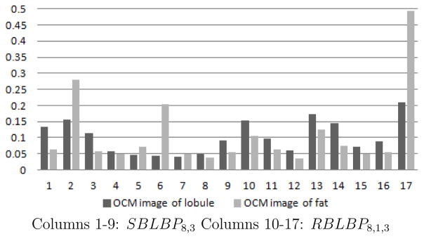
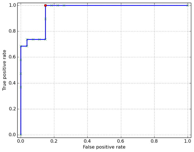
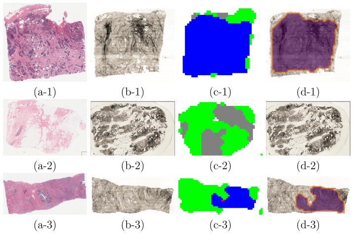
This paper proposes a texture analysis technique that can effectively classify different types of human breast tissue imaged by Optical Coherence Microscopy (OCM). OCM is an emerging imaging modality for rapid tissue screening and has the potential to provide high resolution microscopic images that approach those of histology. OCM images, acquired without tissue staining, however, pose unique challenges to image analysis and pattern classification. We examined multiple types of texture features and found Local Binary Pattern (LBP) features to perform better in classifying tissues imaged by OCM. In order to improve classification accuracy, we propose novel variants of LBP features, namely average LBP (ALBP) and block based LBP (BLBP). Compared with the classic LBP feature, ALBP and BLBP features provide an enhanced encoding of the texture structure in a local neighborhood by looking at intensity differences among neighboring pixels and among certain blocks of pixels in the neighborhood. Fourty-six freshly excised human breast tissue samples, including 27 benign (e.g. fibroadenoma, fibrocystic disease and usual ductal hyperplasia) and 19 breast carcinoma (e.g. invasive ductal carcinoma, ductal carcinoma in situ and lobular carcinoma in situ) were imaged with large field OCM with an imaging area of 10 × 10 mm2 (10, 000 × 10, 000 pixels) for each sample. Corresponding H&E histology was obtained for each sample and used to provide ground truth diagnosis. 4310 small OCM image blocks (500 × 500 pixels) each paired with corresponding H&E histology was extracted from large-field OCM images and labeled with one of the five different classes: adipose tissue (n = 347), fibrous stroma (n = 2,065), breast lobules (n = 199), carcinomas (pooled from all sub-types, n = 1,127), and background (regions outside of the specimens, n = 572). Our experiments show that by integrating a selected set of LBP and the two new variant (ALBP and BLBP) features at multiple scales, the classification accuracy increased from 81.7% (using LBP features alone) to 93.8% using a neural network classifier. The integrated feature was also used to classify large-field OCM images for tumor detection. A receiver operating characteristic (ROC) curve was obtained with an area under the curve value of 0.959. A sensitivity level of 100% and specificity level of 85.2% was achieved to differentiate benign from malignant samples. Several other experiments also demonstrate the complementary nature of LBP and the two variants (ALBP and BLBP features) and the significance of integrating these texture features for classification. Using features from multiple scales and performing feature selection are also effective mechanisms to improve accuracy while maintaining computational efficiency.
Keywords: Local binary patterns; Optical coherence microscopy; Texture features; Tissue classification.
Copyright © 2017 Elsevier B.V. All rights reserved.
Figures
Similar articles
-
Visualization and tissue classification of human breast cancer images using ultrahigh-resolution OCT.Lasers Surg Med. 2017 Mar;49(3):258-269. doi: 10.1002/lsm.22654. Epub 2017 Mar 6. Lasers Surg Med. 2017. PMID: 28264146 Free PMC article.
-
Integrated optical coherence tomography and microscopy for ex vivo multiscale evaluation of human breast tissues.Cancer Res. 2010 Dec 15;70(24):10071-9. doi: 10.1158/0008-5472.CAN-10-2968. Epub 2010 Nov 5. Cancer Res. 2010. PMID: 21056988 Free PMC article.
-
Differentiation of fat-poor angiomyolipoma from clear cell renal cell carcinoma in contrast-enhanced MDCT images using quantitative feature classification.Med Phys. 2017 Jul;44(7):3604-3614. doi: 10.1002/mp.12258. Epub 2017 Jun 9. Med Phys. 2017. PMID: 28376281
-
Role of texture analysis in breast MRI as a cancer biomarker: A review.J Magn Reson Imaging. 2019 Apr;49(4):927-938. doi: 10.1002/jmri.26556. Epub 2018 Nov 3. J Magn Reson Imaging. 2019. PMID: 30390383 Free PMC article. Review.
-
An overview of optical coherence tomography for ovarian tissue imaging and characterization.Wiley Interdiscip Rev Nanomed Nanobiotechnol. 2015 Jan-Feb;7(1):1-16. doi: 10.1002/wnan.1306. Epub 2014 Oct 20. Wiley Interdiscip Rev Nanomed Nanobiotechnol. 2015. PMID: 25329515 Free PMC article. Review.
Cited by
-
Features extraction using encoded local binary pattern for detection and grading diabetic retinopathy.Health Inf Sci Syst. 2022 Jun 29;10(1):14. doi: 10.1007/s13755-022-00181-z. eCollection 2022 Dec. Health Inf Sci Syst. 2022. PMID: 35782197 Free PMC article.
-
Deep learning radiomics based prediction of axillary lymph node metastasis in breast cancer.NPJ Breast Cancer. 2024 Mar 12;10(1):22. doi: 10.1038/s41523-024-00628-4. NPJ Breast Cancer. 2024. PMID: 38472210 Free PMC article.
-
Involvement of Machine Learning for Breast Cancer Image Classification: A Survey.Comput Math Methods Med. 2017;2017:3781951. doi: 10.1155/2017/3781951. Epub 2017 Dec 31. Comput Math Methods Med. 2017. PMID: 29463985 Free PMC article. Review.
-
Automated assessment of breast margins in deep ultraviolet fluorescence images using texture analysis.Biomed Opt Express. 2022 Aug 30;13(9):5015-5034. doi: 10.1364/BOE.464547. eCollection 2022 Sep 1. Biomed Opt Express. 2022. PMID: 36187258 Free PMC article.
-
Optical coherence tomography and computer-aided diagnosis of a murine model of chronic kidney disease.J Biomed Opt. 2017 Dec;22(12):1-11. doi: 10.1117/1.JBO.22.12.121706. J Biomed Opt. 2017. PMID: 29197178 Free PMC article.
References
-
- Ahonen Timo, Hadid Abdenour, Pietikäinen Matti. Computer Vision-ECCV 2004. Springer; 2004. Face recognition with local binary patterns; pp. 469–481.
-
- Ahonen Timo, Hadid Abdenour, Pietikainen Matti. Face description with local binary patterns: Application to face recognition. Pattern Analysis and Machine Intelligence, IEEE Transactions on. 2006;28(12):2037–2041. - PubMed
-
- Ahsen Osman O, Tao Yuankai K, Potsaid Benjamin M, Sheikine Yuri, Jiang James, Grulkowski Ireneusz, Tsai Tsung-Han, Jayaraman Vijaysekhar, Kraus Martin F, Connolly James L, et al. Swept source optical coherence microscopy using a 1310 nm vcsel light source. Optics express. 2013;21(15):18021–18033. - PMC - PubMed
MeSH terms
Grants and funding
LinkOut - more resources
Full Text Sources
Other Literature Sources
Medical
Research Materials
Miscellaneous
