. 2017 Mar 22:7:45029.
doi: 10.1038/srep45029.

Hypoxia induces H19 expression through direct and indirect Hif-1α activity, promoting oncogenic effects in glioblastoma

Affiliations

Hypoxia induces H19 expression through direct and indirect Hif-1α activity, promoting oncogenic effects in glioblastoma

Weining Wu et al. Sci Rep. .

Abstract

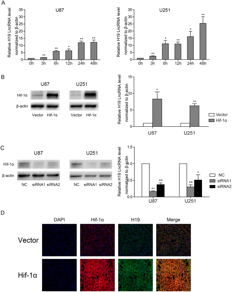
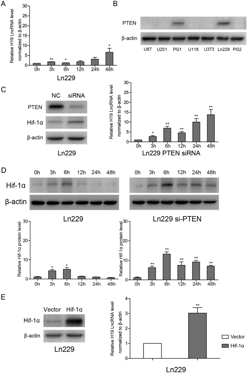
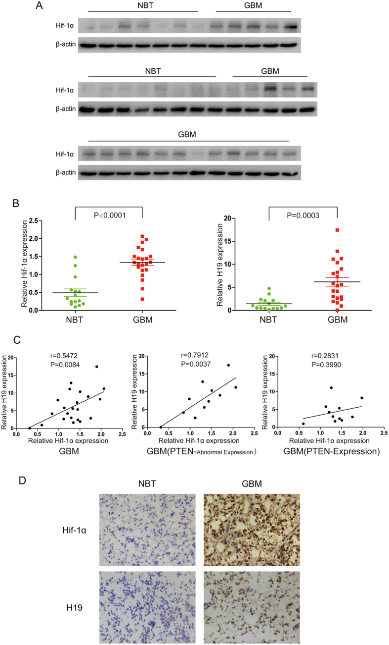
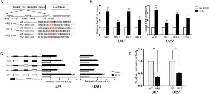
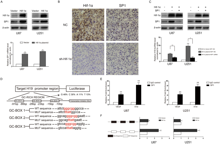
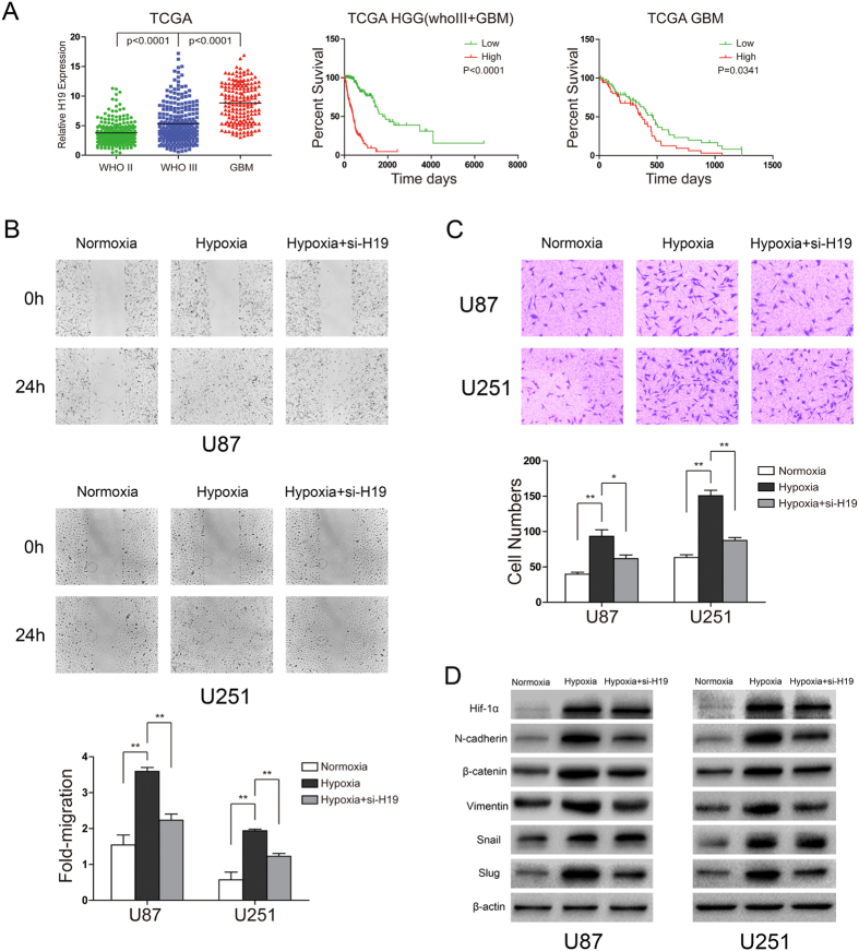
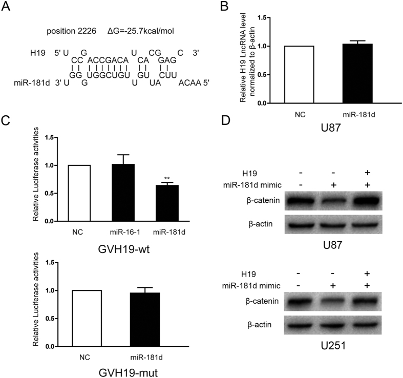
H19 expression is elevated in many human tumors including glioblastomas, suggesting an oncogenic role for the long noncoding RNA; yet the upregulation of H19 in glioblastomas remains unclear. Here we report that hypoxia significantly stimulated H19 expression in glioblastoma cell lines, which was related to hypoxia-inducible factors 1α (Hif-1α). Hif-1α promoted H19 expression in U87 and U251 cells. Meanwhile PTEN is an advantageous factor to affect H19 expression, through attenuating Hif-1α stability. Hif-1α also positively correlates with H19 in human glioblastoma samples depending on PTEN status. ChIP and luciferase reporter assays showed that Hif-1α induced H19 transcription through directly binding to the H19 promoter. Furthermore, Hif-1α upregulated specific protein 1 (SP1) expression in glioblastomas cells in vitro and in vivo, and SP1 also strongly interacted with the H19 promoter to promote H19 expression under hypoxia. We also showed that H19 acts as a molecular sponge that binds miR-181d, relieving inhibition of β-catenin expression. Therefore, H19 participates in hypoxia-driven migration and invasion in glioblastoma cells. In summary, our results uncover the mechanisms that stimulate H19 expression under hypoxia to promote malignant effects in glioblastomas and suggest H19 might be a promising therapeutic target.


Conflict of interest statement

The authors declare no competing financial interests.

Figures

Figure 1
Figure 1. H19 expression is elevated under hypoxia and Hif-1α is a critical factor responsible for the induction of H19 RNA in U87 and U251 cells.
The levels of H19 RNA normalized to β-actin were quantified by qPCR. Protein levels were detected by western blot and normalized to β-actin levels. (A) U87 and U251 cells were exposed to hypoxia (2% O2) for the indicated times. H19 levels at each time point (bar graphs) were normalized to normoxic controls. (B) U87 and U251 cells were transfected with Hif-1α expression plasmid or empty vector before culture under hypoxia for 24 h. (C) U87 and U251 cells were transfected with a control siRNA, or Hif-1α siRNA and cultured under hypoxia for 24 h. All experiments were repeated three times with similar results. (*p < 0.05, **p < 0.01). (D) Immunofluorescent double staining was performed on xenograft tumors after U87 cells stably expressing Hif-1α or control vector be subcutaneously injected. The positive expression of Hif-1α and H19 were overlapped in the same section of U87 xenograft tumors.
Figure 2
Figure 2. H19 induction is dependent on PTEN status at the early stages of hypoxia in Ln229.
PTEN interferes with H19 induction by promoting Hif-1α degradation under hypoxia. (A) Ln229 cells were exposed to hypoxia (2% O2) for the indicated time periods. (B) PTEN protein levels analyzed by western blot in U87, U251, Ln229, U373, U118, GP1 and GP2 cells. (C) PTEN siRNA and control siRNA treatment of Ln229 cell before hypoxic cultivation. Western blots show that PTEN inhibited Hif-1α expression in Ln229 cells under hypoxia at 6 h. (D) Hypoxia induced different effects on Hif-1α protein levels between control siRNA-treated Ln229 cells and PTEN siRNA-treated Ln229 cells. (E) Ln229 cells transfected with an exogenous Hif-1α expression plasmid or an empty vector. All experiments were repeated three times with similar results. (*p < 0.05, **p < 0.01).
Figure 3
Figure 3. Expression levels of Hif-1α and H19 in human glioblastoma (GBM) and adjacent normal brain tissues (NBT).
(A) The levels of Hif-1α in GBM and NBT were detected by western blot. The expression of Hif-1α was normalized to β-actin. (B) The levels of H19 in GBM and NBT were detected by QPCR. The expression of H19 was normalized to β-actin. (C) The correlation between Hif-1α and H19 in GBM specimens or in the subgroup on the basis of PTEN status. Pearson’s correlation coefficient was applied. (D) Immunohistochemistry analysis of Hif-1α and H19 in two representative tissues (one NBT specimen and one GBM specimen).
Figure 4
Figure 4. Hypoxia response elements (HREs) in the H19 promoter are not the only mechanism to mediate Hif-1α-induced H19 expression.
(A) Sequences and locations of the three HREs within 2 kb before the transcription start site in the H19 promoter. The wild-type HRE consensus sequence of designed luciferase report construct was 5′-ACGTG-3′, and the mutant HRE sequence was 5′-AATAT-3′. (B) Chromatin immunoprecipitation assays were performed to indicate binding of Hif-1α to the HREs located in the H19 and VEGF promoters. Mouse anti-Hif-1α antibody or control mouse IgG was used for immunoprecipitation with DNA isolated from U87 and U251 cells. The immunoprecipitate was amplified by qPCR using primers targeting the HREs. The results were normalized to the negative control IgG. The HRE in the VEGF promoter was used as positive control. (C) Five luciferase reporter plasmids were designed containing different combinations of wild type and mutated HREs (schematized on the left). Relative luciferase activity was observed for each reporter construct following transfection of empty vector or Hif-1α (horizontal bars, right) in U87 and U251 cells with 24 h hypoxia treatment. Renilla activity was used as an internal loading control. (D) The relative luciferase activity for each reporter construct following transfection with the same Hif-1α plasmid normalized to the WT group. (*p < 0.05, **p < 0.01).
Figure 5
Figure 5. Hif-1α indirectly stimulates H19 expression through SP1 regulation and SP1 regulates H19 by directly binding to the SP1 binding sites in the H19 promoter region in U87 and U251 cells.
(A) U87 and U251 cells transfected with the Hif-1α expression plasmid or empty vector and cultured under hypoxia for 24 h. (B) IHC analysis of consecutive sections of Hif-1α and SP1 expression in subcutaneous xenografts originated from U87 cells after Hif-1α sh-RNA treatment. (C) U87 and U251 cells co-transfected with the Hif-1α expression plasmid and SP1 siRNA and cultured under hypoxia for 24 h. (D) The locations of the putative SP1 binding sites within 100 bp upstream of the transcription start site in the H19 promoter. These binding sites constituted a GC-rich region. The wild-type sequence of reporter construct was 5′-GGGCGG-3′, and the mutant one was 5′-TTTATT-3′. (*p < 0.05, **p < 0.01). (E) ChIP analysis confirmed SP1 directly bound to the H19 promoter through the three GC-boxes. GC-boxes from the VEGF promoter were used as positive controls. (F) Two reporter plasmids harboring wild type or mutant GC-boxes were co-transfected with the SP1 expression plasmid or empty vector and the Renilla plasmid into U87 and U251 cells. Relative luciferase activity was analyzed after 24 h treatment. (*p < 0.05, **p < 0.01).
Figure 6
Figure 6. H19 plays an important role in hypoxia-driven migration and invasion in U87 and U251 cells.
H19 regulates EMT-related protein expression, affecting migration and invasion in U87 and U251 cells. U87 and U251 cells were transfected with control siRNA or H19 siRNA. The samples are derive from the same experiment and that blots were processed in parallel. (A) Increased H19 expression correlates with glioma grade and confers a poor prognosis in high grade glioma (HGG) and GBM patients. Levels of H19 were analyzed in different glioma tissues of TCGA data. Kaplan–Meier survival curves for H19 expression in HGG and GBM patients of TCGA data. (B) The scratch-wound gap was photographed before and after 24 h incubation under normoxia and hypoxia. After performing the wound healing test. The results were analyzed by measuring the range of migrating cells from 3 different fields for each wound. (C) Transwell migration assays were used to evaluate glioblastoma cell invasion. Migrated cells with normoxic or hypoxic treatment for 24 h were stained with crystal violet and counted for statistical analysis. (D) EMT-related proteins in the three cell groups were examined by western blot and normalized to β-actin. (*p < 0.05, **p < 0.01). The samples are derived from the same experiment and that blots were processed in parallel.
Figure 7
Figure 7. H19 affects β-catenin expression by regulating miR-181d activity.
(A) Bioinformatics predicted a miR-181d binding site in human H19. (B) Ectopic miR-181d expression by miRNA mimics did not affect the H19 levels in U87 cells. (C) U87 cells were co-transfected with miR-181d mimics or miR-16-1 mimics and luciferase reporters harboring the miR-181d binding site sequence or a mutated one from H19. Luciferase activities were normalized to the activities in cells expressing scrambled miRNA mimics. (D) U87 and U251 cells were co-transfected with miR-181d mimics and H19 expression plasmids, β-catenin protein levels were examined by western blot. (*p < 0.05, **p < 0.01). The samples are derived from the same experiment and that blots were processed in parallel.

Similar articles

Cited by

References

    1. Jensen R. L. Hypoxia in the tumorigenesis of gliomas and as a potential target for therapeutic measures. Neurosurg Focus 20, E24 (2006). - PubMed
    1. Evans S. M. et al.. Hypoxia is important in the biology and aggression of human glial brain tumors. Clin Cancer Res 10, 8177–84 (2004). - PubMed
    1. Li S., Meng W., Guan Z., Guo Y. & Han X. The hypoxia-related signaling pathways of vasculogenic mimicry in tumor treatment. Biomed Pharmacother 80, 127–35 (2016). - PubMed
    1. Thambi T., Park J. H. & Lee D. S. Hypoxia-responsive nanocarriers for cancer imaging and therapy: recent approaches and future perspectives. Chem Commun (Camb) 52, 8492–500 (2016). - PubMed
    1. Wan J., Wu W. & Zhang R. Local recurrence of small cell lung cancer following radiofrequency ablation is induced by HIF-1alpha expression in the transition zone. Oncol Rep 35, 1297–308 (2016). - PMC - PubMed

Publication types

MeSH terms