. 2017 Nov;72(11):1666-1676.
doi: 10.1111/all.13166. Epub 2017 Apr 20.

BTK inhibition is a potent approach to block IgE-mediated histamine release in human basophils

Affiliations

BTK inhibition is a potent approach to block IgE-mediated histamine release in human basophils

D Smiljkovic et al. Allergy. 2017 Nov.

Abstract

Background: Recent data suggest that Bruton's tyrosine kinase (BTK) is an emerging therapeutic target in IgE receptor (IgER)-cross-linked basophils.

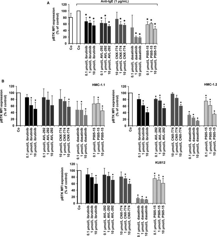
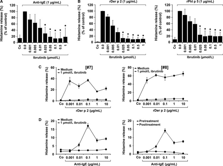
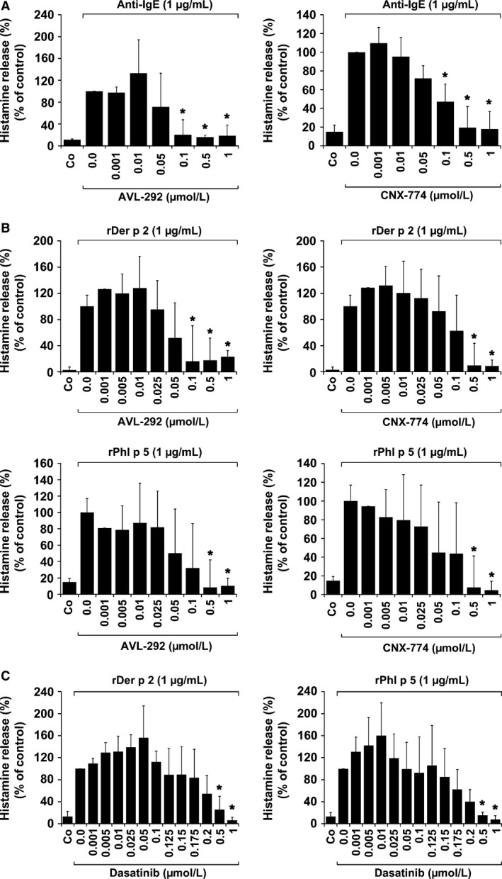
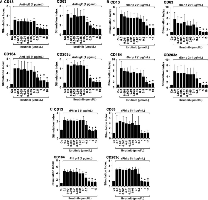
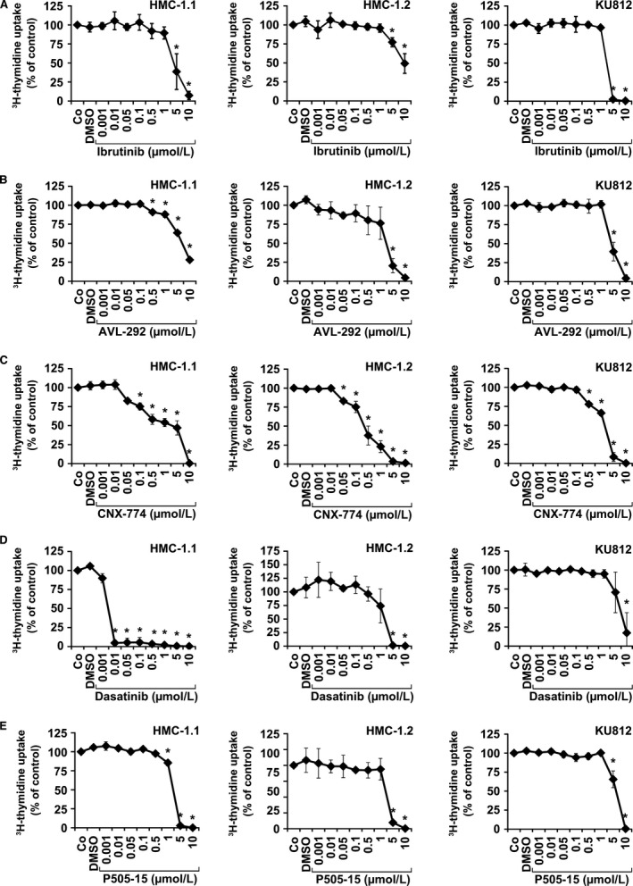
Methods: We examined the effects of four BTK inhibitors (ibrutinib, dasatinib, AVL-292, and CNX-774) on IgE-dependent activation and histamine release in blood basophils obtained from allergic patients (n=11) and nonallergic donors (n=5). In addition, we examined the effects of these drugs on the growth of the human basophil cell line KU812 and the human mast cell line HMC-1.

Results: All four BTK blockers were found to inhibit anti-IgE-induced histamine release from basophils in nonallergic subjects and allergen-induced histamine liberation from basophils in allergic donors. Drug effects on allergen-induced histamine release were dose dependent, with IC50 values ranging between 0.001 and 0.5 μmol/L, and the following rank order of potency: ibrutinib>AVL-292>dasatinib>CNX-774. The basophil-targeting effect of ibrutinib was confirmed by demonstrating that IgE-dependent histamine release in ex vivo blood basophils is largely suppressed in a leukemia patient treated with ibrutinib. Dasatinib and ibrutinib were also found to counteract anti-IgE-induced and allergen-induced upregulation of CD13, CD63, CD164, and CD203c on basophils, whereas AVL-292 and CNX-774 showed no significant effects. Whereas dasatinib and CNX-774 were found to inhibit the growth of HMC-1 cells and KU812 cells, no substantial effects were seen with ibrutinib or AVL-292.

Conclusions: BTK-targeting drugs are potent inhibitors of IgE-dependent histamine release in human basophils. The clinical value of BTK inhibition in the context of allergic diseases remains to be determined.

Keywords: IgE receptor; allergy; signaling molecules; targeted drugs.


Figures

Figure 1
Figure 1
Effects of ibrutinib on expression of pBTK in primary activated human basophils (BA), HMC‐1 cells, and KU812 cells. (A) BA‐containing mononuclear cells (MNC) were incubated in control medium (Co) or in medium containing dasatinib, ibrutinib, AVL‐292, CNX‐774, or P505‐15 (each 0.1‐10 μmol/L) at 37°C for 15 minutes. Then, cells were incubated with anti‐IgE at 37°C for 15 minutes and incubated with a PE‐labeled mAb against CD203c for 15 minutes. Then, cells were permeabilized and stained with an antibody against phosphorylated (p) BTK (pBTK) (phosphorylation site: Y223) as described in the text. Expression of intracellular targets was quantified by multicolor flow cytometry on a FACSCalibur. BA were identified as CD203c‐positive cells. Results show the mean fluorescence intensity (MFI) of pBTK expression (% of control) and represent mean±SD from three independent experiments. Asterisk (*) P<0.05 by Student's t test with Bonferroni correction. (B) HMC‐1.1 cells (upper left panel), HMC‐1.2 cells (upper right panel), and KU812 cells (lower panel) were incubated with control medium (Co) or medium containing ibrutinib, AVL‐292, CNX‐774, dasatinib, or P505‐15 (0.1‐10 μmol/L) at 37°C for 4 hours. Thereafter, cells were permeabilized and stained with an antibody against pBTK (Y223). Expression of phosphorylated (p) signaling molecules in HMC‐1 and KU812 cells was determined by flow cytometry. Results show MFI values expressed as percentage of control and represent the mean±SD from three independent experiments. Asterisk (*): P<0.05 by Student's t test with Bonferroni correction
Figure 2
Figure 2
Effects of ibrutinib on IgE‐mediated histamine release in human basophils. Basophils (BA) obtained from three nonallergic donors (A) or patients allergic to Der p 2 (n=3) or Phl p 5 (n=3) (B) were preincubated in control medium (Co) or various concentrations of ibrutinib (0.001‐1 μmol/L) at 37°C for 30 minutes. Then, cells were exposed to anti‐IgE (1 μg/mL; nonallergic donors) or recombinant allergens (1 μg/mL of rDer p 2 or rPhl p 5; allergic patients) at 37°C for 30 minutes. After centrifugation, histamine concentrations were determined in supernatants and cell lysates. Histamine release is expressed as percentage of total histamine. Results show the percentage of control and represent mean±SD of three independent experiments. Asterisk (*): P<0.05 by Student's t test. (C) BA from two patients allergic to Der p 2 (left and right panel) were incubated in control medium (Co) or 1 μmol/L ibrutinib at 37°C for 30 minutes. Then, cells were incubated in histamine release buffer (HRB) in the absence or presence of rDer p 2 (0.001‐10 μg/mL) at 37°C for 30 minutes. After incubation, cells were centrifuged at 4°C and cell‐free supernatants and cell suspensions analyzed for histamine content. Histamine release is expressed as percentage of total histamine. Results represent the mean±SD of triplicates. (D) Left panel: BA from a patient with chronic lymphocytic leukemia (CLL) were incubated in control medium (Co) or 1 μmol/L ibrutinib at 37°C for 30 minutes. Thereafter, cells were incubated in HRB in the absence or presence of anti‐IgE (0.001‐10 μg/mL) at 37°C for 30 minutes. After incubation, cells were centrifuged at 4°C and cell‐free supernatants and cell suspensions analyzed for histamine content. Histamine release is expressed as percentage of total histamine. Results represent the mean±SD of triplicates. Right panel: In the same patient with CLL, BA were obtained before therapy with ibrutinib (pretreatment; ■‐■) and 14 days after treatment with 280 mg/day ibrutinib (post‐treatment; ●‐●). Cells were incubated in HRB in the absence or presence of anti‐IgE antibody E‐124.2.8 (0.001‐10 μg/mL) for 30 minutes. Then, histamine release was measured as described above. Histamine release is expressed as percentage of total histamine. Results represent the mean±SD of triplicates.
Figure 3
Figure 3
Effects of AVL‐292, CNX‐774 and dasatinib on IgE‐mediated histamine release in human basophils. Basophils (BA) obtained from three nonallergic donors (A), three patients allergic to Der p 2, and three patients allergic to Phl p 5 (B and C) were preincubated in control medium (Co) or medium containing various concentrations of AVL‐292, CNX‐774, or dasatinib (0.001‐1 μmol/L) at 37°C for 30 minutes. Then, cells were exposed to anti‐IgE antibody E‐124.2.8 (1 μg/mL; nonallergic donors) or recombinant allergens (1 μg/mL of rDer p 2 or rPhl p 5 in allergic patients) at 37°C for 30 minutes. After centrifugation, histamine concentrations were determined in cell‐free supernatants and cell lysates. Histamine release is expressed as percentage of total histamine. Results show the percentage of control and represent mean±SD from three independent experiments (three donors). Asterisk (*): P<0.05 by Student's t test with Bonferroni correction
Figure 4
Figure 4
Effects of ibrutinib on expression of activation‐linked cell surface antigens on human blood basophils. Basophils (BA) obtained from three nonallergic donors (A), three patients allergic to Der p 2 (B), and three allergic to Phl p 5 (C) were preincubated in control medium (Co) or in medium containing various concentrations of ibrutinib (0.001‐10 μmol/L) at 37°C for 30 minutes. Then, cells were exposed to anti‐IgE antibody E‐124.2.8 (1 μg/mL; healthy donors) or recombinant allergens (1 μg/mL of rDer p 2 or rPhl p 5; allergic patients) for another 15 minutes (37°C). Thereafter, cells were stained with monoclonal antibodies against CD13, CD63, CD164, or CD203c and analyzed by multicolor flow cytometry as described in the text. BA were defined as CD203c+ cells. Anti‐IgE‐ or allergen‐induced upregulation of CD antigens was determined from mean fluorescence intensities (MFI) obtained with stimulated (MFIstim) and unstimulated (MFIcontrol) cells and expressed as stimulation index (SI=MFIstim: MFIcontrol). Results show SI values and represent the mean±SD from three donors in each experiment. Asterisk (*): P<0.05 by Student's t test with Bonferroni correction
Figure 5
Figure 5
Effects of ibrutinib, AVL‐292, and CNX‐774 on proliferation in HMC‐1 cells and KU812 cells. HMC‐1.1 cells, HMC‐1.2 cells, and KU812 cells were cultured in control medium (Co), control medium containing DMSO 1:1000 (DMSO), or with increasing concentrations of ibrutinib (0.001‐10 μmol/L) (A), AVL‐292 (0.001‐10 μmol/L) (B), CNX‐774 (0.001‐10 μmol/L) (C), dasatinib (0.000001‐10 μmol/L) (D), or P505‐15 (0.001‐10 μmol/L) (E) at 37°C for 48 hours. Thereafter, ³H‐thymidine uptake was measured. Results show the percentage of ³H‐thymidine uptake compared to control (Co) and represent the mean±SD of three independent experiments in each cell line. Asterisk (*): P<0.05 by Student's t test after Bonferroni correction

Similar articles

Cited by

References

    1. Valent P, Bettelheim P. Cell surface structures on human basophils and mast cells: biochemical and functional characterization. Adv Immunol. 1992;52:333‐423. - PubMed
    1. Falcone FH, Haas H, Gibbs BF. The human basophil: a new appreciation of its role in immune responses. Blood. 2000;96:4028‐4038. - PubMed
    1. Metcalfe DD. Mast cells and mastocytosis. Blood. 2008;112:946‐956. - PMC - PubMed
    1. Kinet JP. The high‐affinity IgE receptor (Fc epsilon RI): from physiology to pathology. Annu Rev Immunol. 1999;17:931‐972. - PubMed
    1. Nadler MJ, Matthews SA, Turner H, Kinet JP. Signal transduction by the high‐affinity immunoglobulin E receptor Fc epsilon RI: coupling form to function. Adv Immunol. 2000;76:325‐355. - PubMed

MeSH terms