Review
. 2017 Mar 22;13(4):350-359.
doi: 10.1038/nchembio.2337.

Versatile modes of cellular regulation via cyclic dinucleotides

Affiliations
Review

Versatile modes of cellular regulation via cyclic dinucleotides

Petya Violinova Krasteva et al. Nat Chem Biol. .

Abstract

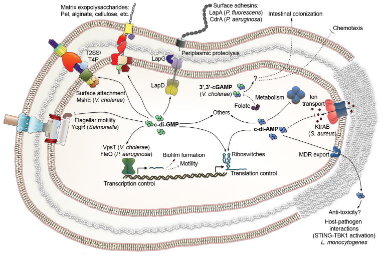
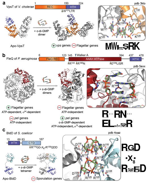
Since the discovery of c-di-GMP almost three decades ago, cyclic dinucleotides (CDNs) have emerged as widely used signaling molecules in most kingdoms of life. The family of second messengers now includes c-di-AMP and distinct versions of mixed cyclic GMP-AMP (cGAMP) compounds. In addition to these nucleotides, a vast number of proteins for the production and turnover of these molecules have been described, as well as effectors that translate the signals into physiological responses. The latter include, but are not limited to, mechanisms for adaptation and survival in prokaryotes, persistence and virulence of bacterial pathogens, and immune responses to viral and bacterial invasion in eukaryotes. In this review, we will focus on recent discoveries and emerging themes that illustrate the ubiquity and versatility of cyclic dinucleotide function at the transcriptional and post-translational levels and, in particular, on insights gained through mechanistic structure-function analyses.


Conflict of interest statement

Competing financial interests: The authors declare no competing financial interests.

Figures

Figure 1
Figure 1. Cyclic dinucleotide (CDN) signaling
a. Structural overview of the four prevalent cyclic dinucleotides. b. Regulation of CDN signaling. A generic route for the synthesis and degradation of CDNs is shown. Regulatory feedback loops controlling cellular CDN levels have been described in some instances, e.g. for I-site-containing GGDEF domain proteins or the effect of linear di-GMP (pGpG) on PDE-A activity. Note that the listed receptor/effector classes are common examples and not mutually exclusive. Enzymatic activities arise from stand-alone, single-domain proteins or, more typically, from multi-domain proteins, including proteins with both cyclase (i.e. GGDEF) and PDE (i.e. EAL) domains. Regulatory domains (X) determine the mode of enzyme regulation (e.g. environmental sensing, canonical two-component regulation, etc.). Known stimuli include allosteric ligands, post-translational modifications, light, gases, and mechanotransduction. c. Prevalence, biosynthesis and degradation pathways for the four major CDNs.
Figure 2
Figure 2. Bacterial CDNs and representative regulatory mechanisms
Solid lines show direct binding or transport; dashed lines illustrate indirect effects. The prevalent distribution of c-di-GMP/cGAMP and c-di-AMP in Gram-negative and Gram-positive bacteria, respectively, is illustrated by differences in the cell envelope, with a thick peptidoglycan layer illustrating the Gram-positive cell wall. Abbreviations are as follows: T2SS - Type 2 secretion system; T4P - type 4 pili; LmPC - L. monocytogenes pyruvate carboxylase.
Figure 3
Figure 3. Conformational adaptability and mechanism of action of CDNs
a. Using c-di-GMP as an example, CDN conformations found in protein co-crystal structures are depicted. Several of these conformations have also been shown to be sampled in solution. b. Representative cases of protein regulation via CDNs, exemplified by c-di-GMP binding modules, are shown. Cartoons depict concepts derived from available crystal structures (or modeling in the case CLP). A detailed description of these modes of action are provided in the main text.
Figure 4
Figure 4. Tripartite transmembrane signaling through HAMP domain-containing proteins with active or degenerate GGDEF and EAL domains
a. Inside-out signaling via c-di-GMP. A model of ‘inside-out’ signaling through the LapADG system in P. fluorescens is shown integrating crystallographic snapshots of the LapDG components. Conformational changes upon c-di-GMP recognition via the LapDEAL module lead to release of autoinhibitory intramolecular interactions, transmembrane signal transmission to the periplasmic output domain, and LapG protease sequestration. LapG recruitment to LapD• c-di-GMP prevents the protease reaching its substrate, LapA, at the outer membrane surface. As a result, LapA is retained at the cell surface, constituting an important regulatory step in biofilm formation. b. ‘Outside-in’ signaling via the YfiBNR system in P. aeruginosa. YfiN is a HAMP domain-containing DGC. Cell wall stress can release YfiN inhibition through the sequestration of the inhibitory YfiR partner by the outer membrane component YfiB. Conformational changes are transmitted through the membrane and a membrane-proximal HAMP module to activate intracellular c-di-GMP production.
Figure 5
Figure 5. Structures and nucleotide recognition of c-di-GMP-regulated transcription factors
Domain organizations, crystal structures, c-di-GMP-dependent changes in oligomerization, CDN-binding sites, consensus binding motifs, and observed physiological effects are shown for VpsT of V. cholerae (a), FleQ of P. aeruginosa (b), and BldD of S. coelicor (c). Key binding site side chains, as well as bound c-di-GMP molecules, are shown as sticks. Abbreviations are as introduced in the text: REC, receiver domain; HTH, helix-turn-helix motif; CTD, C-terminal domain.
Figure 6
Figure 6. CDN-dependent regulation of ion transport
a. Crystal structures of the KtrAB duo and mode of CDN recognition. A cartoon representation of the ATP-bound KtrAB crystal structure is shown (left panel). Transparent surface representation is included for the transmembrane KtrB dimer, ATP is shown as spheres, and a dimer of KtrA protomers is shown in color. The right panel depicts an full-length KtrA dimer extracted from the structure shown on the left (top) and the c-di-AMP-bound RCK_C module of a homolog (bottom). Conformational changes in the RKC-C dimer interface upon c-di-AMP recognition could be transmitted to the KtrB-proximal, ATP-binding RCK_N modules. b. A schematic representation of the Hyperpolarization-activated cyclic nucleotide-gated channel 4 (HCN4) tetramer and its antagonistic regulation by cyclic mono- and dinucleotides.
Figure 7
Figure 7. C-di-GMP dependent regulation of exopolysaccharide secretion in biofilms
a. Examples of synthase-dependent systems subject to direct c-di-GMP regulation. Color-coding for functionally homologous proteins is shown on the right and c-di-GMP binding is indicated by yellow stars. b. Cartoon representation of the crystal structure and topology of a catalytic BcsAB complex from R. sphaeroides. C. Crystallographic snapshots of the active site pocket in different CDN-free and bound states. Gating loop residues (dark blue), the cellulose product (light grey), and bound c-di-GMP (olive green) are shown as sticks, the UDP-glucose substrate as spheres, and inhibitory salt bridge interactions are indicated by dotted lines.
Figure 8
Figure 8. CDN recognition by STING
Crystal structures of human STING bound to c-di-GMP (a) and cGAMP (b) are shown. The bottom panels depict different views of the nucleotide binding pocket showing key CDN-coordinating side chains and bound ligands as sticks.

References

    1. Ross P, et al. Regulation of cellulose synthesis in Acetobacter xylinum by cyclic diguanylic acid. Nature. 1987;325:279–281. Seminal work on the discovery of c-di-GMP as an allosteric regulator of bacterial cellulose synthesis that laid the foundation of CDN signaling research. - PubMed
    1. Weinhouse H, et al. c-di-GMP-binding protein, a new factor regulating cellulose synthesis in Acetobacter xylinum. FEBS Lett. 1997;416:207–211. - PubMed
    1. Tal R, et al. Three cdg operons control cellular turnover of cyclic di-GMP in Acetobacter xylinum: genetic organization and occurrence of conserved domains in isoenzymes. J Bacteriol. 1998;180:4416–4425. - PMC - PubMed
    1. Paul R, et al. Cell cycle-dependent dynamic localization of a bacterial response regulator with a novel di-guanylate cyclase output domain. Genes Dev. 2004;18:715–727. - PMC - PubMed
    1. Christen M, Christen B, Folcher M, Schauerte A, Jenal U. Identification and characterization of a cyclic di-GMP-specific phosphodiesterase and its allosteric control by GTP. J Biol Chem. 2005;280:30829–30837. - PubMed

Publication types

Substances