

Human Immunodeficiency Virus Type 1 DNA Decay Dynamics With Early, Long-term Virologic Control of Perinatal Infection
- PMID: 28329153
- PMCID: PMC5434384
- DOI: 10.1093/cid/cix192
Human Immunodeficiency Virus Type 1 DNA Decay Dynamics With Early, Long-term Virologic Control of Perinatal Infection
Erratum in
-
Erratum.Clin Infect Dis. 2017 Oct 15;65(8):1431-1433. doi: 10.1093/cid/cix563. Clin Infect Dis. 2017. PMID: 29017252 Free PMC article. No abstract available.
Abstract
Background.: Early antiretroviral therapy (ART) limits proviral reservoirs, a goal for human immunodeficiency virus type 1 (HIV-1) remission strategies. Whether this is an immediate or long-term effect of virologic suppression (VS) in perinatal infection is unknown.
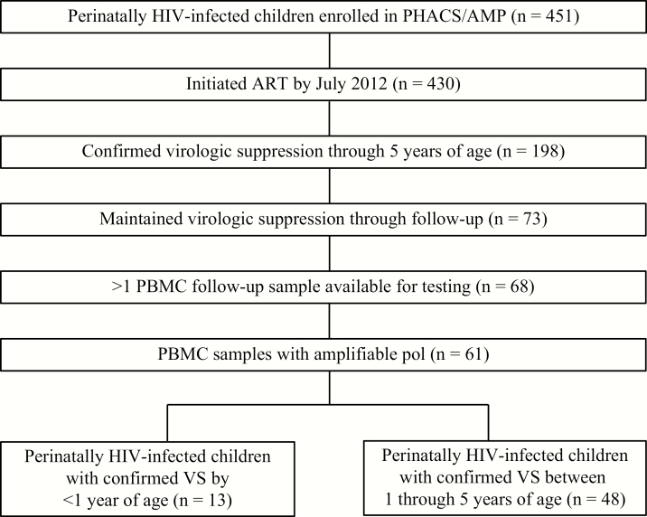
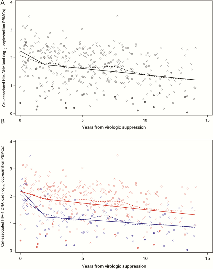
Methods.: We quantified HIV-1 DNA longitudinally for up to 14 years in peripheral blood mononuclear cells (PBMCs) among 61 perinatally HIV-1-infected youths in the Pediatric HIV/AIDS Cohort Study who achieved VS at different ages. Participants in group 1 (n = 13) were <1 year of age and in group 2 (n = 48) from 1 through 5 years of age at VS. Piecewise linear mixed-effects regression models assessed the effect of age at VS on HIV-1 DNA trajectories during VS.
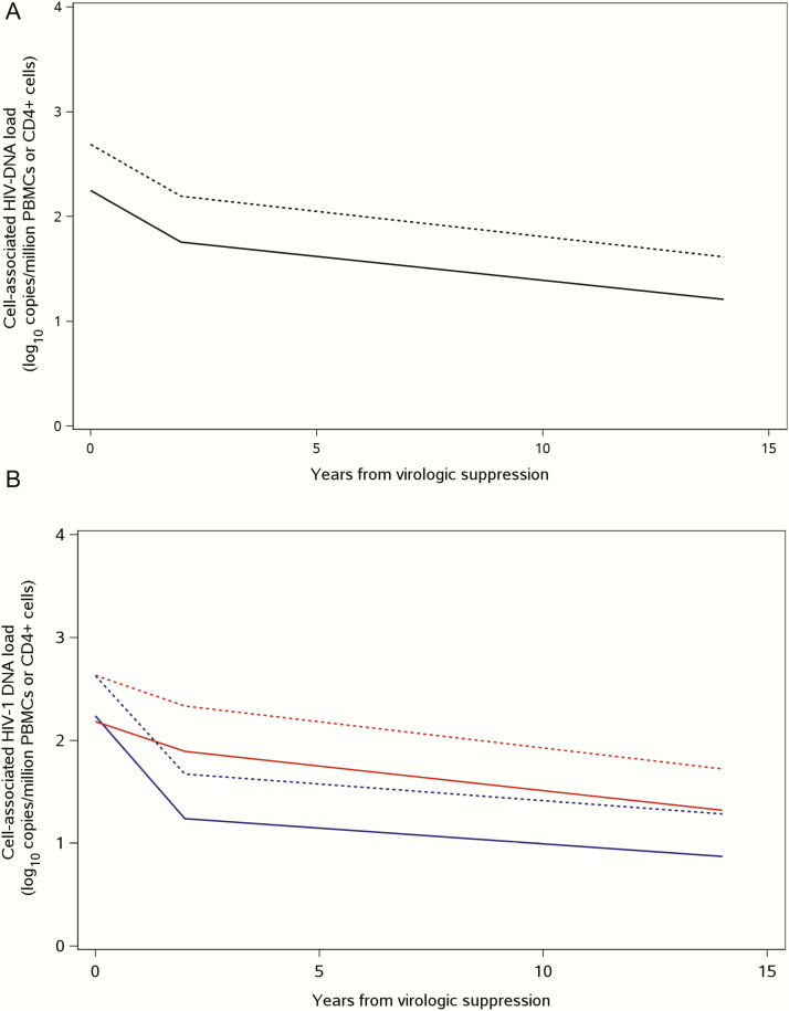
Results.: In the first 2 years following VS, HIV-1 DNA levels decreased by -0.25 (95% confidence interval [CI], -.36 to -.13) log10 copies/million PBMCs per year and was faster with early VS by age 1 year compared with after age 1 (-0.50 and -0.15 log10 copies/million PBMCs per year, respectively). Between years 2 and 14 from VS, HIV-1 DNA decayed by -0.05 (95% CI, -.06 to -.03) log10 copies/million PBMCs per year and was no longer significantly different between groups. The estimated mean half-life of HIV-1 DNA from VS was 15.9 years and was shorter for group 1 compared to group 2 at 5.9 years and 18.8 years, respectively (P = .09). Adjusting for CD4 cell counts had no effect on decay estimates.
Conclusions.: Early effective, long-term ART initiated from infancy leads to decay of HIV-1-infected cells to exceedingly low concentrations desired for HIV-1 remission strategies.
Keywords: HIV-1 DNA decay; early ART.; perinatal HIV-1 infection.
© The Author 2017. Published by Oxford University Press for the Infectious Diseases Society of America. All rights reserved. For permissions, e-mail: journals.permissions@oup.com.
Figures
References
-
- Chun TW, Carruth L, Finzi D, et al. Quantification of latent tissue reservoirs and total body viral load in HIV-1 infection. Nature 1997; 387:183–8. - PubMed
-
- Finzi D, Hermankova M, Pierson T, et al. Identification of a reservoir for HIV-1 in patients on highly active antiretroviral therapy. Science 1997; 278:1295–300. - PubMed
-
- Siliciano JD, Kajdas J, Finzi D, et al. Long-term follow-up studies confirm the stability of the latent reservoir for HIV-1 in resting CD4+ T cells. Nat Med 2003; 9:727–8. - PubMed
MeSH terms
Substances
Grants and funding
LinkOut - more resources
Full Text Sources
Other Literature Sources
Medical
Research Materials
