. 2017 Oct 1;38(37):2838-2849.
doi: 10.1093/eurheartj/ehx081.

Effects of noise on vascular function, oxidative stress, and inflammation: mechanistic insight from studies in mice

Affiliations

Effects of noise on vascular function, oxidative stress, and inflammation: mechanistic insight from studies in mice

Thomas Münzel et al. Eur Heart J. .

Abstract

Aims: Epidemiological studies indicate that traffic noise increases the incidence of coronary artery disease, hypertension and stroke. The underlying mechanisms remain largely unknown. Field studies with nighttime noise exposure demonstrate that aircraft noise leads to vascular dysfunction, which is markedly improved by vitamin C, suggesting a key role of oxidative stress in causing this phenomenon.

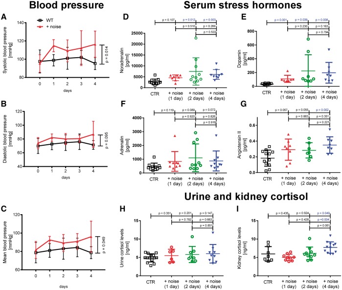
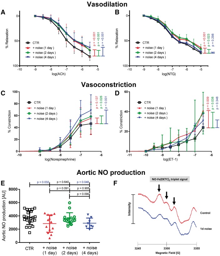
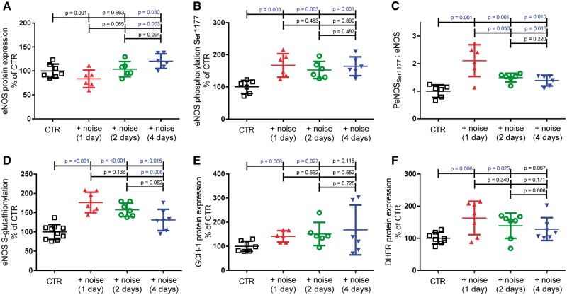
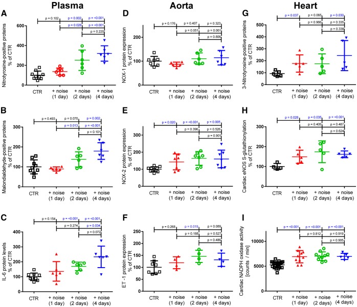
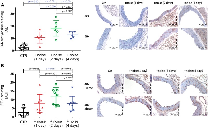
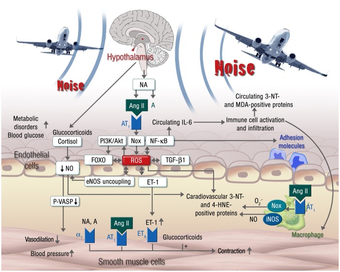
Methods and results: We developed a novel animal model to study the vascular consequences of aircraft noise exposure. Peak sound levels of 85 and mean sound level of 72 dBA applied by loudspeakers for 4 days caused an increase in systolic blood pressure, plasma noradrenaline and angiotensin II levels and induced endothelial dysfunction. Noise increased eNOS expression but reduced vascular NO levels because of eNOS uncoupling. Noise increased circulating levels of nitrotyrosine, interleukine-6 and vascular expression of the NADPH oxidase subunit Nox2, nitrotyrosine-positive proteins and of endothelin-1. FACS analysis demonstrated an increase in infiltrated natural killer-cells and neutrophils into the vasculature. Equal mean sound pressure levels of white noise for 4 days did not induce these changes. Comparative Illumina sequencing of transcriptomes of aortic tissues from aircraft noise-treated animals displayed significant changes of genes in part responsible for the regulation of vascular function, vascular remodelling, and cell death.

Conclusion: We established a novel and unique aircraft noise stress model with increased blood pressure and vascular dysfunction associated with oxidative stress. This animal model enables future studies of molecular mechanisms, mitigation strategies, and pharmacological interventions to protect from noise-induced vascular damage.

Keywords: Endothelial dysfunction; Environmental stressor; NADPH oxidase; Noise exposure; Oxidative stress; Vascular inflammation; eNOS uncoupling.


Figures

Figure 1
Figure 1
Effects of noise for 1, 2, and 4 days on blood pressure and stress hormone release. Noise increased significantly systolic and mean arterial (A, C) but not diastolic (B) blood pressure. Noise increased noradrenalin, dopamine and angiotensin II levels significantly and adrenalin by trend (DG). Cortisol levels in urine and kidney showed a weak trend for an increase under noise exposure, which was significant for kidney cortisol on day 4 (H, I). Data are mean ± SD from n = 8–16 mice/day (A–C), 8–11 (D), 7–8 (E), 9–11 (F), 7–12 (G), 6–14 (H) and 7–12 (I) mice/group.
Figure 2
Figure 2
Effects of noise on vascular function, sensitivity to vasoconstrictors and vascular NO production. Relaxation by the endothelium-dependent and -independent vasodilators acetylcholine (ACh, A) and nitroglycerin (NTG, B) were impaired by noise exposure. The sensitivity of the aorta to vasoconstrictors like norepinephrine (C) and endothelin-1 (D) was increased upon noise exposure. Noise exposure for 1 and 4 days significantly reduced the aortic NO production and bioavailability measured by EPR. (E) Quantification of the NO signal detected by Fe(DECT)2 spin trapping. (F) Representative NO traces of mice not exposed (Ctr.) and exposed to noise. For detailed statistical analysis see Supplementary material online, Tables 2S and 3S. Data are mean ± SD from n = 13–26 (A, B), 8–22 (C, D), 8–22 (E) mice/group.
Figure 3
Figure 3
Effects of noise for 1, 2, and 4 days on endothelial NO-synthase protein expression and activity. (AC) Endothelial NO synthase protein expression is increased after 4 days noise exposure and activating phosphorylation at Ser1177 is increased significantly at all time points measured. (D) eNOS S-glutathionylation as a surrogate marker for uncoupling of the protein was increased significantly at all time points of noise exposure. (E, F) Noise increased GTP-cyclohydrolase I (GCH-I) and dihydrofolate reductase (DHFR) expression leading to increased synthesis of the eNOS cofactor tetrahydrobiopterin (BH4) responsible for the coupling of the enzyme. Data are mean ± SD from n = 6–8 samples (pooled from 2 to 3 mice per sample) (A–C, E, F) and 7–10 samples (pooled from 2 mice per sample) (D).
Figure 4
Figure 4
Effects of noise (1, 2, and 4 days) on oxidative stress markers in mouse plasma and vascular tissue. (A, B) Levels of 3-nitrotyrosine- and malondialdehyde-positive proteins, surrogate markers for oxidative stress, were significantly increased in mouse plasma. (C) IL-6 levels were significantly elevated in mouse plasma compatible with increased inflammation. (D, E) The vascular NADPH oxidase subunit NOX-2 was significantly up-regulated after noise exposure, while NOX-1 was not changed. (F) Endothelin-1 expression was increased in mouse aorta upon noise exposure. Cardiac 3-nitrotyrosine-positive proteins were significantly increased (G). Cardiac S-glutathionylation of eNOS, the marker for eNOS uncoupling, was increased on all days of noise exposure (H), which also correlated with cardiac NADPH oxidase activity as measured by NADPH (200 µM)-stimulated lucigenin (5 µM) ECL in heart membrane fractions (I). Data are mean ± SD from n = 6–9 (AC), 4–9 samples (pooled from 2 to 3 mice per sample) (DF), 4–5 samples/group (pooled from 2 to 3 mice per sample) (G, H) and n = 22 (Ctr), 10 (1 + 2 days), 5 (4 days) mice/group (I).
Figure 5
Figure 5
Effects of 4 days noise on vascular 3-nitrotyrosine and ET-1 levels as assessed by immunohistochemistry. (A) Noise markedly increased immunostaining of 3-nitrotyrosine of intact aortic rings (brown color). The densitometric analysis revealed a significant increase in all noise-exposed groups. (B) Endothelin-1 immunostaining (brown color) is markedly increased in response to noise. The densitometric analysis showed significant progression of ET-1 protein expression on day 2 of noise exposure. Data are mean ± SD from n = 6–8 mice/group.
Figure 6
Figure 6
Proposed mechanisms of aircraft noise-induced vascular dysfunction. Noise exposure leads to an over-activation of the sympathetic system, resulting in elevated levels of noradrenalin (NA), adrenalin (A) and angiotensin II (Ang II) and subsequently cortisol. Ang II, in turn, activates endothelial NADPH oxidase causing oxidative stress, which may induce direct scavenging of nitric oxide (NO) and eNOS uncoupling through oxidation of BH4 and eNOS S-glutathionylation. Reactive oxygen species (ROS) play a key role in linking different pathways, including PI3K/Akt signaling, the FOXO transcription factors, TGF-β1 and NF-κB signaling as well as the endothelin-1 (ET-1) system (see text for details), increasing the circulating levels of IL-6 and the expression of vascular adhesion molecules. Superoxide and nitric oxide produced by infiltrating immune cells (neutrophils, NK cells and monocytes/macrophages) promotes the formation of 3-nitrotyrosine-, malondialdehyde- and 4-hydroxynonenal-positive proteins and inflicts additional cellular oxidative damage. The uncoupling of eNOS not only reduces NO production, but also potentiates the pre-existing oxidative stress. Endothelial NO production is further reduced by glucocorticoids like cortisol, leading to impaired vasodilation and increased blood pressure. The overproduction of NA, A and ET-1 enhances contraction, which is further potentiated by glucocorticoids. All of these vascular alterations support the development of metabolic disorders as envisaged by increased blood glucose levels.

Comment in

References

    1. Munzel T, Gori T, Babisch W, Basner M.. Cardiovascular effects of environmental noise exposure. Eur Heart J 2014;35:829–836. - PMC - PubMed
    1. Munzel T, Sorensen M, Gori T, Schmidt FP, Rao X, Brook FR, Chen LC, Brook RD, Rajagopalan S.. Environmental stressors and cardio-metabolic disease: part II-mechanistic insights. Eur Heart J 2017;38:557–564. - PMC - PubMed
    1. Munzel T, Sorensen M, Gori T, Schmidt FP, Rao X, Brook J, Chen LC, Brook RD, Rajagopalan S.. Environmental stressors and cardio-metabolic disease: part I-epidemiologic evidence supporting a role for noise and air pollution and effects of mitigation strategies. Eur Heart J 2017;38:550–556. - PubMed
    1. Babisch W. Stress hormones in the research on cardiovascular effects of noise. Noise Health 2003;5:1–11. - PubMed
    1. Babisch W, Ising H, Gallacher JE.. Health status as a potential effect modifier of the relation between noise annoyance and incidence of ischaemic heart disease. Occup Environ Med 2003;60:739–745. - PMC - PubMed