. 2017 Mar 21;18(12):2825-2835.
doi: 10.1016/j.celrep.2017.02.071.

Lineage-Biased Stem Cells Maintain Estrogen-Receptor-Positive and -Negative Mouse Mammary Luminal Lineages

Affiliations

Lineage-Biased Stem Cells Maintain Estrogen-Receptor-Positive and -Negative Mouse Mammary Luminal Lineages

Chunhui Wang et al. Cell Rep. .

Abstract

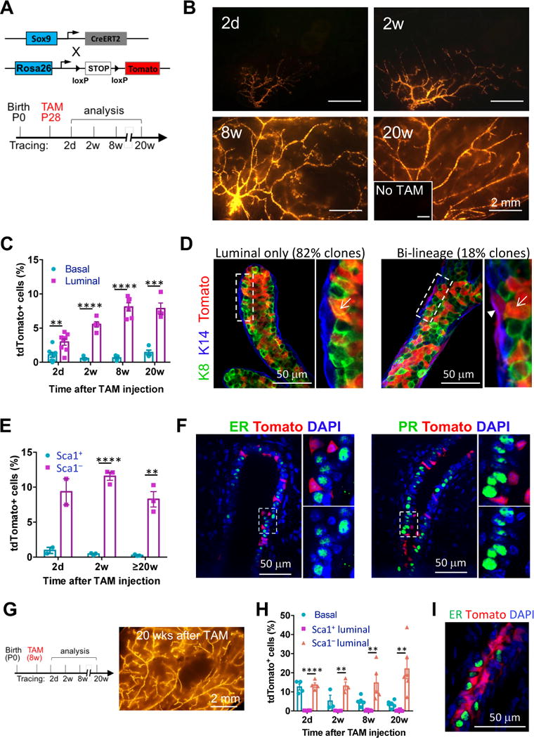
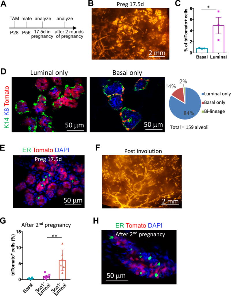
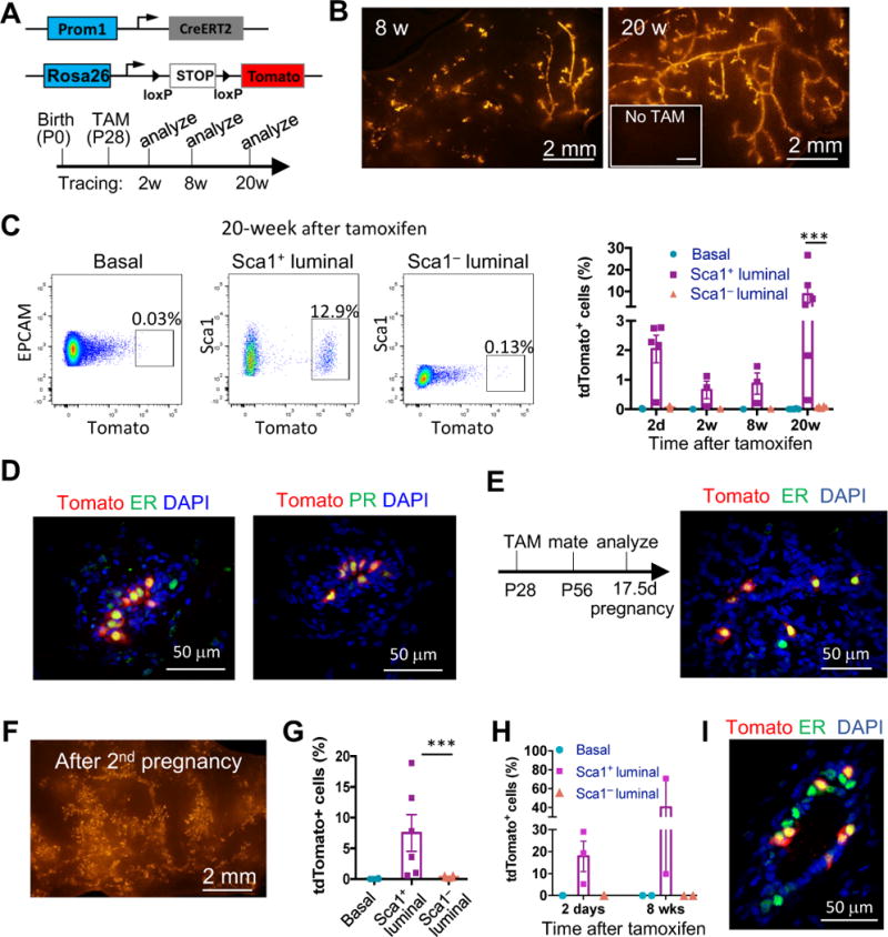
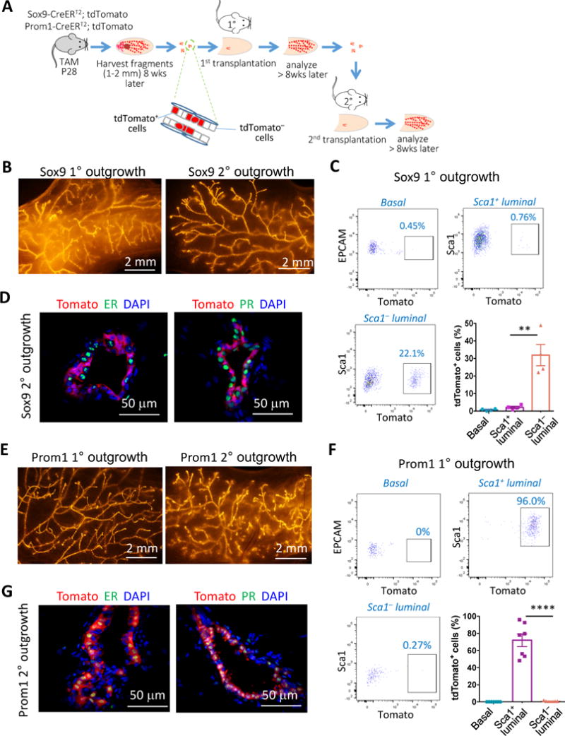
Delineating the mammary differentiation hierarchy is important for the study of mammary gland development and tumorigenesis. Mammary luminal cells are considered a major origin of human breast cancers. However, how estrogen-receptor-positive (ER+) and ER- luminal cells are developed and maintained remains poorly understood. The prevailing model suggests that a common stem/progenitor cell generates both cell types. Through genetic lineage tracing in mice, we find that SOX9-expressing cells specifically contribute to the development and maintenance of ER- luminal cells and, to a lesser degree, basal cells. In parallel, PROM1-expressing cells give rise only to ER+ luminal cells. Both SOX9+ and PROM1+ cells specifically sustain their respective lineages even after pregnancy-caused tissue remodeling or serial transplantation, demonstrating characteristic properties of long-term repopulating stem cells. Thus, our data reveal that mouse mammary ER+ and ER- luminal cells are two independent lineages that are maintained by distinct stem cells, providing a revised mammary epithelial cell hierarchy.

Keywords: breast cancer; cancer cell-of-origin; estrogen receptor negative cancer; estrogen receptor positive cancer; lineage tracing; mammary differentiation; mammary gland development; mammary gland regeneration; mammary stem cells; stem cell hierarchy.


Figures

Figure 1
Figure 1. SOX9+ cells are long-term repopulating and maintain the ER luminal and basal lineages in the postnatal mammary gland
(A) The mouse models (upper) and the experimental schedule (lower) for lineage-tracing experiments shown in Figure 1B to 1F. (B) Representative tdTomato whole-mount images of Sox9-CreERT2; R26R-tdTomato mammary glands at the indicated time points after tamoxifen treatment. Mice without tamoxifen treatment (No TAM) at 24 weeks of age were used as the negative control (inset). Minimum 3 animals were examined for each time point. (C) Percentages of tdTomato+ cells in the basal and luminal populations at the indicated time points, as determined by flow cytometry (mean ± SEM, n = 3–8). (D) K8 and K14 immunostaining of mammary gland sections for characterizing the tdTomato+ cells after 20 weeks of lineage tracing. Representative luminal only and bi-lineage tdTomato+ ducts are shown. The arrow head and arrows point to basal and luminal cells, respectively. Total 57 tdTomato+ cell clusters in 3 mice were counted. (E) Percentages of tdTomato+ cells in the Sca1+ and Sca1 luminal populations at the indicated time points, as determined by flow cytometry (mean ± SEM, n = 2–3). (F) ER and PR immunostaining of mammary gland sections 2 weeks after tamoxifen treatment. Magnification of the selected areas is shown on the right of each panel. 98% of tdTomato+ cells were ER and PR. (G) Experimental schedule (left) for adult Sox9-CreERT2 lineage-tracing experiments as shown in Figure 1G to 1I, and a representative tdTomato whole-mount image (right) of Sox9-CreERT2; R26R-tdTomato mammary glands 20 weeks after the tamoxifen treatment. (H) Percentages of tdTomato+ cells in the basal, Sca1+ and Sca1 luminal populations at indicated time points after the tamoxifen treatment, as determined by flow cytometry (mean ± SEM, n = 3–6). (I) ER immunostaining of mammary glands after 20 weeks of lineage tracing of adult SOX9+ cells. 99.5% tdTomato+ cells were ER. *P < 0.05, **P < 0.01, ***P<0.001, and ****P < 0.0001. See also Figure S1.
Figure 2
Figure 2. SOX9+ cells contribute to alveologenesis and persist after repeated pregnancy
(A) The experimental schedule for lineage-tracing studies shown in Figure 2. (B) A representative tdTomato whole-mount image of Sox9-CreERT2; R26R-tdTomato mammary glands on day 17.5 of pregnancy (Preg17.5d). (C) Percentages of tdTomato+ cells in the basal and luminal populations of Preg17.5d mice, as determined by flow cytometry (mean ± SEM, n = 3, paired t-test). (D) K8 and K14 immunostaining of Preg17.5d mammary gland sections for characterizing the tdTomato+ cells. Examples of alveoli with either tdTomato-labeled luminal or basal cells were shown. The graph on the right shows percentage of alveoli containing the indicated types of tdTomato+ cells. (E) ER immunostaining of Preg17.5d mammary gland sections. 99% tdTomato+ cells were ER. (F) A representative tdTomato whole-mount image of Sox9-CreERT2; R26R-tdTomato mammary glands at least 3 weeks after weaning of the second litter. (G) Percentage of tdTomato+ cells in the basal, Sca1+ and Sca1 luminal populations 3 weeks after weaning of the second litter (mean ± SEM, n = 6). (H) ER immunostaining of mammary glands after the 2nd pregnancy. 99.5% of tdTomato+ cells were ER. *P < 0.05 and **P < 0.01. See also Figure S2.
Figure 3
Figure 3. PROM1+ cells are long-term populating and specifically maintain the ER+ lineage
(A) The mouse models (upper) and the experimental schedule (lower) for PROM1+ cell lineage-tracing experiments as shown in Figure 3A–3D. (B) Representative tdTomato whole-mount images of Prom1-CreERT2; R26R-tdTomato mammary glands at the indicated time points after tamoxifen treatment. Mice without tamoxifen treatment (No TAM) at 24 weeks of age were used as the negative control. Minimum 3 animals were examined for each time point. (C) Representative flow cytometric profiles of the basal, Sca1+ and Sca1 luminal populations showing percentages of tdTomato+ cells after 20 weeks of lineage tracing. Percentages of tdTomato+ cells in specific cell populations are quantified on the right (mean ± SEM, n = 3–6, paired t-test). (D) ER and PR immunostaining of mammary gland sections after 20 weeks of lineage tracing. 97% and 100% of tdTomato+ cells are ER+ and PR+, respectively. (E) The experimental schedule for lineage-tracing studies of Prom1-CreERT2; R26R-tdTomato mice during pregnancy (left), and ER immunostaining of mammary gland sections at Preg17.5d (right). All tdTomato+ cells were ER+. (F) A representative tdTomato whole-mount image of Prom1-CreERT2; R26R-tdTomato mammary glands 3 weeks after 2 rounds of pregnancy and lactation. (G) Percentages of tdTomato+ cells in the indicated mammary cell types 3 weeks after 2 rounds of pregnancy and lactation, as determined by flow cytometry (mean ± SEM, n = 6, paired t-test). (H) Percentages of tdTomato+ cells in the indicated mammary cell types at the indicated time points of adult PROM1+ cell lineage tracing (n = 2–3). (I) ER immunostaining of mammary gland sections after 8 weeks of adult PROM1+ cell lineage tracing. 96% tdTomato+ cells were ER+. ***P<0.001. See also Figure S3.
Figure 4
Figure 4. ER and ER+ luminal lineages are regenerated by different unipotent stem cells during serial mammary gland transplantation
(A) A schematic diagram of the serial transplantation experiments. Sox9-CreERT2; R26R-tdTomato or Prom1-CreERT2; R26R-tdTomato mice were treated with tamoxifen at P28. Small mammary ductal fragments were isolated ≥8 weeks later and used for cleared mammary fat pad transplantation. The fragments of primary outgrowths were then used for secondary transplantation. (B) Representative tdTomato whole-mount images of the primary and secondary outgrowths regenerated by Sox9-CreERT2; R26R-tdTomato mammary ductal fragments. (C) Representative flow cytometric profiles and quantification of percentages of tdTomato+ cells in the indicated cell types of Sox9-CreERT2; R26R-tdTomato primary outgrowths (mean ± SEM, n = 4). (D) ER and PR immunostaining of the secondary outgrowths from Sox9-CreERT2; R26R-tdTomato ductal fragments. 97% and 99% of tdTomato+ cells were ER and PR, respectively. (E) Representative tdTomato whole-mount images of the primary and secondary outgrowths derived from Prom1-CreERT2; R26R-tdTomato mammary ductal fragments. (F) Representative flow cytometric profiles and quantification of frequency of tdTomato+ cells in the indicated cell types of Prom1-CreERT2; R26R-tdTomato primary outgrowths (mean ± SEM, n = 7). (G) ER and PR immunostaining of the secondary outgrowths from Prom1-CreERT2; R26R-tdTomato ductal fragments. 97% and 98% of tdTomato+ cells were ER+ and PR+, respectively. **P < 0.01 and ****P < 0.0001. See also Figure S4.

References

    1. Cowin P, Wysolmerski J. Molecular mechanisms guiding embryonic mammary gland development. Cold Spring Harb Perspect Biol. 2010;2:a003251. - PMC - PubMed
    1. de Visser KE, Ciampricotti M, Michalak EM, Tan DW, Speksnijder EN, Hau CS, Clevers H, Barker N, Jonkers J. Developmental stage-specific contribution of LGR5(+) cells to basal and luminal epithelial lineages in the postnatal mammary gland. J Pathol. 2012;228:300–309. - PubMed
    1. Eaves CJ. Hematopoietic stem cells: concepts, definitions, and the new reality. Blood. 2015;125:2605–2613. - PMC - PubMed
    1. Giraddi RR, Shehata M, Gallardo M, Blasco MA, Simons BD, Stingl J. Stem and progenitor cell division kinetics during postnatal mouse mammary gland development. Nat Commun. 2015;6:8487. - PMC - PubMed
    1. Guo W, Keckesova Z, Donaher JL, Shibue T, Tischler V, Reinhardt F, Itzkovitz S, Noske A, Zurrer-Hardi U, Bell G, et al. Slug and Sox9 cooperatively determine the mammary stem cell state. Cell. 2012;148:1015–1028. - PMC - PubMed

Publication types