. 2017 Mar 21;46(3):379-392.
doi: 10.1016/j.immuni.2017.02.017.

The IFN-λ-IFN-λR1-IL-10Rβ Complex Reveals Structural Features Underlying Type III IFN Functional Plasticity

Affiliations

The IFN-λ-IFN-λR1-IL-10Rβ Complex Reveals Structural Features Underlying Type III IFN Functional Plasticity

Juan L Mendoza et al. Immunity. .

Abstract

Type III interferons (IFN-λs) signal through a heterodimeric receptor complex composed of the IFN-λR1 subunit, specific for IFN-λs, and interleukin-10Rβ (IL-10Rβ), which is shared by multiple cytokines in the IL-10 superfamily. Low affinity of IL-10Rβ for cytokines has impeded efforts aimed at crystallizing cytokine-receptor complexes. We used yeast surface display to engineer a higher-affinity IFN-λ variant, H11, which enabled crystallization of the ternary complex. The structure revealed that IL-10Rβ uses a network of tyrosine residues as hydrophobic anchor points to engage IL-10 family cytokines that present complementary hydrophobic binding patches, explaining its role as both a cross-reactive but cytokine-specific receptor. H11 elicited increased anti-proliferative and antiviral activities in vitro and in vivo. In contrast, engineered higher-affinity type I IFNs did not increase antiviral potency over wild-type type I IFNs. Our findings provide insight into cytokine recognition by the IL-10R family and highlight the plasticity of type III interferon signaling and its therapeutic potential.


Figures

Figure 1
Figure 1. Engineering a High-Affinity IFN-λ
(A) Wild-type IFN-λ3 (Gad et al., 2009) was displayed on-yeast and stained with 1 μM monomer (left) or 400 nM streptavidin tetramers of IL-10Rβ (Yoon et al.,2010) labeled with Alexa 647 (middle and right panel) (x axis) in the presence (right panel) or absence (middle panel) of 50 nM streptavidin tetramers of IFN-λR1(Miknis et al., 2010) labeled with phycoerythrin (PE) (y axis). (B) Histogram plot of fluorescence (Alexa 647) for the naive and evolved first-generation (error-prone) and second-generation (shuffled) IFN-λ3 yeast displayedlibraries stained with 1 μM IL-10Rβ monomers in the presence IFN-λR1. (C) Sequences and on-yeast affinity measurements of evolved IFN-λ mutants (H11 mutations are colored in light red) compared to wild-type (light blue). (D) IL-10Rβ affinity for the wild-type IFN-λR1/IFN-λ3 or H11-containing binary complexes was determined by surface plasmon resonance. KD values weredetermined by fitting to a first order equilibrium binding model. See also Figure S1.
Figure 2
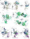
Figure 2. Structure of the IFN-λ Ternary Complex
(A) The structure of the ternary complex reveals the mechanism of IL-10Rβ (gold) recognition of IFN-λ3 H11 (blue) and IFN-λR1 (gray). The IL-10Rβ makesextensive and continuous contacts with H11 at sites 2a and 2b, and site 3 makes stem-stem contacts with the IFN-λR1. Unique to the IFN-λ structure, a largesurface area of the cytokine remains surface exposed in the ternary complex. (B) Structure of the IL-10/IL-10R1 partial signaling complex (PDB: 1J7V) (Josephson et al., 2001). (C) Structure of a type I IFN receptor complex (PDB: 3SE4) (Thomas et al., 2011). See also Table S1.
Figure 3
Figure 3. Specific Interactions Mediating the Stability of the Ternary Complex
For a Figure360 author presentation of Figure 3, see http://dx.doi.org/10.1016/j.immuni.2017.02.017#mmc3. (A) Overview of the IFN-λ3 H11 (blue)/IFN-λR1 (gray)/IL-10Rβ (gold) ternary complex. (B) Detailed views of the site 2a and site 2b contacts between IFN-λ3 H11 and IL-10Rβ tyrosines 59, 82, and 140. Hydrogen bonds are indicated by dashedblack lines. (C) Shared receptor use of aromatic residues to bind cytokines. (D) IL-10Rβ uses Tyr82 residue as a hotspot of binding like other shared receptor systems. (E) Y103 of γc bound to IL-2 (PDB: 3QAZ) (Levin et al., 2012). (F) F169 of gp130 bound to IL-6 (PDB: 1P9M) (Boulanger et al., 2003b). See also Figure S2.
Figure 4
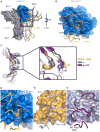
Figure 4. Structural and Chemical Conservation of IL-10Rβ Binding in the IL-10 Superfamily
(A) Structures of IL-10 (green) (PDB: 1J7V) (Josephson et al., 2001) and IL-22 (orange) (PDB: 3DLQ) (Bleicher et al., 2008) were structurally aligned to IFN-λ3 H11(blue ribbon) in the ternary complex structure, highlighting conserved features of IL-10Rβ (gold surface) recognition. IL-10Rβ surface residues within contactdistance of H11 are shaded red. Residues at which alanine mutations have been shown to negatively impact IL-10Rβ binding to IL-10 (green), IL-22 (orange), andIFN-λ3 (blue) are indicated as sticks (right panel). (B) View of the IL-10Rβ binding interface of IFN-λ3 H11 mapped onto the structures of other IL-10 superfamily members. Residues that impact IL-10Rβ bindingidentified through mutagenesis analysis and residues that share hydrogen bonds with IL-10Rβ in the IFN-λ ternary complex structure are shown as sticks. (C) View ofthe IL-10Rβ binding interface modeled on the face ofIL-10 and IL-22 using the IFN-λ3 H11 ternary complex. Cytokine residues at the IL-10Rβ interfaceare colored by chemical properties (red for polar/charged or white for hydrophobic) to highlight the conserved “hydrophobic” pockets in which the IL-10Rβtyrosines likely dock. See also Figure S3.
Figure 5
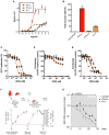
Figure 5. Functional Characterization of an Engineered IFN-λ3 Variant
IFN-λ3 H11 (orange) was compared to the wild-type IFN-λ3 (black) and the type I IFN-ω (red) in several functional assays. (A) STAT1 activation in Hap1 cells. Curves were fit to a first-order logistic model. Error bars represent ± SEM (n = 3). (B) Induction of ISG15 in Hap1 cells treated with 5 pM with each interferon for 6 hr as determined byqPCR. Error bars represent 95% confidence intervals (n = 3). (C) Antiviral activity of IFNs in Huh7.5 cells infected with HCV. Error bars represent ± SEM (n = 4). (D) Antiproliferative activity of IFNs in Huh7.5 cells. (E) Antiproliferative activity of IFNs is enhanced in Huh7.5 cells overexpressing IFN-λR1. Error bars represent ± SD (n = 6) (D and E). (F) Human-liver chimeric mice were generated by injecting human hepatoblasts into Fah−/−Rag2−/−Il2rgnull (FRG) mice and infected with 2 × 10⁸ DNA copies ofvirus obtained from a genotype C eAg negative patient. Human albumin was tracked by ELISA (red circles) and HBV DNA quantified by qPCR (green circles) over220 days prior to IFN treatment. (G) Mice were treated daily with 10 μg/kg of body weight for 4 weeks with vehicle, IFN-λ3, or IFN-λ3H11. The engineered IFN-λ3H11 has improved in vivo activityover the wild-type IFN-λ3 (p = 0.02). Error bars represent ± SEM, where n = 2 for the Control and n = 3 for WT or H11 treated mice. See also Figure S4.
Figure 6
Figure 6. Probing the Molecular Mechanism of Type I IFN Functional Differences from Type III IFN via Cytokine Engineering and High-Throughput Functional Screening
(A) A site-directed IFN-ω library designed to mutagenize the low affinity IFN-αR1 site was combined with a rationally designed affinity-enhancing Lys152Argmutation at the IFN-αR2 interface on the cytokine. The SD4 domain of IFN-αR1 was modeled to the complex (PDB: 3SE4) (Thomas et al., 2011) using the mouseIFN-αR1 structure (PDB: 3WCY). (B) 288 clones were screened for activity usingahigh-throughput functional screen. Displayed IFNs were cleaved from yeast by the site-specific3C protease. Thefiltered supernatants were then used to treat VPN53 (Moraga et al., 2009) cells to measure antiproliferative activity and HCV-infected Huh-7.5 cells to measureantiviral activity. (C) Functional characterization of phospo-STAT1 signaling, antiviral and antiproliferative potencies as a function of complex stability. Potencies of IFN-mediatedactivation of STAT1 on Jurkat cells, viral clearance in HCV-infected Huh7.5 cells, and proliferation inhibition of WISH cells were used to determine EC50s andplotted as a function of complex stability. (D) Fold ISG induction as measured by qPCR relative to complex stability. See also Table S2.

Similar articles

Cited by

References

    1. Andrus L, Marukian S, Jones CT, Catanese MT, Sheahan TP, Schoggins JW, Barry WT, Dustin LB, Trehan K, Ploss A, et al. Expression of paramyxovirus V proteins promotes replication and spread of hepatitis C virus in cultures of primary human fetal liver cells. Hepatology. 2011;54:1901–1912. - PMC - PubMed
    1. Ank N, West H, Bartholdy C, Eriksson K, Thomsen AR, Paludan SR. Lambda interferon (IFN-λambda), a type III IFN, is induced by viruses and IFNs and displays potent antiviral activity against select virus infections in vivo. J Virol. 2006;80:4501–4509. - PMC - PubMed
    1. Azuma H, Paulk N, Ranade A, Dorrell C, Al-Dhalimy M, Ellis E, Strom S, Kay MA, Finegold M, Grompe M. Robust expansion of human hepatocytes in Fah-/-/Rag2-/-/Il2rg-/- mice. Nat Biotechnol. 2007;25:903–910. - PMC - PubMed
    1. Baldridge MT, Nice TJ, McCune BT, Yokoyama CC, Kambal A, Wheadon M, Diamond MS, Ivanova Y, Artyomov M, Virgin HW. Commensal microbes and interferon-λ determine persistence of enteric murine norovirus infection. Science. 2015;347:266–269. - PMC - PubMed
    1. Billerbeck E, Mommersteeg MC, Shlomai A, Xiao JW, Andrus L, Bhatta A, Vercauteren K, Michailidis E, Dorner M, Krishnan A, et al. Humanized mice efficiently engrafted with fetal hepatoblasts and syn-geneic immune cells develop human monocytes and NK cells. J Hepatol. 2016;65:334–343. - PMC - PubMed

Publication types