. 2017 Apr 6;544(7648):105-109.
doi: 10.1038/nature21706. Epub 2017 Mar 22.

The lung is a site of platelet biogenesis and a reservoir for haematopoietic progenitors

Affiliations

The lung is a site of platelet biogenesis and a reservoir for haematopoietic progenitors

Emma Lefrançais et al. Nature. .

Abstract

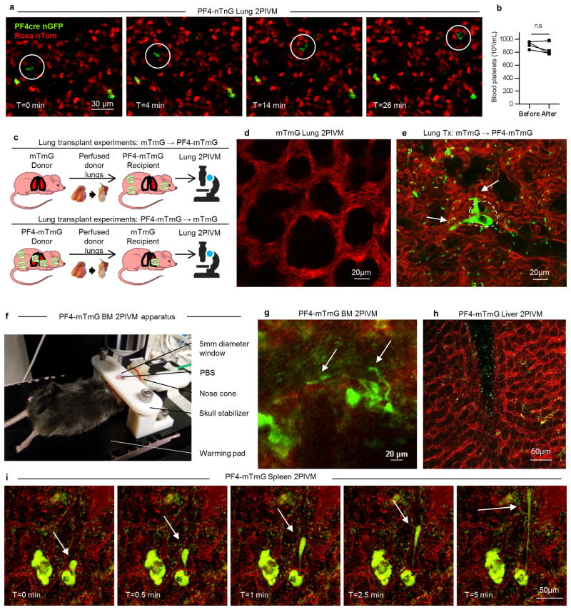
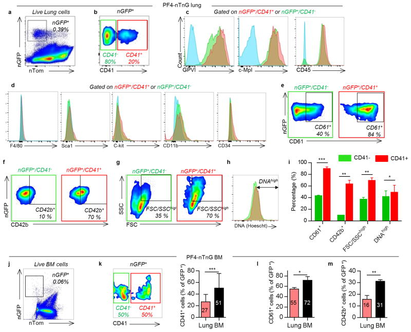
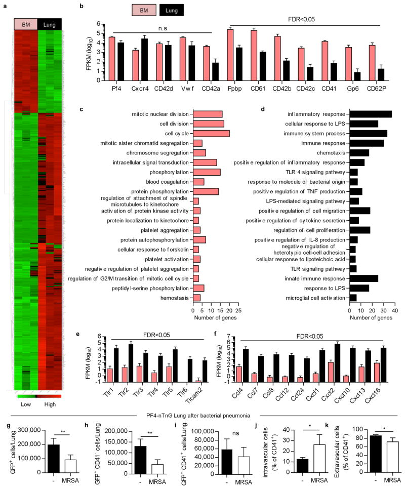
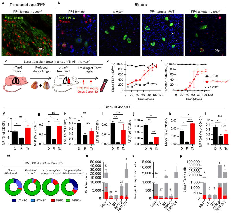
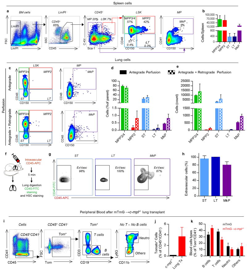
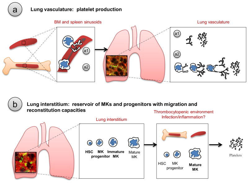
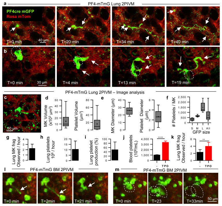
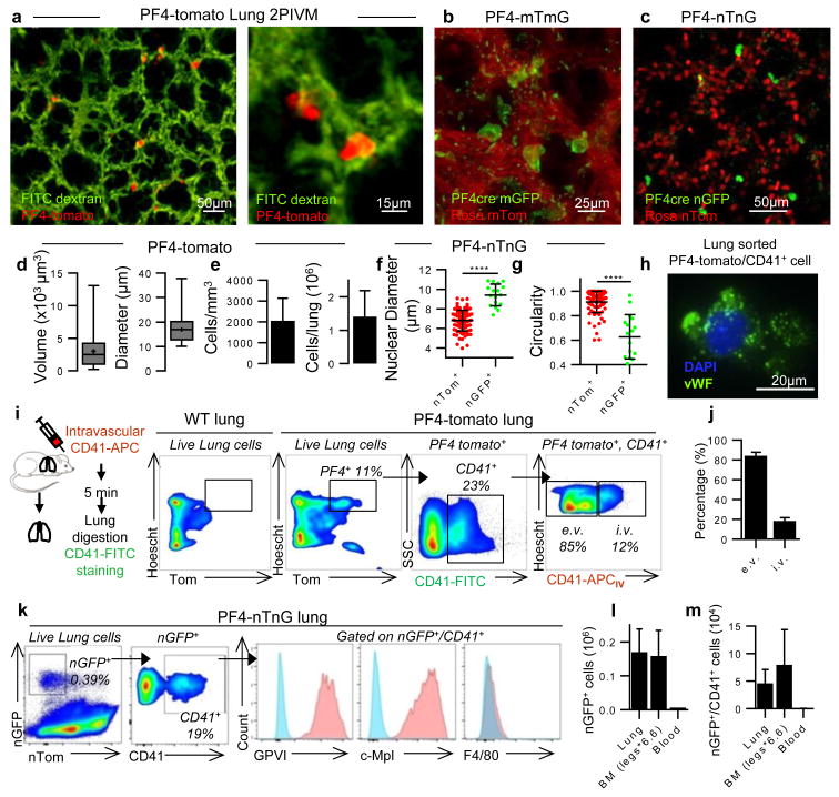
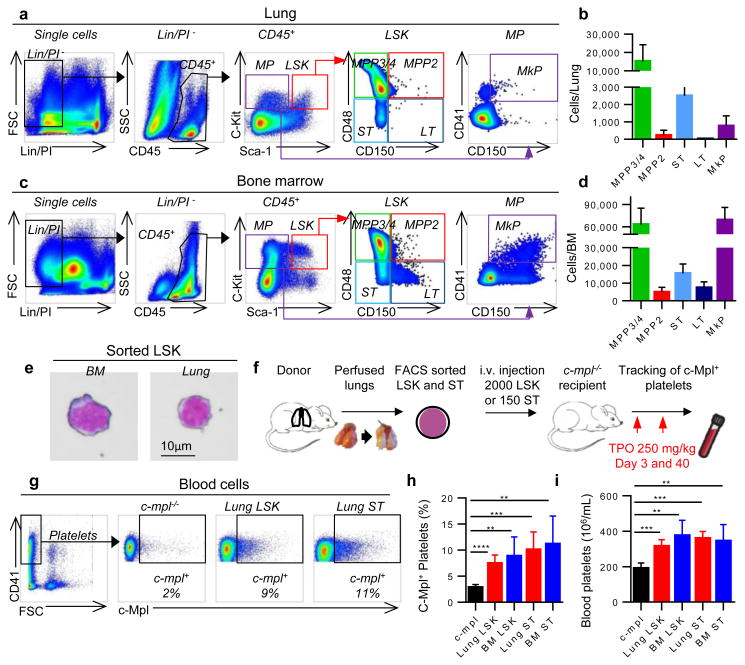
Platelets are critical for haemostasis, thrombosis, and inflammatory responses, but the events that lead to mature platelet production remain incompletely understood. The bone marrow has been proposed to be a major site of platelet production, although there is indirect evidence that the lungs might also contribute to platelet biogenesis. Here, by directly imaging the lung microcirculation in mice, we show that a large number of megakaryocytes circulate through the lungs, where they dynamically release platelets. Megakaryocytes that release platelets in the lungs originate from extrapulmonary sites such as the bone marrow; we observed large megakaryocytes migrating out of the bone marrow space. The contribution of the lungs to platelet biogenesis is substantial, accounting for approximately 50% of total platelet production or 10 million platelets per hour. Furthermore, we identified populations of mature and immature megakaryocytes along with haematopoietic progenitors in the extravascular spaces of the lungs. Under conditions of thrombocytopenia and relative stem cell deficiency in the bone marrow, these progenitors can migrate out of the lungs, repopulate the bone marrow, completely reconstitute blood platelet counts, and contribute to multiple haematopoietic lineages. These results identify the lungs as a primary site of terminal platelet production and an organ with considerable haematopoietic potential.


Figures

Extended Data Figure 1
Extended Data Figure 1. MKs and proplatelets observed in lung circulation are from an extrapulmonary source
(a) Lung 2PIVM of a PF4-nTnG mouse (nuclear GFP). The presence of the mobile GFP+ nucleated cells (circled) indicates the presence of a nucleus in circulating MKs. (b) Platelet counts in the blood before and after imaging. (c) Experimental schema of mTmG (perfused donor lung) to PF4-mTmG (recipient mouse) and vice-versa lung transplantation followed by 2PIVM. (d) 2PIVM of a mTmG mouse lung showing no GFP signal. (e) 2PIVM of a mTmG mouse lung transplanted into a PF4-mTmG recipient mouse showing GFP+ cells from recipient origin and platelet production in the lung. (f) BM 2PIVM apparatus. (g) Representative image of proplatelet release in the BM sinusoids (arrows). (h) Liver 2PIVM of PF4-mTmG mouse. Small platelets (GFP, green) were seen in the circulation but neither resident nor circulating MKs or proplatelets were observed. (i) Spleen 2PIVM of PF4-mTmG mouse. Sequential images show resident MKs (GFP, green) releasing proplatelets (arrows) in the spleen vasculature (in red).
Extended Data Figure 2
Extended Data Figure 2. Resident MKs in lung and other organs
(a) Survey of PF4-tomato mouse lung visualized by 2PIVM. PF4-tomato expressing cells (in red) are found in high numbers in the lung. Lung vasculature is labeled by intravascular injection of FITC dextran (green). A total area of 2.49 mm2 (1.6 mm × 1.6 mm) was imaged. (b) Resident (static) GFP+ cells are found in PF4-mTmG (donor lung) to mTmG (recipient) transplanted mouse. (c) BM and (d) spleen 2PIVM images of PF4-mTmG mice. (c,d) Large-sized MKs (GFP, green) are found in the BM and spleen in large numbers. (e) Size characterization of resident (static) GFP+ cells by image analysis of PF4-mTmG lungs, BM, and spleen. Min-to-max boxplots are presented: the line in the middle of the box is plotted at the median, the box extends from the 25th to 75th percentiles and the whiskers range from the smallest to the largest values. The + indicates the mean.
Extended Data Figure 3
Extended Data Figure 3. Surface expression of lung MKs compared to BM MKs
(a) Flow cytometric analysis of nGFP+ cells from PF4-nTnG lungs. (b) CD41 expression defines two populations of MKs: CD41+ (red) and CD41- (green). (c) Positive surface expression was detected for the following markers: GPVI, c-Mpl and CD45 in both populations. Unstained cells are plotted in blue. (d) Negative surface expression was detected for the following markers: F4/80, CD34, CD11b, Sca-1, c-Kit. (e) The CD41+ population has a higher percentage of CD61+ cells, (f) CD42b+ cells, (g) larger cells, and (h) higher DNA content, and summarized in (i). (j) Flow cytometric analysis of nGFP+ cells from PF4-nTnG BM. Compared to the lung, the BM nGFP+ population has a higher percentage of (k) CD41+ cells, (l) CD61+ cells, and (m) CD42b+ cells. Data are representative of three or more replicates. Mean ± SD are presented. Unpaired t-test: *P < 0.05, **P < 0.01, ***P < 0.001.
Extended Data Figure 4
Extended Data Figure 4. Gene expression analysis of lung vs BM MKs and bacterial pneumonia experiment reveal an immature profile and a potential role in immunity for lung MKs
MKs (nGFP+/CD41+) were sorted from PF4-nTnG lungs and BM followed by mRNA isolation and sequencing. (a) Relative mRNA expression is shown on a scale from low (green) to high (red). 3 independent experiments (4 mice each) were used for statistical analysis. Heatmap of all significant differentially expressed genes (false discovery rate (FDR) <0.05). (b) Read counts for MK-related genes, which are found in both lung and BM MKs, but some are underrepresented in the lung MKs. (c,d) Gene ontology biological processes analysis related to the genes downregulated (c) or upregulated (d) in the lung MKs. Top 20 biological processes are shown. Vertical axis represents gene ontology categories, while horizontal axis indicates the number of genes in each ontology category. (e) Read counts for TLR gene pathways and (f) chemokines overexpressed in the lung MKs. FPKM: Fragments per kilobase of exon per million fragments mapped. (g-k) Lung MKs and progenitor populations are altered during infection. Flow cytometric analysis of nGFP+ cells from PF4-nTnG lungs, 24 hours after intratracheal administration of Staphylococcus aureus (MRSA, 5 × 10 cfu). CD41-APC was injected i.v before lung digestion and staining with CD41-FITC. Number of cells in normal or infected lung are shown; all nGFP+ cells (g), less mature cells (nGFP+/CD41-) (h), and mature cells (nGFP+/CD41+) (i). Percentage of intravascular MKs (j) and extravascular MKs (k) in the mature population (nGFP+/CD41+). n= 4-6 mice per group.
Extended Data Figure 5
Extended Data Figure 5. Platelet reconstitution after lung transplantation ± TPO injection
Blood was collected from the mandibular vein every week following lung transplant. After transplant, a group of mice represented in red and orange received TPO injection (250mg/kg, day 3 and 40). The other group was left untreated and is represented in blue and purple. A fraction of mice per group showed a sustained platelet production for more than 3 months (blue and red). In the other fraction (purple and orange), the platelet production was lower and transient (less than 3 months). The fractions of mice in each group are indicated. Data from individual mice (a,c,e,g) or group average (b,d,f,h) are plotted. (a-d,i,j) Percentage of donor-derived platelets. Percentage was analyzed by FACS, counting the tomato+ platelets (CD41+/FSCsmall gate). (e-h,k) Overall platelet counts in the peripheral blood. (j,k) Plots from mice with 10 months of sustained platelet production are shown. (i) Percentage of tomato+ platelets in control lung transplants. (l) Color code for the different lung transplant groups according to lung origin (donor), recipient mouse, treatment received (±TPO) and observed response (sustained or transient).
Extended Data Figure 6
Extended Data Figure 6. Characterization of platelets produced after lung transplantation
(a,b) Flow cytometric analysis of tomato+ platelets observed in the blood and stained with antibodies against CD41, CD42d, GPVI and c-Mpl. Blood from (a) PF4-tomato or (b) PF4-tomato→c-mpl-/- lung transplants. (c) Platelet activation experimental schema. (d) Flow cytometric analysis of tomato+ platelets after stimulation with thrombin (10 nM) and stained with antibody against CD62P. (e) Percentage of CD62P+ platelets before and after thrombin activation. Ctrl = PF4-tomato, Lung tx = PF4-tomato→c-mpl-/-. Mean ± SD are presented (n=2-3 mice per group). Unpaired t-test: **P < 0.01, ***P < 0.001
Extended Data Figure 7
Extended Data Figure 7. Lung and BM analysis of transplanted mice
Mice with sustained production of lung-derived platelets were sacrificed at least 3 months after lung transplantation. (a) Representative 2PIVM image of a lung after PF4-tomato→c-mpl-/- transplant. (b) Representative 2PIVM image of flushed BM cells from PF4-tomato and c-mpl-/- mice, and PF4-tomato→WT and PF4-tomato→c-mpl-/- lung transplants. (c) Experimental schema of mTmG→c-mpl-/- lung transplants. Blood was collected from the mandibular vein every two weeks to test for donor-derived platelets (tomato+ platelets) and overall blood platelet counts. (d) Percentage of donor-derived platelets was analyzed by FACS counting of tomato+ events in the CD41+/FSCsmall gate. (e) The platelet count in peripheral blood was determined by CBC. (d,e) Mean ± S.E.M. are presented. (f-l) BM cells from donor (mTmG, D), recipient (c-mpl-/-, R), or mTmG→c-mpl-/- lung transplants (Tx) with 3 months sustained donor-derived platelet production were analyzed. Population frequencies within the CD45+ compartment: (f) Myeloerythroid progenitors (MP: Lin-/Sca-1-/c-Kit+), (g) LSK (Lin-/Sca-1+/c-Kit+), (h) MkP, (i) LT, (j) ST, (k) MPP2, and (l) MPP3/4. (m) Population frequencies within the LSK compartment from indicated groups. (n-p) Total cell populations (grey bars), donor origin tomato+ cells (red bars), and %tomato+ cells (above bars) in (n) BM, (o) recipient native lung (right lung) and (p) spleen. Mean ± SD are presented (n=2-3 mice per group). Unpaired t-test: *P < 0.05, **P < 0.01, ***P < 0.001.
Extended Data Figure 8
Extended Data Figure 8. Lung hematopoietic progenitors are extravascular and have multi-lineage capabilities
(a) Representative spleen FACS plots of hematopoietic progenitors within the LSK compartment (LSK: Lin-,/Sca-1+/c-Kit+) and the myeloerythroid progenitors compartment (MP: Lin-/Sca-1-/c-Kit+). (b) Cell counts of hematopoietic progenitor populations in the spleen. (c-e) Lungs were perfused before digestion. (c) Representative FACS plot of LSK and MP compartments with antegrade perfusion +/- retrograde perfusion. (d) Frequencies and (e) cell counts of lung hematopoietic progenitor populations. (f-h) CD45-APC mAb was injected i.v. via the tail vein 5 min before lung digestion and staining with CD45-FITC mAb. (g) Hematopoietic progenitor populations were examined for labeling with injected CD45 mAb by flow cytometry. (h) Percentage of CD45-FITC+ cells positive or negative (extravascular cells) for the i.v. CD45-APC mAb. (i-k) Peripheral blood was analyzed 2-3 months after mTmG→c-mpl-/- lung transplants (i) Representative FACS plot of blood cell analysis. (j) Percentage of lung-derived cells (Tom+/CD41-) in the blood after transplant. (k) Percentage of B cells (CD19+), T cells (CD3+) or neutrophils (CD11b+, Ly6G+) in the lung-derived cells (Tom+/CD41-). Mean ± SEM are presented (n=5 mice per group).
Extended Data Figure 9
Extended Data Figure 9. Proposed schema of lung involvement in platelet biogenesis
The role of the lung in platelet biogenesis is two-fold and occurs in two different compartments. (a) Platelet production in the lung vasculature. After being released from the BM or the spleen, proplatelets (a1) and MKs (a2) are retained in the lung vasculature, the first capillary bed encountered by any cell leaving the BM, where proplatelet formation and extension and final platelet release is observed. (b) Presence of MKs and hematopoietic progenitors in the lung interstitium. Mature and immature MKs along with hematopoietic progenitors are found in the lung interstitium. In thrombocytopenic environments, hematopoietic progenitors from the lung migrate and restore BM hematopoietic deficiencies.
Figure 1
Figure 1. The lung is an important site of MK circulation and platelet production
(a-c) Visualization of MKs and platelet production in the lung circulation by 2-photon intravital microscopy (2PIVM) in PF4-mTmG mice. (a,b) Sequential images show large-sized MK (green) in the lung capillaries (red) where it undergoes proplatelet formation (arrows). (b) Dark hole in the cytoplasm (circled) indicates the nucleus. Time elapsed is indicated. (c-f) Characterization of PF4+ events by image analysis. (c) Representative image of surface analysis of the GFP channel. (d) Volume distribution and (e) equivalent diameter of MKs and platelets. (f) Number of platelets produced by one MK according to its size: Small (<500 platelets, n=18), Medium (500-1000 platelets, n=7), Large (>1000 platelets, n=10). (d-f) Min-to-max boxplots are presented. (g-i) Quantification of lung platelet production. (g) Number of MKs releasing platelets observed per hour in imaged volume of lung (2 hour movies, n=10). (h) Estimation of the number and (i) the percentage of platelets produced by the lung. (j) Platelet counts in the blood and (k) number of MKs releasing platelets in the lung 5 days after TPO treatment. n≥5 mice per group. Unpaired t-test: ****P < 0.0001, **P < 0.005. (g-k) Mean ± S.D. are presented. (l,m) Visualization of proplatelet release (arrow) and MK migration (circled) in the BM sinusoids by 2PIVM in PF4-mTmG mice.
Figure 2
Figure 2. Resident MKs are present in the extravascular spaces of the lung
(a-c) Visualization of resident/static MKs in the lung by 2PIVM of (a) PF4-tomato, (b) PF4-mTmG, or (c) PF4-nTnG mice. (d) Size characterization of PF4+ cells (red, >10μm) by quantitative image analysis of PF4-tomato lungs. Min-to-max boxplots are presented. (e) Quantification of PF4+ cells (red, >10μm). (f) Comparison of nuclear size and (g) circularity between PF4+ cells (green) and all other lung cells (red) by quantitative image analysis of PF4-nTnG lungs. Unpaired t-test: ****P < 0.0001. (h) Representative immunofluorescence images of PF4 and CD41+ cells sorted from perfused PF4-tomato lung and stained with anti-vWF (green) and DAPI (blue). (i,j) Intravascular (i.v.) or extravascular (e.v.) localization of PF4+ and CD41-FITC+ cells: (i) experimental schema, representative FACS plots, and (j) percentage (mean of 4 experiments, n=8 mice). (k) FACS gating strategy and surface expression of nucleated PF4+ and CD41+ cells from PF4-nTnG lungs. (l) FACS quantification of nucleated PF4+ and (m) nucleated PF4+/CD41+ cells present in PF4-nTnG whole lung, BM (2 femurs, 2 tibias × 6.6) and blood (1.5mL). (e-g,l,m) Mean ± S.D. are presented.
Figure 3
Figure 3. Lung-derived progenitors reconstitute platelet counts and hematopoietic stem cell deficiency in thrombocytopenic mice
(a-f) PF4-tomato to c-mpl-/- lung transplants. (a) Experimental schema. (b) Blood platelet counts. (c,d) %donor-derived platelets was analyzed by counting tomato+ events in the CD41+/FSClow gate. (b,d) Mean ± S.E.M. are presented. (e,f) BM cells from donor (PF4-tom), recipient (c-mpl-/-), or transplanted mice with 10 months sustained donor-derived platelet production (Lung Tx) were analyzed. (e) FACS analysis of BM cells reveals Tomato+ cells (CD41+ and CD41- populations). Percentage of lineage negative BM cells positive for Tomato and CD41. (f) Representative immunofluorescence image of Tomato+ cell (red) in BM of a transplanted mouse stained with anti-CD41 (green) and Syto60 nucleic acid stain (blue). (g-n) mTmG to c-mpl-/- lung transplants. BM cells from donor (D, mTmG), recipient (R, c-mpl-/-), or transplanted (Tx) mice with sustained donor-derived platelet production (3 months) were analyzed. (g) Representative FACS analysis of the myeloerythroid progenitor compartment (MP) and the MK progenitor population (MkP). (h) Percentage of the MkP population within the MP compartment. (i,j) Percentage of donor origin Tomato+ cells in the MkP population. (k) Representative FACS analysis of the LSK compartment (LSK). Multipotent Progenitors 2 (MPP2), Multipotent Progenitors 3 and 4 (MPP3/4), Short-term HSC (ST) and Long-term HSC (LT) population frequencies within the LSK compartment. (l) Percentage of the LT HSC population within the LSK compartment. (m,n) Percentage of donor origin Tomato+ cells in the LT population. (h,j,l,n) Mean ± S.D. are presented (n=2-4 mice per group). Unpaired t-test: **P < 0.01, *P < 0.05
Figure 4
Figure 4. The lung contains hematopoietic progenitors including MK progenitors
Representative (a) lung and (c) BM FACS plots of hematopoietic progenitors within the LSK compartment (LSK) and the myeloerythroid progenitors compartment (MP). (b,d) Cell counts of hematopoietic progenitor populations in the whole lung and the BM (legs), respectively. MPP2, MPP3/4, ST-HSC, LT HSC, and MkP. (e) Representative image of Wright-Giemsa staining of LSK cells sorted from BM or lung. (f) Experimental schema. (g,h) Percentage of donor-derived platelets was analyzed by FACS counting of c-Mpl+ events in the CD41+/FSCsmall gate. (i) Blood platelet counts. (h,i) Mean ± S.D. are presented (n=4-5 mice per group). Unpaired t-test: **P < 0.01, ***P < 0.001, ****P < 0.0001

Comment in

References

    1. Semple JW, Italiano JE, Jr, Freedman J. Platelets and the immune continuum. Nat Rev Immunol. 2011;11:264–274. doi: 10.1038/nri2956. - DOI - PubMed
    1. Versteeg HH, Heemskerk JW, Levi M, Reitsma PH. New fundamentals in hemostasis. Physiol Rev. 2013;93:327–358. doi: 10.1152/physrev.00016.2011. - DOI - PubMed
    1. Machlus KR, Italiano JE., Jr The incredible journey: From megakaryocyte development to platelet formation. J Cell Biol. 2013;201:785–796. doi: 10.1083/jcb.201304054. - DOI - PMC - PubMed
    1. Howell WH, Donahue DD. The Production of Blood Platelets in the Lungs. J Exp Med. 1937;65:177–203. - PMC - PubMed
    1. Levine RF, et al. Circulating megakaryocytes: delivery of large numbers of intact, mature megakaryocytes to the lungs. European journal of haematology. 1993;51:233–246. - PubMed

Publication types