. 2017 Sep;66(3):758-771.
doi: 10.1002/hep.29177. Epub 2017 Aug 1.

Ginsenoside Rg3 restores hepatitis C virus-induced aberrant mitochondrial dynamics and inhibits virus propagation

Affiliations

Ginsenoside Rg3 restores hepatitis C virus-induced aberrant mitochondrial dynamics and inhibits virus propagation

Seong-Jun Kim et al. Hepatology. 2017 Sep.

Abstract

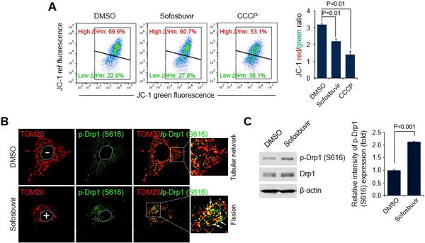
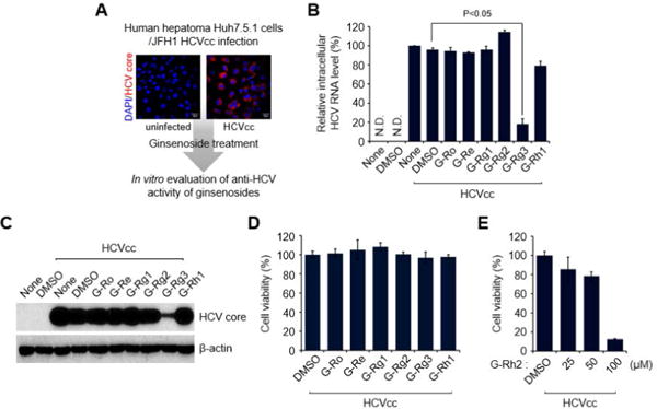
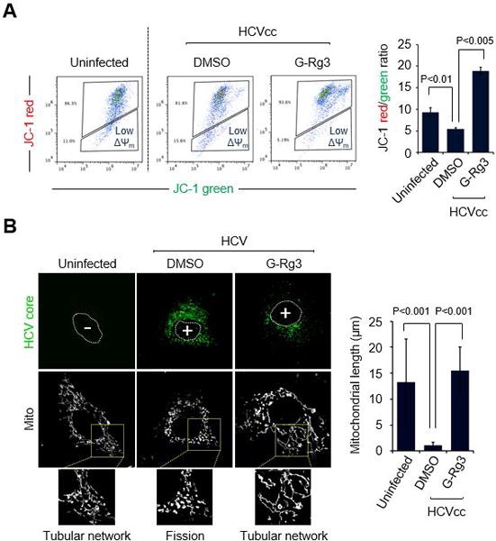
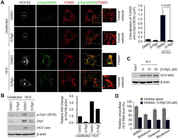
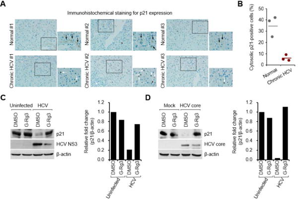
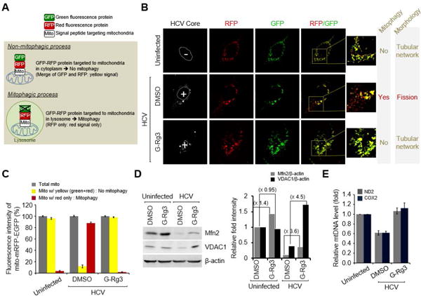
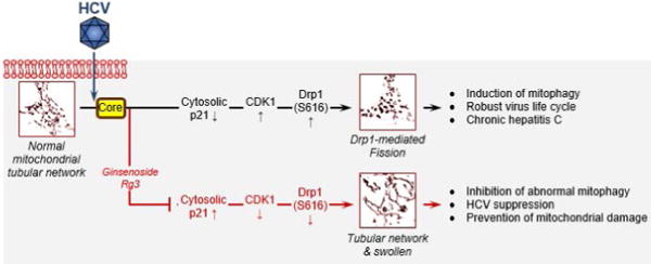
Hepatitis C virus (HCV) alters mitochondrial dynamics associated with persistent viral infection and suppression of innate immunity. Mitochondrial dysfunction is also a pathologic feature of direct-acting antiviral (DAA) treatment. Despite the high efficacy of DAAs, their use in treating patients with chronic hepatitis C in interferon-sparing regimens occasionally produces undesirable side effects such as fatigue, migraine, and other conditions, which may be linked to mitochondrial dysfunction. Here, we show that clinically prescribed DAAs, including sofosbuvir, affect mitochondrial dynamics. To counter these adverse effects, we examined HCV-induced and DAA-induced aberrant mitochondrial dynamics modulated by ginsenoside, which is known to support healthy mitochondrial physiology and the innate immune system. We screened several ginsenoside compounds showing antiviral activity using a robust HCV cell culture system. We investigated the role of ginsenosides in antiviral efficacy, alteration of mitochondrial transmembrane potential, abnormal mitochondrial fission, its upstream signaling, and mitophagic process caused by HCV infection or DAA treatment. Only one of the compounds, ginsenoside Rg3 (G-Rg3), exhibited notable and promising anti-HCV potential. Treatment of HCV-infected cells with G-Rg3 increased HCV core protein-mediated reduction in the expression level of cytosolic p21, required for increasing cyclin-dependent kinase 1 activity, which catalyzes Ser616 phosphorylation of dynamin-related protein 1. The HCV-induced mitophagy, which follows mitochondrial fission, was also rescued by G-Rg3 treatment.

Conclusion: G-Rg3 inhibits HCV propagation. Its antiviral mechanism involves restoring the HCV-induced dynamin-related protein 1-mediated aberrant mitochondrial fission process, thereby resulting in suppression of persistent HCV infection. (Hepatology 2017;66:758-771).


Figures

Fig. 1
Fig. 1. Sofosbuvir induces mitochondrial damage and Drp1-mediated mitochondrial fission
(A) FACS analysis showing decrease in ΔΨm in the presence of Sofosbuvir. At 24 hours after treatment with Sofosbuvir (100 nM), Huh7 cells were stained with JC-1 dye and then analyzed on a flow cytometer. The mitochondrial uncoupler CCCP (5 μM) was used as a control. The accompanying graph indicates that Sofosbuvir induced decreases in the level of ΔΨm, as indicated by the JC-1 red/green ratio (right panel). (B) Representative confocal microscope image showing phosphorylated Drp1 translocation to mitochondria and mitochondrial fission in the presence of Sofosbuvir. At 24 hours after treatment with Sofosbuvir (100 nM), Huh7 cells were immunostained with antibodies against TOM20 (red) and p-Drp1 (S616) (green). Nuclei are demarcated with white dotted circles. Treated (+) and untreated (–) cells are marked. In the zoomed images, the arrows indicate the colocalization of TOM20 and p-Drp1 (S616) in Sofosbuvir-treated cells (yellow spots). (C) Western blot analysis of p-Drp1 (S616) and Drp1 expression in Sofosbuvir-treated cells. Whole-cell lysates extracted from Huh7 cells treated with Sofosbuvir (100 nM) for 24 hours were analyzed by immunoblotting with antibodies specific to p-Drp1 (S616) and Drp1 proteins. β-actin was used as an internal loading control. The accompanying graph indicates that Sofosbuvir stimulates Drp1 phosphorylation (right panel).
Fig. 2
Fig. 2. G-Rg3 inhibits HCV propagation
(A) A strategy for screening ginsenosides that show antiviral effects during HCV propagation. Huh7.5.1 cells infected with JFH1 HCVcc for 1 day at an MOI of 5 were treated with various ginsenosides at 100 μM. At 2 days posttreatment, cells were harvested and used for analyses of intracellular HCV RNA (B) and protein expression (C). Confocal-microscope images show HCV core protein expression (red) in uninfected (left) and infected (right) cells. Nuclei are immunostained with DAPI (blue). (B) Intracellular HCV RNA levels were analyzed by real-time qRT-PCR as described in the Materials and Methods. GAPDH was used as the control for determining the normalized changes in HCV RNA expression. (C) Western blot analysis showing the reduction in HCV core protein expression induced by G-Rg3 treatment. Whole-cell lysates extracted from HCV-infected cells were analyzed by immunoblotting with an antibody specific to HCV core protein. (D) MTT assay data showing the viability of HCV-infected cells treated with ginsenosides for 2 days. Cell viability was measured as described in the Materials and Methods. (E) Viability of HCV-infected cells treated with G-Rh2.
Fig. 3
Fig. 3. G-Rg3 inhibits HCV-induced mitochondrial fission
(A) Representative FACS analysis showing restoration of HCV-induced reduction of ΔΨm in the presence of G-Rg3. Cells infected with HCVcc for 1 day were treated with G-Rg3 (100 μM). At 1 day posttreatment, HCV-infected Huh7 cells were stained with JC-1 dye and then analyzed on a flow cytometer. The accompanying graph indicates that G-Rg3 restores the HCV-induced decrease in the level of ΔΨm, as indicated by the JC-1 red/green ratio (right panel). Data is average of two independent experiments. (B) Representative confocal images showing the mitochondrial tubular network in HCV-infected cells treated with G-Rg3. Huh7 cells infected with HCVcc at an MOI of 1 were treated with G-Rg3 (100 μM). At 1 day posttreatment, cells prestained with MitoTracker (Mito, white) were immunostained with HCV core antibody (green). Nuclei are demarcated with white dots. Infected (+) and uninfected (–) cells are marked. In the zoomed images, uninfected (left panel) and HCV-infected/G-Rg3 (right panel) cells show the typical tubular mitochondrial network, whereas HCV infected cells (middle panel) display a fragmented mitochondrial structure. The accompanying graph is quantitative analysis of the mitochondrial length in left confocal images (mean ± SEM; n = 10 mitochondria, two independent experiments).
Fig. 4
Fig. 4. G-Rg3 inhibits HCV-induced mitochondrial recruitment of Drp1
(A) Confocal-microscope images showing inhibition of Drp1 translocation to mitochondria in HCV-infected cells in the presence of G-Rg3. At 2 days after treatment with G-Rg3 (100 μM), Huh7 cells infected with HCVcc were immunostained with antibodies against TOM20 (red), p-Drp1 (S616) (green) and HCV E2 protein (white). Nuclei are demarcated with white dotted circles. Infected (+) and uninfected (–) cells are marked. In the zoomed images, the arrows indicate the colocalization of TOM20 and p-Drp1 (S616) in HCV-infected cells (yellow spots). The accompanying graph is quantitative analysis of the colocalization of TOM20 and p-Drp1 (S616) in left panel. (mean ± SEM; n = 10 mitochondria, two independent experiments). (B) Western blot analysis of p-Drp1 (S616) and Drp1 expression in HCV-infected cells treated with G-Rg3. Whole-cell lysates extracted from HCV-infected cells treated with G-Rg3 (100 μM) for 2 days were analyzed by immunoblotting with antibodies specific to p-Drp1 (S616), Drp1, and HCV core protein. β-actin was used as an internal loading control. The accompanying graph is quantitation of Western blot data in left panel. (C) Western blot analysis showing the antiviral effect of G-Rg3 in R-1 HCV subgenomic replicon cells. Whole-cell lysates of R-1 cells treated with G-Rg3 were analyzed by immunoblotting with antibodies specific to HCV NS3 protein. β-actin was used as an internal loading control. (D) HCV-infected Huh7 cells were treated with various HCV inhibitors at 10 nM (representing an approximate 10-fold dilution of EC50) or in combination with G-Rg3 (50 μM). At 1 day posttreatment, cells were harvested and used for analyses of intracellular HCV RNA by real-time qRT-PCR as described in the Materials and Methods. GAPDH was used as the control for determining the normalized changes in HCV RNA expression.
Fig. 5
Fig. 5. G-Rg3 restores HCV-induced decrease in p21 expression
(A) Immunohistochemical analysis showing the reduction of cytosolic p21 expression in liver biopsy materials from patients with CHC. The immunohistochemical assay was performed with a specific antibody for p21 protein (dark brown). In the zoomed images, black arrows (dark brown) indicate the cytosolic expression of p21 protein, whereas white arrows indicate p21-positive staining in nuclei in HCV-infected tissue. (B) Quantitative analyses of p21-positive signals targeting the cytoplasm in panel A. (C and D) Western blot analyses showing rescue of the decreased cytosolic p21 expression level in G-Rg3-treated HCV-infected cells (C) and HCV-core-protein-transfected cells (D). The crude cytosolic fractions isolated from G-Rg3-treated HCV-infected cells (C) and HCV-core-protein-transfected cells (D) were analyzed by immunoblotting with antibodies specific to p21 and HCV core protein. β-actin was used as an internal loading control. Accompanying graphs are quantitation of Western blot data in left panel, respectively.
Fig. 6
Fig. 6. G-Rg3 Inhibits HCV-induced mitophagy
(A) A system for monitoring the mitophagosomal maturation process by which mitophagosomes are delivered to lysosomes (mitophagy) using a dual fluorescence reporter/sensor, p-mito-mRFP-EGFP. Lysosomal delivery of the tandem fusion protein mito-mRFP-EGFP targeting entire mitochondria results in differential quenching and degradation of the two individual fluorochromes, thereby allowing for visual analysis of mitophagic flux. (B) Confocal microscope images showing G-Rg3-mediated inhibition of HCV-induced mitophagy. HCV-infected cells transiently expressing mito-RFP-GFP were treated with G-Rg3 (100 μM) for 48 hours and then immunostained with anti-HCV core antibody (white). Nuclei are demarcated with white dotted circles. Infected (+) and uninfected (–) cells are marked. The fluorescence signals in the zoomed images indicate the expression of mito-RFP-GFP targeting mitochondria: yellow color, no mitophagy; red color, mitophagy. (C) Quantitative analyses of the fluorescence signals targeting mitochondria in panel A. (D) Western blot analyses of Mfn2 and VDAC1 expression in HCV-infected cells treated with G-Rg3. Whole-cell lysates extracted from HCV-infected cells treated with G-Rg3 (100 μM) for 2 days were analyzed by immunoblotting with antibodies specific to Mfn2 and VDAC1 protein. β-actin was used as an internal loading control. (E) Real-time qRT-PCR analysis of mitochondrial DNA level in HCV-infected cells treated with G-Rg3. Mitochondrial ND2 and COX2 DNAs isolated from HCV-infected cells treated with G-Rg3 (100 μM) for 2 days were analyzed by real-time qRT-PCR with primers specific to ND2 and COX2 gene. β-actin was used for normalization.
Fig. 7
Fig. 7. Schematic of the function of G-Rg3 in mitochondrial dynamics perturbed by HCV infection
HCV infections induce endoplasmic reticulum stress and trigger calcium leakage from the endoplasmic reticulum, resulting in mitochondrial oxidative stress and an altered membrane potential, which in turn causes mitochondrial depolarization and induces mitochondrial fission initiated by the mitochondrial translocation of p-Drp1. HCV core protein induces degradation of cytosolic p21 expression that leads to up-regulation in CDK1 and p-Drp1 that can be modulated by G-Rg3 treatment.

References

    1. Rose L, Bias TE, Mathias CB, Trooskin SB, Fong JJ. Sofosbuvir: A Nucleotide NS5B Inhibitor for the Treatment of Chronic Hepatitis C Infection. Ann Pharmacother. 2014;48:1019–1029. - PubMed
    1. Kiser JJ, Flexner C. Direct-acting antiviral agents for hepatitis C virus infection. Annu Rev Pharmacol Toxicol. 2013;53:427–449. - PMC - PubMed
    1. Elbaz T, El-Kassas M, Esmat G. New era for management of chronic hepatitis C virus using direct antiviral agents: A review. J Adv Res. 2015;6:301–310. - PMC - PubMed
    1. Cho Y, Cho EJ, Lee JH, Yu SJ, Yoon JH, Kim YJ. Sofosbuvir-based therapy for patients with chronic hepatitis C: Early experience of its efficacy and safety in Korea. Clin Mol Hepatol. 2015;21:358–364. - PMC - PubMed
    1. Suen DF, Norris KL, Youle RJ. Mitochondrial dynamics and apoptosis. Genes Dev. 2008;22:1577–1590. - PMC - PubMed

Publication types

MeSH terms