. 2017 Mar 22;5(1):24.
doi: 10.1186/s40478-017-0427-7.

Calcium dysregulation, functional calpainopathy, and endoplasmic reticulum stress in sporadic inclusion body myositis

Affiliations

Calcium dysregulation, functional calpainopathy, and endoplasmic reticulum stress in sporadic inclusion body myositis

David R Amici et al. Acta Neuropathol Commun. .

Abstract

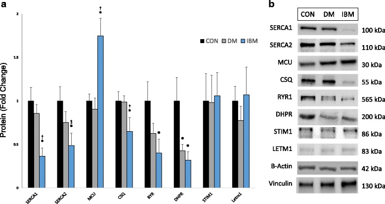
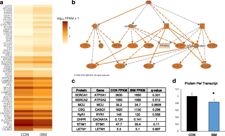
Sporadic inclusion body myositis (IBM) is the most common primary myopathy in the elderly, but its pathoetiology is still unclear. Perturbed myocellular calcium (Ca2+) homeostasis can exacerbate many of the factors proposed to mediate muscle degeneration in IBM, such as mitochondrial dysfunction, protein aggregation, and endoplasmic reticulum stress. Ca2+ dysregulation may plausibly be initiated in IBM by immune-mediated membrane damage and/or abnormally accumulating proteins, but no studies to date have investigated Ca2+ regulation in IBM patients. We first investigated protein expression via immunoblot in muscle biopsies from IBM, dermatomyositis, and non-myositis control patients, identifying several differentially expressed Ca2+-regulatory proteins in IBM. Next, we investigated the Ca2+-signaling transcriptome by RNA-seq, finding 54 of 183 (29.5%) genes from an unbiased list differentially expressed in IBM vs. controls. Using an established statistical approach to relate genes with causal transcription networks, Ca2+ abundance was considered a significant upstream regulator of observed whole-transcriptome changes. Post-hoc analyses of Ca2+-regulatory mRNA and protein data indicated a lower protein to transcript ratio in IBM vs. controls, which we hypothesized may relate to increased Ca2+-dependent proteolysis and decreased protein translation. Supporting this hypothesis, we observed robust (4-fold) elevation in the autolytic activation of a Ca2+-activated protease, calpain-1, as well as increased signaling for translational attenuation (eIF2a phosphorylation) downstream of the unfolded protein response. Finally, in IBM samples we observed mRNA and protein under-expression of calpain-3, the skeletal muscle-specific calpain, which broadly supports proper Ca2+ homeostasis. Together, these data provide novel insight into mechanisms by which intracellular Ca2+ regulation is perturbed in IBM and offer evidence of pathological downstream effects.

Keywords: Calcium; Calpain; Inclusion body; Muscular diseases; Myositis; Unfolded protein response.


Figures

Fig. 1
Fig. 1
Altered Ca2+-regulatory protein expression in IBM. a Protein levels of pre-specified panel of proteins, as assessed by immunoblot, expressed as mean + SEM. N = 5, 4, and 7 for non-myositis controls (CON), DM, and IBM, respectively. b Representative immunoblots. *P < 0.05 vs CON; P < 0.05 vs DM; P = 0.07 vs DM
Fig. 2
Fig. 2
Ca2+ signaling transcriptome perturbations in IBM and related post-hoc analyses. a Heat map of differentially expressed (q < 0.05) genes in IBM (n = 9) versus controls (CON; n = 7) from the KEGG Ca2+ signaling pathway. b Representative network image showing the relationship between Ca2+ signaling and transcriptomic regulators; Ca2+ abundance was considered a significant (q < 0.01) upstream regulator of observed changes. Orange nodes indicate activation consistent with Ca2+ abundance. c Expression of mRNA (FPKM) for the genes encoding previously immunoblotted Ca2+-regulatory proteins, demonstrating no significant (q < 0.05) changes in expression. d Reduced protein to transcript ratio amongst Ca2+-regulatory proteins in IBM, expressed as mean + SEM. *P < 0.05 versus CON
Fig. 3
Fig. 3
Calpain-1 autolysis and calpain-3 reduction in IBM. a Representative immunoblot demonstrating prominent autolysis of 80 kDa native calpain-1 to proteolytically active calpain isoforms. b Quantification of 78 and 76 kDa calpain-1 band areas divided by total calpain-1. c Representative western blot of native calpain-3 expression. d Quantified expression of native calpain-3 protein levels. e Transcript expression of CAPN3 gene as determined by RNA-seq. Graphed data expressed as mean + SEM. N of 5, 4, and 7 for non-myositis controls (CON), DM, and IBM, respectively; *P < 0.05 vs CON; P < 0.05 vs DM
Fig. 4
Fig. 4
Activation of the UPR and downstream translational attenuation in IBM. a XBP1 transcript levels (RNA-seq). b Isoform-specific expression of ubiquitous (U) and spliced (S) XBP1 transcript (RNA-seq). c Representative immunoblots of UPR effector molecules d Quantification of UPR effector protein levels. e Relative phosphorylation of eIF2α, a marker of translational attenuation. Graphed data expressed as mean + SEM. N of 5, 4, and 7 for non-myositis controls (CON), DM, and IBM, respectively; *P < 0.05 vs CON; P < 0.05 vs DM
Fig. 5
Fig. 5
A theoretical mechanism of progressive Ca2+ dysregulation in IBM and downstream pathological relevance. Stage 1: An initial insult to Ca2+ regulation may come from immune-mediated membrane injury (pictured: cytotoxic T-lymphocyte), permeabilization of Ca2+ channels by abnormal proteins, and/or reduction in calpain-3. Stage 2: Ca2+ elevations in the cytosol are then propagated through ROS production, SR Ca2+ leak, and calpain proteolysis of Ca2+-regulatory proteins. Stage 3: Prolonged Ca2+ dyshomeostasis causes and/or exacerbates the inter-related phenomena of mitochondrial dysfunction, UPR signaling, protein aggregate formation, and contractile dysfunction

References

    1. Aggad D, Vérièpe J, Tauffenberger A, Parker JA. TDP-43 toxicity proceeds via calcium dysregulation and necrosis in aging Caenorhabditis elegans motor neurons. J Neurosci. 2014;34:12093–12103. doi: 10.1523/JNEUROSCI.2495-13.2014. - DOI - PMC - PubMed
    1. Ahmed M, Machado PM, Miller A, Spicer C, Herbelin L, He J, Noel J, Wang Y, McVey AL, Pasnoor M, et al. Targeting protein homeostasis in sporadic inclusion body myositis. Sci Transl Med. 2016;8:331ra341. doi: 10.1126/scitranslmed.aad4583. - DOI - PMC - PubMed
    1. Baki A, Tompa P, Alexa A, Molnár O, Friedrich P. Autolysis parallels activation of mu-calpain. Biochem J. 1996;318(Pt 3):897–901. doi: 10.1042/bj3180897. - DOI - PMC - PubMed
    1. Benjamini Y, Hochberg Y. Controlling the false discovery rate: a practical and powerful approach to multiple testing. J R Statists Soc. 1995;57:289–300.
    1. Benveniste O, Stenzel W, Hilton-Jones D, Sandri M, Boyer O, van Engelen BG. Amyloid deposits and inflammatory infiltrates in sporadic inclusion body myositis: the inflammatory egg comes before the degenerative chicken. Acta Neuropathol. 2015;129:611–624. doi: 10.1007/s00401-015-1384-5. - DOI - PMC - PubMed

Publication types