. 2017 Apr 11;8(4):977-990.
doi: 10.1016/j.stemcr.2017.02.010. Epub 2017 Mar 16.

Efficient Ex Vivo Engineering and Expansion of Highly Purified Human Hematopoietic Stem and Progenitor Cell Populations for Gene Therapy

Affiliations

Efficient Ex Vivo Engineering and Expansion of Highly Purified Human Hematopoietic Stem and Progenitor Cell Populations for Gene Therapy

Erika Zonari et al. Stem Cell Reports. .

Abstract

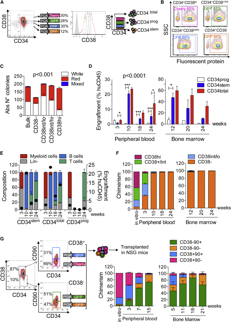
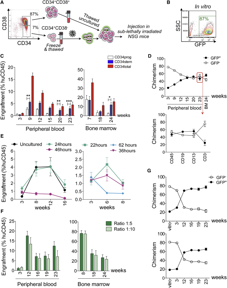
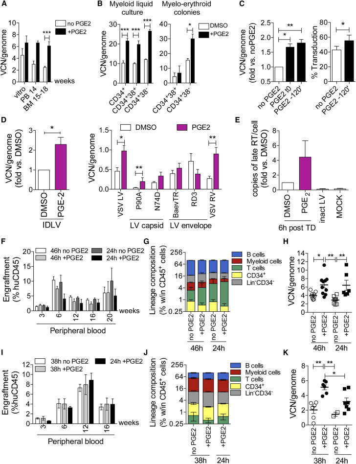
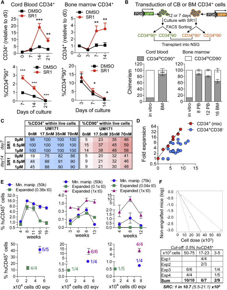
Ex vivo gene therapy based on CD34+ hematopoietic stem cells (HSCs) has shown promising results in clinical trials, but genetic engineering to high levels and in large scale remains challenging. We devised a sorting strategy that captures more than 90% of HSC activity in less than 10% of mobilized peripheral blood (mPB) CD34+ cells, and modeled a transplantation protocol based on highly purified, genetically engineered HSCs co-infused with uncultured progenitor cells. Prostaglandin E2 stimulation allowed near-complete transduction of HSCs with lentiviral vectors during a culture time of less than 38 hr, mitigating the negative impact of standard culture on progenitor cell function. Exploiting the pyrimidoindole derivative UM171, we show that transduced mPB CD34+CD38- cells with repopulating potential could be expanded ex vivo. Implementing these findings in clinical gene therapy protocols will improve the efficacy, safety, and sustainability of gene therapy and generate new opportunities in the field of gene editing.

Keywords: HSC expansion; HSC gene therapy; UM171; lentiviral vector transduction; prostaglandin E2; purified HSCs.


Figures

Figure 1
Figure 1
In Vivo Tracking of Hematopoietic Reconstitution by CD34+ mPB Subpopulations (A) Four mPB CD34+ cell subpopulations differing in CD38 expression levels were sorted and transduced with LVs expressing the indicated FPs (PGK, phosphoglycerate kinase promoter). Subpopulation frequency within total CD34+ cells and CD38 expression upon post-sorting re-analysis are shown (histogram plot). The scheme on the right shows how transduced subpopulations were mixed in the three xenotransplantation groups, maintaining their relative frequency at sorting. (B) Representative transduction efficiency of the indicated subpopulations, as measured in the myeloid colonies after 14 days of culture. (C) Clonogenic potential of the transduced subpopulations (n = 4 per group). Statistics by one-way ANOVA with Bonferroni's multiple comparison test: white colonies, CD38 versus CD38int/lo∗∗, CD38 versus CD38hi; for red colonies, CD38 versus Bulk ∗∗∗, CD38 versus CD38int/lo∗∗, CD38 versus CD38int/hi∗∗∗, CD38 versus CD38hi∗∗∗. (D) Human CD45+ cell engraftment in PB and BM at the indicated number of weeks post-transplant (n = 4 mice per group). Data were analyzed by two-way ANOVA with Bonferroni's multiple comparison test, significant differences with respect to the CD34stem group are shown. (E) Lineage composition (left axis) of the human CD45+ cell graft in the PB at the indicated number of weeks post-transplantation. Black dots indicate huCD45+ cell engraftment levels (right axis). B cells, CD19+; myeloid cells, CD13+, T cells, CD3+; Lin, CD19, CD13, CD3 cells. (F) Distribution of the four FPs in the CD34total group before injection (in vitro) or within human CD45+ cells sampled from the PB and BM at the indicated number of weeks post-transplantation allows back-tracking the origin of hematopoietic reconstitution to the originally transplanted cell subpopulations indicated in the legend. (G) Four subpopulations differing in CD38 and CD90 expression levels were sorted, transduced with the indicated LVs, mixed back maintaining their relative frequency, and xenografted (n = 9 mice, representative experiment). Hematologic reconstitution at the indicated number of weeks post-transplantation in PB and BM is back-tracked to the population of origin, as in (F). Error bars represent SEM. See also Figures S1–S3.
Figure 2
Figure 2
Co-transplantation of Gene-Modified, Long-Term Repopulating Cells with Uncultured Short-Term Progenitors (A) Batches of CD34+CD38+ and CD34+CD38 cells were generated and cryopreserved. As shown in the scheme, CD34+CD38 (CD34stem) cells were LV transduced and xenotransplanted with or without uncultured CD34+CD38+ (CD34prog) cells. (B) Representative transduction efficiency of CD34+CD38 cells, as measured in the myeloid progeny after 14 days of culture. (C) Human CD45+ cell engraftment in PB and BM following transplantation of 50,000 GFP LV-transduced CD34stem cells (46 hr culture protocol; n = 5 mice), 350,000 uncultured CD34prog cells (n = 5 mice), or a mix of both (CD34total cells, stem:prog ratio 1:8; n = 6 mice). Significant differences with respect to the CD34stem group are shown (two-way ANOVA with Bonferroni's multiple comparison test). (D) Top: GFP+ and GFP cell chimerism in PB and BM within the human CD45+ cell compartment following transplantation of CD34total cells. Red arrow: GFP+/GFP cell chimerism in CD19+ B cells, CD13+ myeloid cells, and CD3+ T cells (PB, 23 weeks post-transplant). (E) Left: mice were xenotransplanted with CD34+CD38+ cells that were uncultured (n = 4 mice), or cultured for 24 hr (n = 5 mice) or 46 hr (n = 4 mice) under standard conditions. Human CD45+ cell engraftment in the PB is shown. Right (replicate experiment): PB engraftment of CD34+CD38+ cells cultured for 22 hr (n = 4 mice), 36 hr (n = 6 mice), or 62 hr (n = 3). (F) Mice were xenotransplanted with CD34total cells composed of 50,000 GFP LV-transduced CD34stem cells (24 hr ex vivo protocol, no IL-3) and 200,000 uncultured CD34prog cells (ratio 1:5, n = 5 mice) or 450,000 uncultured CD34prog cells (ratio 1:10, n = 6 mice). Human CD45+ cell engraftment in PB and BM is shown. (G) GFP+ and GFP cell chimerism of the transplanted CD34total cells (in vitro) or the resulting human CD45+ cell graft in the PB at the indicated time points post-transplant for the 1:5 (top) or 1:10 (bottom) stem:prog ratio. Error bars represent SEM.
Figure 3
Figure 3
Prostaglandin E2 Enables Efficient HSC Transduction with Shortened Ex Vivo Protocols (A) CB CD34+ cells were transduced with LVs in the presence or absence of 10 μM PGE2. VCN was measured in the progeny after >7 days culture (n = 5) or in the human CD45+ cell xenograft harvested from the PB (n = 2 experiments, blood from individual mice was pooled) or BM (2 experiments, n = 13 mice in the no PGE2 group, n = 10 mice in PGE2 group, statistics by Student's t test). (B) BM CD34+ subpopulations (n = 3 donors) were transduced with GFP LV (MOI = 100) + 10 μM PGE2 or DMSO, and VCN was measured in the myeloid progeny after 14 days of culture or on myelo-erythroid colonies obtained from clonogenic assay. Statistics by two-way ANOVA with Bonferroni's multiple comparison test. (C) mPB CD34+ cells (n = 8 donors) were transduced with GFP LV (38 hr protocol) ± 10 μM PGE2 added at the beginning of culture (t0) or during pre-stimulation, 120 min before LV addition (−120′). Transduction was evaluated after 14 days in culture. Left: VCN relative to the no PGE2 group (statistics by Wilcoxon signed rank test). Right: transduction efficiency measured by flow cytometry (n = 6) (statistics by Student's t test). (D) mPB CD34+ cells (n = 3 donors, with two replicates each) were transduced with GFP-expressing LV variants after adding PGE2 or DMSO (−120′). IDLV, integrase-deficient, VSV-G pseudotyped LV; VSV LV, integration-competent, VSV-G-pseudotyped LV; P90A and N74D, VSV-G-pseudotyped LV with capsid mutants; BaEV-TR and RD114-TR, integration-competent LV pseudotyped with the BaEV-TR and RD114-TR envelopes, respectively. VCN were measured by specific droplet digital PCR assays. Statistics were performed by Mann-Whitney test. (E) Quantification of late reverse-transcription DNA intermediates (late-RT) in mPB CD34+ cells (n = 6 donors), 6 hr after transduction (TD) in PGE2 or DMSO. (F–H) mPB CD34+ cells were transduced with gp91phox-expressing LVs (MOI = 100) using a 24 or 46 hr ex vivo protocol ± 10 μM PGE2, and xenotransplanted with the day 0 equivalent of 500,000 cells (n = 6–9 mice/group). (F) Human CD45+ cell engraftment in the PB. (G) Lineage composition of the human CD45+ cell graft in the BM at 20 weeks post-xenotransplantation. B cells, CD19+; myeloid cells: CD33+; T cells: CD3+; CD34+: HSPCs; LinCD34: CD19 CD33CD3CD34 cells. (H) VCN in the human graft recovered from the BM at 20 weeks post-xenotransplantation. Statistics by one-way ANOVA with Bonferroni's multiple comparison test. (I–K) mPB CD34+CD38 cells were transduced with purified Globe LV (MOI = 100) using a 24 or 38 hr ex vivo protocol ±10 μM PGE2, and xenotransplanted with the day 0 equivalent of 50,000 cells (experiment 1: 38 hr no PGE2, n = 4; 38 hr + PGE2, n = 4; 24 hr + PGE2, n = 5; experiment 2: 38 hr no PGE2, n = 4; 38 hr + PGE2, n = 4; 24 hr no PGE2, n = 5; 24 hr + PGE2, n = 4). (I) Human CD45+ cell engraftment in the PB. (J) Lineage composition of the human CD45+ cell graft in the BM at 20 weeks post-xenotransplantation (representative mice from experiment 1). (K) VCN in the human graft recovered from the BM at 18 weeks (experiment 2) or 20 weeks (experiment 1) post-xenotransplantation. Statistics by one-way ANOVA with Bonferroni's multiple comparison test. Error bars represent SEM. See also Figure S4.
Figure 4
Figure 4
Ex Vivo Expansion of Gene-Modified HSCs Supported by Small-Molecule Compounds (A) LV-transduced CB (left panel, n = 6 donors) or BM (right panel, n = 5 donors) CD34+ HSPCs were expanded ex vivo in the presence of vehicle (DMSO) or StemRegenin 1 (SR1, 1 μM), as described by Boitano et al. (2010). The fraction of CD34highCD90+ cells in the culture at the indicated number of days is shown (statistics by matched two-way ANOVA with Bonferroni's multiple comparison test). (B) CB or BM CD34+ cells were divided into equal fractions and transduced with GFP- or OFP- expressing LVs. GFP+ and OFP+ cells were expanded for 12 days (CB) and 7 days (BM) in the presence of SR1, followed by sorting of each group into a CD34highCD90+ and CD34+CD90 fractions. Each GFP+ fraction was then re-combined with the complementary OFP+ CD34+ cell fraction and xenotransplanted. The fraction of CD34hiCD90+ cells in vitro after re-combining, as well as the in vivo output deriving from this fraction tracked by expression of the associated FP (CB CD34+ cells: BM at 20 weeks post-xenografting; BM CD34+ cells: PB and BM up to 16 weeks) are shown. CB, n = 6 mice; BM, n = 6 mice. (C) mPB CD34+CD38 cells were expanded ex vivo in the presence of increasing concentrations of SR1, UM171 or combinations of both. Reported are the percentages of CD34+ and CD90+ cells in the culture at 7 and 14 days. (D) Growth kinetics of total CD34+ versus CD34+CD38 mPB cells expanded in the presence of UM171 (35 nM). (E) mPB CD34+CD38 cells were LV transduced and xenotransplanted after <36 hr ex vivo (minimally manipulated cells, 50,000–75,000 cells/mouse) or after an additional 7–8 days of expansion in 35 nM UM171. Injected cell dose is indicated relative to the minimally manipulated cell dose (t0 equivalent). Upper graphs show the percentage of huCD45+ cells in the PB. Lower graphs show BM engraftment relative to administered cell dose (t0 cell equivalents) at the experimental endpoint, 20 weeks (left) or 13–14 weeks (middle and right graphs) post-transplant. The number of engrafted mice (>0.5% huCD45+ and both myeloid and lymphoid reconstitution in BM at the endpoint) is indicated. (F) Estimation of SRC frequency in ex-vivo-expanded CD34+CD38 cells by extreme limiting dilution statistics, considering the t0 cell dose equivalents (62,500–20,000–4,000) transplanted in four independent experiments (Exp). Error bars represent SEM.

References

    1. Aiuti A., Biasco L., Scaramuzza S., Ferrua F., Cicalese M.P., Baricordi C., Dionisio F., Calabria A., Giannelli S., Castiello M.C. Lentiviral hematopoietic stem cell gene therapy in patients with Wiskott-Aldrich syndrome. Science. 2013;341:1233151. - PMC - PubMed
    1. Baldwin K., Urbinati F., Romero Z., Campo-Fernandez B., Kaufman M.L., Cooper A.R., Maziuk K., Hollis R.P., Kohn D.B. Enrichment of human hematopoietic stem/progenitor cells facilitates transduction for stem cell gene therapy. Stem Cells. 2015;33:1532–1542. - PMC - PubMed
    1. Biasco L., Pellin D., Scala S., Dionisio F., Basso-Ricci L., Leonardelli L., Scaramuzza S., Baricordi C., Ferrua F., Cicalese M.P. In vivo tracking of human hematopoiesis reveals patterns of clonal dynamics during early and steady-state reconstitution phases. Cell Stem Cell. 2015;19:107–119. - PMC - PubMed
    1. Biffi A., Montini E., Lorioli L., Cesani M., Fumagalli F., Plati T., Baldoli C., Martino S., Calabria A., Canale S. Lentiviral hematopoietic stem cell gene therapy benefits metachromatic leukodystrophy. Science. 2013;341:1233158. - PubMed
    1. Boitano A.E.A., Wang J., Romeo R., Bouchez L.C., Parker A.E., Sutton S.E., Walker J.R., Flaveny C.A., Perdew G.H., Denison M.S. Aryl hydrocarbon receptor antagonists promote the expansion of human hematopoietic stem cells. Science. 2010;329:1345–1348. - PMC - PubMed

Publication types