

Pericardial, But Not Hepatic, Fat by CT Is Associated With CV Outcomes and Structure: The Multi-Ethnic Study of Atherosclerosis
- PMID: 28330662
- PMCID: PMC5591038
- DOI: 10.1016/j.jcmg.2016.10.024
Pericardial, But Not Hepatic, Fat by CT Is Associated With CV Outcomes and Structure: The Multi-Ethnic Study of Atherosclerosis
Abstract
Objectives: The study sought to determine the associations between local (pericardial) fat and incident cardiovascular disease (CVD) events and cardiac remodeling independent of markers of overall adiposity.
Background: The impact of pericardial fat-a local fat depot encasing the heart-on myocardial function and long-term CV prognosis independent of systemic consequences of adiposity or hepatic fat is an area of active debate.
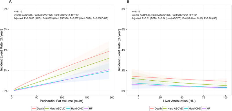
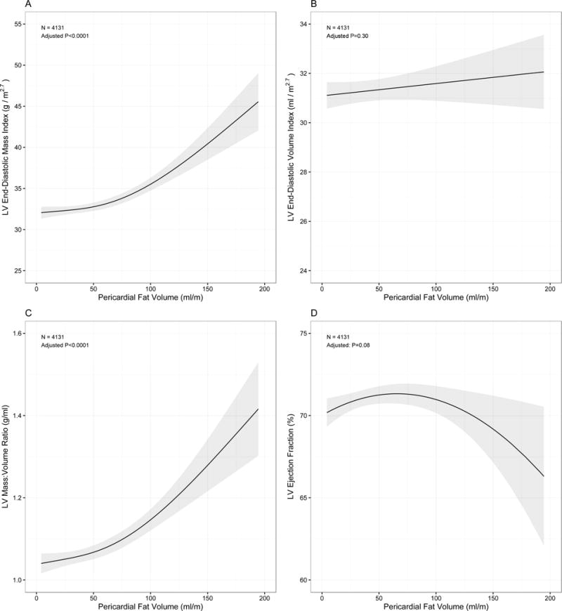
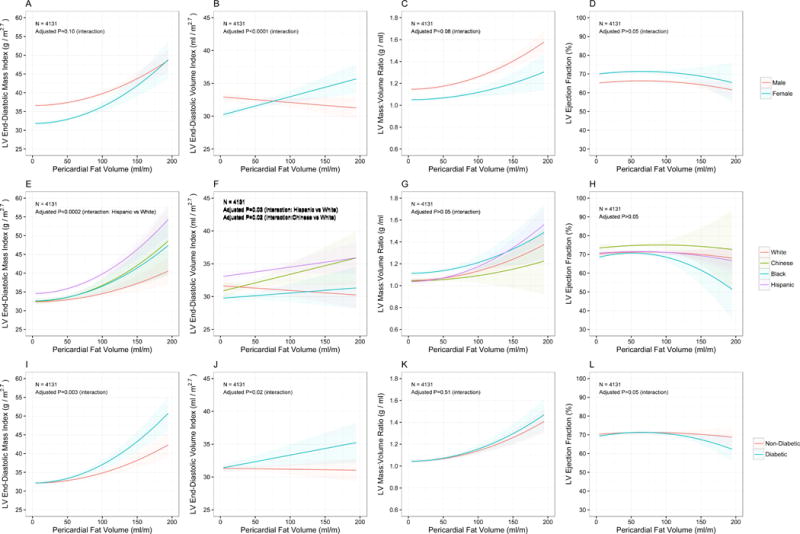
Methods: We studied 4,234 participants enrolled in the MESA (Multi-Ethnic Study of Atherosclerosis) study with concomitant cardiac magnetic resonance imaging and computed tomography (CT) measurements for pericardial fat volume and hepatic attenuation (a measure of liver fat). Poisson and Cox regression were used to estimate the annualized risk of incident hard atherosclerotic CVD (ASCVD), all-cause death, heart failure, all-cause CVD, hard coronary heart disease, and stroke as a function of pericardial and hepatic fat. Generalized additive models were used to assess the association between cardiac magnetic resonance indices of left ventricular (LV) structure and function and pericardial fat. Models were adjusted for relevant clinical, demographic, and cardiometabolic covariates.
Results: MESA study participants with higher pericardial and hepatic fat were more likely to be older, were more frequently men, and had a higher prevalence of cardiometabolic risk factors (including dysglycemia, dyslipidemia, hypertension), as well as adiposity-associated inflammation. Over a median 12.2-year follow-up (interquartile range: 11.6 to 12.8 years), pericardial fat was associated with a higher rate of incident hard ASCVD (standardized hazard ratio: 1.22; 95% confidence interval: 1.10 to 1.35; p = 0.0001). Hepatic fat by CT was not significantly associated with hard ASCVD (standardized hazard ratio: 0.96; 95% confidence interval: 0.86 to 1.08; p = 0.52). Higher pericardial fat was associated with greater indexed LV mass (37.8 g/m2.7 vs. 33.9 g/m2.7, highest quartile vs. lowest quartile; p < 0.01), LV mass-to-volume ratio (1.2 vs. 1.1, highest quartile vs. lowest quartile; p < 0.01). In adjusted models, a higher pericardial fat volume was associated with greater LV mass (p < 0.0001) and concentricity (p < 0.0001).
Conclusions: Pericardial fat is associated with poorer CVD prognosis and LV remodeling, independent of insulin resistance, inflammation, and CT measures of hepatic fat.
Keywords: cardiac magnetic resonance imaging; hepatic fat; obesity; pericardial fat; remodeling.
Copyright © 2017 American College of Cardiology Foundation. Published by Elsevier Inc. All rights reserved.
Figures
Comment in
-
Pericardial Fat and CVD: Is All Fat Created Equally?JACC Cardiovasc Imaging. 2017 Sep;10(9):1028-1030. doi: 10.1016/j.jcmg.2016.11.018. Epub 2017 Mar 15. JACC Cardiovasc Imaging. 2017. PMID: 28330656 No abstract available.
References
-
- Powell BD, Redfield MM, Bybee KA, Freeman WK, Rihal CS. Association of obesity with left ventricular remodeling and diastolic dysfunction in patients without coronary artery disease. Am J Cardiol. 2006;98:116–20. - PubMed
-
- Peterson LR, Waggoner AD, Schechtman KB, Meyer T, Gropler RJ, Barzilai B, Davila-Roman VG. Alterations in left ventricular structure and function in young healthy obese women: assessment by echocardiography and tissue Doppler imaging. J Am Coll Cardiol. 2004;43:1399–404. - PubMed
-
- Lavie CJ, Milani RV, Ventura HO, Cardenas GA, Mehra MR, Messerli FH. Disparate effects of left ventricular geometry and obesity on mortality in patients with preserved left ventricular ejection fraction. Am J Cardiol. 2007;100:1460–4. - PubMed
-
- Heckbert SR, Post W, Pearson GD, Arnett DK, Gomes AS, Jerosch-Herold M, Hundley WG, Lima JA, Bluemke DA. Traditional cardiovascular risk factors in relation to left ventricular mass, volume, and systolic function by cardiac magnetic resonance imaging: the Multiethnic Study of Atherosclerosis. J Am Coll Cardiol. 2006;48:2285–92. - PMC - PubMed
Publication types
MeSH terms
Grants and funding
- R01 HL071739/HL/NHLBI NIH HHS/United States
- N01 HC095169/HL/NHLBI NIH HHS/United States
- N01 HC095161/HC/NHLBI NIH HHS/United States
- N01 HC095164/HC/NHLBI NIH HHS/United States
- R01 HL085323/HL/NHLBI NIH HHS/United States
- N01 HC095160/HC/NHLBI NIH HHS/United States
- N01 HC095159/HL/NHLBI NIH HHS/United States
- N01 HC095169/HC/NHLBI NIH HHS/United States
- N01 HC095165/HC/NHLBI NIH HHS/United States
- N01 HC095163/HC/NHLBI NIH HHS/United States
- N01 HC095159/HC/NHLBI NIH HHS/United States
- N01 HC095162/HC/NHLBI NIH HHS/United States
- K23 HL127099/HL/NHLBI NIH HHS/United States
- N01 HC095165/HL/NHLBI NIH HHS/United States
LinkOut - more resources
Full Text Sources
Other Literature Sources
Medical
