Clinical Trial
. 2017 Mar 22;9(382):eaaf9194.
doi: 10.1126/scitranslmed.aaf9194.

Influenza vaccines differentially regulate the interferon response in human dendritic cell subsets

Affiliations
Clinical Trial

Influenza vaccines differentially regulate the interferon response in human dendritic cell subsets

Shruti Athale et al. Sci Transl Med. .

Abstract

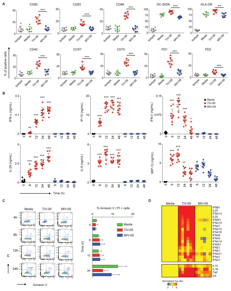
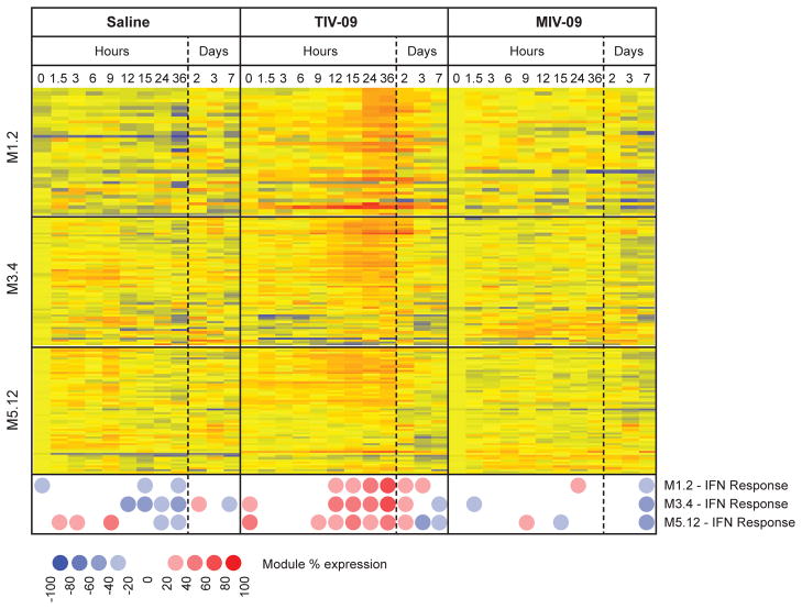
Human dendritic cells (DCs) play a fundamental role in the initiation of long-term adaptive immunity during vaccination against influenza. Understanding the early response of human DCs to vaccine exposure is thus essential to determine the nature and magnitude of maturation signals that have been shown to strongly correlate with vaccine effectiveness. In 2009, the H1N1 influenza epidemics fostered the commercialization of the nonadjuvanted monovalent H1N1 California vaccine (MIV-09) to complement the existing nonadjuvanted trivalent Fluzone 2009-2010 vaccine (TIV-09). In retrospective studies, MIV-09 displayed lower effectiveness than TIV-09. We show that TIV-09 induces monocyte-derived DCs (moDCs), blood conventional DCs (cDCs), and plasmacytoid DCs (pDCs) to express CD80, CD83, and CD86 and secrete cytokines. TIV-09 stimulated the secretion of type I interferons (IFNs) IFN-α and IFN-β and type III IFN interleukin-29 (IL-29) by moDC and cDC subsets. The vaccine also induced the production of IL-6, tumor necrosis factor, and the chemokines IFN-γ-inducible protein 10 (IP-10) and macrophage inflammatory protein-1β (MIP-1β). Conversely, MIV-09 did not induce the production of type I IFNs in moDCs and blood cDCs. Furthermore, it inhibited the TIV-09-induced secretion of type I IFNs by these DCs. However, both vaccines induced pDCs to secrete type I IFNs, indicating that different influenza vaccines activate distinct molecular signaling pathways in DC subsets. These results suggest that subtypes of nonadjuvanted influenza vaccines trigger immunity through different mechanisms and that the ability of a vaccine to induce an IFN response in DCs may offset the absence of adjuvant and increase vaccine efficacy.


Conflict of interest statement

Competing interests: The authors declare no competing interest.

Figures

Figure 1
Figure 1. TIV-09, but not MIV-09, activates moDCs
(A) moDCs were stimulated with TIV-09 (6 μL/mL), MIV-09 (6 μL/mL) or media for 18h. Surface expression of moDC activation markers was assessed by flow cytometry (N=7). (B) moDCs were cultured in the presence of TIV-09 or MIV-09 (6 μL/mL) for 0, 6, 12, 24 and 48h. Supernatants were harvested for quantification of cytokines by Luminex (N=10). Statistics shown represent the comparison between TIV-09 and MIV-09 at each time point. (C) moDCs were stained with Annexin V and PI to examine cell death at 4h, 8h, 12h and 24h post-vaccine treatment (N =6). (D) Transcriptional analysis of IFNs and other cytokines in moDCs cultured with TIV-09 or MIV-09 for 6h. Data were normalized to media control (N =6). (Mann-Whitney test; *: p<0.05; **: p<0.01; ***: p<0.001).
Figure 2
Figure 2. TIV-09 is a strong inducer of type I IFN
(A) moDCs were treated with increasing doses of TIV-09 with or without anti-IFNAR antibody (10 μg/mL) for 8h. Supernatants were assessed by Luminex for the presence of type I IFN and inflammatory cytokines. Statistics shown represent the comparison between TIV-09 and TIV-09 with IFNα/β R2 antibody at each concentration. (Mann-Whitney test; *: p<0.05; **: p<0.01; ***: p<0.001). (B) moDCs were stimulated with TIV-09 (6 μL/mL) with or without anti-IFNAR antibody for 18h. Surface expression of DC markers was assessed by flow cytometry. The figure is representative of experiments in four independent donors.
Figure 3
Figure 3. MIV-09 inhibits type I IFN
(A) moDCs were treated with increasing doses of Influenza vaccines used during the 2006–2007, 2008–2009, 2009–2010 and 2010–2011 seasons. The graph summarizes data from seven donors. (B) moDCs were treated with 25 μL/mL TIV-09 combined with increasing doses of MIV-09 for 8h. Supernatants were harvested for ELISA. The graph summarizes data from seven donors. (C) moDCs were activated with TIV-09 (6 μL/mL), LPS (50 ng/mL), CD40L (1 μg/mL), R848 (3 μg/mL), or CL097 (5 μg/mL) alone or with MIV-09 (10 μL/mL) with or without recombinant IFN-α (2000 U/mL). Supernatant cytokine concentrations were assessed by Luminex. The graph summarizes data from seven donors. (D) moDCs were stimulated with TIV-09, LPS, CD40L, R848 or CL097 with or without MIV-09. The surface expression of moDC activation markers was examined by flow cytometry. The figure is representative of experiments in four independent donors. (E) Ingenuity Pathway Analysis of the transcripts unique to each vaccine and most highly expressed after vaccine exposure. The top three canonical pathways enriched in the transcripts induced by each vaccine are shown (N=3). (Mann-Whitney test; *: p<0.05; **: p<0.01; ***: p<0.001).
Figure 4
Figure 4. TIV-09 activates cDCs
(A) Sorted blood cDCs were stimulated in vitro for 18h with media alone, TIV-09 or MIV-09. Surface expression of activation markers was examined by flow cytometry. The figure is representative of experiments in four independent donors. (B) Transcriptional analysis of IFNs in cDCs stimulated with TIV-09 or MIV-09 for 6h. Data were normalized to medium control (N=3) (C) cDCs were activated in vitro with media, TIV-09 or MIV-09 for 0, 6, 24 and 48h. IP-10 levels were measured by ELISA. The graph summarizes data from six donors (D) cDCs were treated with TIV-09 (6 μL/mL), combined with different doses of MIV-09 for 8h. Supernatants were harvested and IP-10 was measured by ELISA. The graph summarizes data from six donors (E) cDCs were stimulated with TIV-09 in the presence or absence of MIV. Surface expression of CD80, CD86, CD83 and HLA-DR was measured by flow cytometry. The figure is representative of a result from experiments on four independent donors. (Mann-Whitney test; *: p<0.05; **: p<0.01; ***: p<0.001).
Figure 5
Figure 5. TIV-09 activates pDCs
(A) Sorted pDCs were activated in vitro with TIV-09 or MIV-09 or control media for 18h and surface expression of activation markers was measured by flow cytometry. The figure is representative of experiments in four independent donors (B) Transcriptional analysis of IFNs in pDCs stimulated with TIV-09 or MIV-09 for 6h. Data were normalized to medium control (N=3) (C, D) pDCs were activated in vitro with media, TIV-09 or MIV-09 for 0, 6, 24 and 48h. IP-10 and IFN-β levels were measured by ELISA. (E) pDCs were treated with TIV-09 (6 μL/mL), combined with different doses of MIV-09 for 8h. Supernatants were harvested and IP-10 levels were measured by ELISA. Figs 5 C, D and E summarize data from six donors. (F) pDC were stimulated with TIV-09 in the presence or absence of MIV surface expression of surface expression of CD80, CD86, CD83 and HLA-DR, measured by flow cytometry. The figure is representative of a result from experiments on four independent donors. (Mann-Whitney test; *: p<0.05; **: p<0.01; ***: p<0.001).
Figure 6
Figure 6. TIV-09 but not MIV-09 induces an IFN signature in the blood of vaccinated individuals at day 1
Healthy individuals were vaccinated at t=0h with saline, TIV-09 or MIV-09 (N=3 per group), and blood was drawn at 0h, 1.5h, 3h, 6h, 9h, 12h, 15h, 24h, 36h, 2d, 3d and 7d. Data were normalized to the median of the 0h samples across donors. Transcripts from three IFN blood modules (M1.2, M3.4 and M5.12) (36) were selected for analysis. Both transcript-level and module-level analyses are displayed. Module expression is calculated as the percentage of transcripts from a specific module that are over or underexpressed in a specific condition as compared to the 0h reference control.

Similar articles

Cited by

References

    1. Lambert LC, Fauci AS. Influenza vaccines for the future. N Engl J Med. 2010;363:2036–2044. - PubMed
    1. Gerdil C. The annual production cycle for influenza vaccine. Vaccine. 2003;21:1776–1779. - PubMed
    1. Khurana S, Verma N, Yewdell JW, Hilbert AK, Castellino F, Lattanzi M, Del Giudice G, Rappuoli R, Golding H. MF59 adjuvant enhances diversity and affinity of antibody-mediated immune response to pandemic influenza vaccines. Sci Transl Med. 2011;3:85ra48. - PMC - PubMed
    1. Rumke HC, Richardus JH, Rombo L, Pauksens K, Plassmann G, Durand C, Devaster JM, Dewe W, Oostvogels L. Selection of an adjuvant for seasonal influenza vaccine in elderly people: modelling immunogenicity from a randomized trial. BMC Infect Dis. 2013;13:348. - PMC - PubMed
    1. Clark TW, Pareek M, Hoschler K, Dillon H, Nicholson KG, Groth N, Stephenson I. Trial of 2009 influenza A (H1N1) monovalent MF59-adjuvanted vaccine. N Engl J Med. 2009;361:2424–2435. - PubMed

Publication types

LinkOut - more resources