. 2017 May 1;198(9):3576-3587.
doi: 10.4049/jimmunol.1601636. Epub 2017 Mar 22.

Cell-Specific Variation in E-Selectin Ligand Expression among Human Peripheral Blood Mononuclear Cells: Implications for Immunosurveillance and Pathobiology

Affiliations

Cell-Specific Variation in E-Selectin Ligand Expression among Human Peripheral Blood Mononuclear Cells: Implications for Immunosurveillance and Pathobiology

Mariana Silva et al. J Immunol. .

Abstract

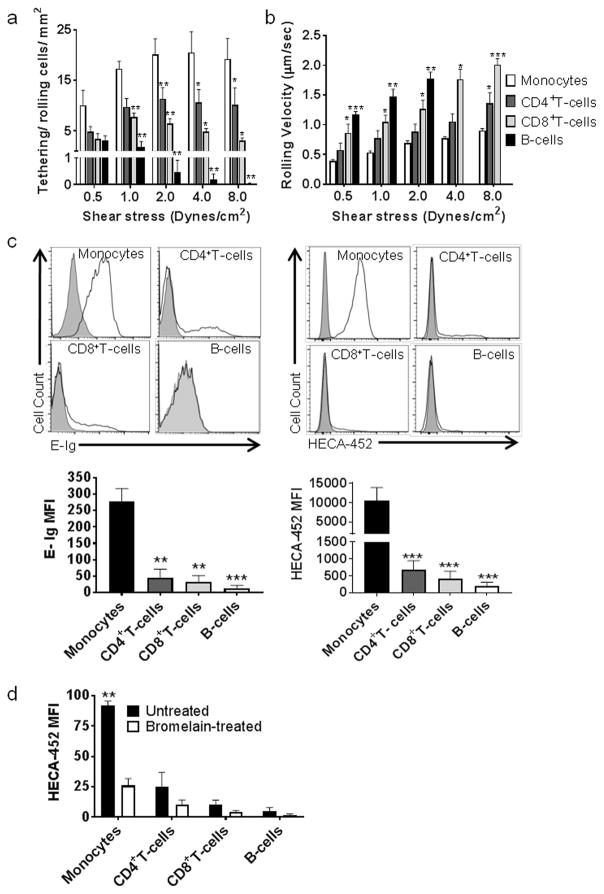
Both host defense and immunopathology are shaped by the ordered recruitment of circulating leukocytes to affected sites, a process initiated by binding of blood-borne cells to E-selectin displayed at target endothelial beds. Accordingly, knowledge of the expression and function of leukocyte E-selectin ligands is key to understanding the tempo and specificity of immunoreactivity. In this study, we performed E-selectin adherence assays under hemodynamic flow conditions coupled with flow cytometry and Western blot analysis to elucidate the function and structural biology of glycoprotein E-selectin ligands expressed on human PBMCs. Circulating monocytes uniformly express high levels of the canonical E-selectin binding determinant sialyl Lewis X (sLeX) and display markedly greater adhesive interactions with E-selectin than do circulating lymphocytes, which exhibit variable E-selectin binding among CD4+ and CD8+ T cells but no binding by B cells. Monocytes prominently present sLeX decorations on an array of protein scaffolds, including P-selectin glycoprotein ligand-1, CD43, and CD44 (rendering the E-selectin ligands cutaneous lymphocyte Ag, CD43E, and hematopoietic cell E-selectin/L-selectin ligand, respectively), and B cells altogether lack E-selectin ligands. Quantitative PCR gene expression studies of glycosyltransferases that regulate display of sLeX reveal high transcript levels among circulating monocytes and low levels among circulating B cells, and, commensurately, cell surface α(1,3)-fucosylation reveals that acceptor sialyllactosaminyl glycans convertible into sLeX are abundantly expressed on human monocytes yet are relatively deficient on B cells. Collectively, these findings unveil distinct cell-specific patterns of E-selectin ligand expression among human PBMCs, indicating that circulating monocytes are specialized to engage E-selectin and providing key insights into the molecular effectors mediating recruitment of these cells at inflammatory sites.


Figures

Figure 1
Figure 1. Native human blood monocytes exhibit greater E-selectin binding activity compared to native human blood lymphocytes
(a and b) Parallel-plate flow chamber analysis of tethering and rolling interactions of PBMCs on E-selectin-expressing endothelial cells. Freshly isolated human blood monocytes, CD4+ and CD8+ T-cells and B-cells were perfused over TNF-α-stimulated HUVECs in flow with shear stress ranging from 0.5 to 8 dynes/cm2. (a) The number of cells rolling on the HUVEC monolayer at shear stress levels of 0.5, 1, 2, 4 and 8 dynes/cm2 was quantified, normalized to 1×10⁶ cells/mL of infusate, and corrected by subtracting the values obtained using a function-blocking mAb to E-selectin. Monocytes and CD4+ T-cells engaged in rolling and adhesive interactions at shear stress levels of up to 8 dynes/cm2, whereas CD8+ showed rolling interactions most efficiently at 1–2 dynes/cm2, and B-cells engaged endothelium only at 0.5–1.0 dynes/cm2 shear stress. The number of rolling cells for monocytes was ~2-fold greater than CD4+ T-cells, >3-fold greater than CD8+ T-cells, and >10-fold greater than B-cells. (b) Rolling velocities of PBMCs on TNF-α activated HUVEC cells were calculated as the distance traveled divided by the time period of observation. Blood monocytes exhibited marked slower rolling velocities than CD4+ T-cells, CD8+ T-cells and B-cells. Statistical significance (*p<0.05; **p<0.01; ***p<0.001) was determined using paired t-test and refers to the difference in E-selectin-dependent rolling interactions between monocytes and the other PBMC subsets (either CD4+ T, CD8+ T or B-cells). Values are mean ± SEM of 5 independent experiments. (c) Representative flow cytometry histograms of E-Ig (left) and HECA-452 (right) staining of monocytes, CD4+ and CD8+ T-cells and B-cells. As shown, human monocytes express the highest levels of E-selectin ligands, followed by CD4+ and CD8+ T-cells, whereas B-cells lack E-Ig and HECA-452 reactivity. Filled histograms represent incubation with E-selectin-Ig in the absence of Ca2+ (EDTA treatment) or with isotype control, and open histograms represent incubation with E-Ig (in presence of 2 mM Ca2+) or HECA-452. Statistical significance (**p<0.01; ***p<0.001) was determined using paired t-test and refers to the difference between monocytes and the other PBMC subsets (either CD4+ T, CD8+ T or B-cells). Graph values are mean ± SEM of 4 (E-Ig) and 7 (HECA-452) independent experiments. (d) Effects of protease treatment on sLeX expression of native human PBMCs. Untreated (black bars) or bromelain-treated (white bars) cells were stained with HECA-452 mAb and analyzed by flow cytometry. Bromelain digestion decreased HECA-452 reactivity in all PBMC subsets tested. Statistical significance (**p<0.01) was determined using paired t-test and refers to the difference in HECA-452 reactivity between bromelain-treated and untreated cells. Data are mean ± SEM of 3 independent experiments.
Figure 2
Figure 2. Identification of glycoprotein E-selectin ligands on native human blood monocytes
(a) Western blot analysis of whole cell lysate of human monocytes stained with E-Ig. E-Ig staining of monocyte lysates resolved under reduced SDS-PAGE conditions revealed three principal bands at ~120–130 kDa, ~80–90 kDa and ~70 kDa. (b) Representative flow cytometry histograms of PSGL-1, CD43 and CD44 expression in human blood monocytes. Filled histograms represent incubation with isotype control and open histograms represent incubation with specific antibodies. As shown, human monocytes uniformly express PSGL-1, CD43 and CD44. Graph values are mean ± SEM of 6 independent experiments. (c) Western blot analysis of glycoprotein E-selectin ligands in human blood monocytes. E-Ig-immunoprecipitated (E-Ig IP) proteins from cell lysates of human blood monocytes were resolved by SDS-PAGE, blotted, and stained with anti-PSGL-1 and anti-CD43 antibodies (Left panel) or with anti-CD44 antibody (Right panel). In addition, CD44 was immunoprecipitated (CD44 IP) from cell lysates of human monocytes, resolved by SDS-PAGE, blotted, and stained with E-Ig (Right panel). Human monocytes express PSGL-1, CD43 and CD44 scaffolds that display E-selectin-reactive epitopes. (d) E-selectin-reactive 65–70 kDa unidentified glycoprotein in monocytes is a cell surface protein. Monocyte surface proteins were labeled with biotin derivative products and lysed. Biotinylated proteins were collected on streptavidin beads, resolved by SDS/PAGE, blotted and stained with E-Ig. Three E-Ig-reactive bands were observed, including the 65–70 kDa unidentified glycoprotein, confirming its expression on the cell surface. (e) Western blot analysis of E-Ig reactivity of myeloperoxidase (MPO) expressed on native human blood monocytes. MPO was immunoprecipitated from monocyte lysates, resolved by SDS-PAGE, blotted, and stained with anti-MPO mAb (Right panel) or E-Ig (Left panel). “Total” represents whole lysate and “Supn” corresponds to the cleared lysate, i.e., following MPO immunoprecipitation (“MPO IP”). As shown, MPO is not an E-selectin ligand in native human blood monocytes. (f) Western blot analysis of L-selectin staining of E-selectin ligands immunoprecipitated from circulating human monocytes. E-selectin ligands were immunoprecipitated from monocyte lysates, resolved by SDS-PAGE under non-reducing conditions, blotted, and stained with HECA-452 mAb (Right panel) or anti-L-selectin mAb (Left panel). “Total” represents whole lysate and “Supn” corresponds to the cleared lysate, i.e., following E-selectin ligand immunoprecipitation (“E-Ig IP”). L-selectin is not an E-selectin ligand on circulating monocytes, as indicated by lack of reactivity with anti-L-selectin mAb on the E-Ig IP product.
Figure 3
Figure 3. Identification of E-selectin ligands expressed on different subsets of lymphocytes
(a) Western blot analysis of E-Ig-reactive proteins in native human blood CD4+ T-cells, CD8+ T-cells and B-cells. Lysates of lymphocytes were resolved by SDS-PAGE electrophoresis, and immunoblotted with E-Ig chimera. CD4+ and CD8+ T-cells show two and one E-Ig reactive band, respectively, whereas B-cells lack E-Ig staining. (b) Flow cytometry analysis of PSGL-1, CD43 and CD44 expression on native human blood lymphocytes. Circulating CD4+ and CD8+ T-cells express high levels of PSGL-1, CD43 and CD44, whereas circulating B-cells only express CD44 and a small amount of CD43. Filled histograms represent incubation with isotype control and open histograms represent incubation with specific antibodies. Statistical significance (*p<0.05; **p<0.01, ***p<0.001) was determined using paired t-test and refers to the difference between B-cells and the other lymphocyte subsets (either CD4+ T or CD8+ T-cells). Graph values are mean ± SEM of 6 independent experiments. (c) Identification of glycoprotein E-selectin ligands expressed on native human blood T lymphocytes. PSGL-1, CD43 and CD44 were immunoprecipitated from whole cell lysates of circulating human CD4+ and CD8+ T-cells. Immunoprecipitates were then resolved by SDS-PAGE, blotted and stained with E-selectin-Ig chimera (E-Ig). Western blots reveal expression of E-selectin ligands CLA, CD43E and HCELL on human CD4+ T-cells, and CLA and CD43E on human CD8+ T-cells.
Figure 4
Figure 4. HCELL is a functional E-selectin ligand on circulating human monocytes and CD4+ T-cells
(a) Circulating human CD4+ T-cells and monocytes express two variant isoforms of CD44. Whole cell lysates of CD4+ T-cells (Left panel) and monocytes (Right panel) were resolved by SDS-PAGE, blotted and stained with CD44 mAb. In addition to the major expression of the standard (“CD44s”) isoform (~90kDa), western blots reveal the expression of two CD44 variant isoforms (~120 and 150 kDa) on native human blood CD4+ T-cells and monocytes. (b) CD44 expressed by human blood monocytes displays E-selectin binding determinants on O-glycans while CD44 expressed by human CD4+ T-cells displays E-selectin binding determinants on N-glycans. CD44 was immunoprecipitated from whole cell lysates of CD4+ T-cells (Left panel) and monocytes (Right panel). CD44 immunoprecipitates were buffer-treated or treated with PNGase-F. Samples were resolved by SDS-PAGE, blotted, and stained with either E-Ig (Top panel) or CD44 (Bottom panel). In contrast to results obtained from PNGase-F digestion of HCELL of circulating human CD4+T-cells, the persistence of E-Ig staining after PNGase-F digestion of HCELL from monocytes shows that CD44 displays sLeX on O-glycans. Black lines represent lanes from the same blot that have been cropped. (c) Blot rolling assay of HCELL isolated from circulating human CD4+ T-cells and monocytes. CD44 was immunoprecipitated from cell lysates of CD4+ T-cells and monocytes. Immunoprecipitates were resolved by SDS-PAGE, blotted and stained with HECA-452 mAb. Functional E-selectin ligand activity was assessed by perfusion of E-selectin-transfected CHO cells (CHO-E) over blots at 0.17 dynes/cm2. Non-specific adhesion was determined by assessing binding of CHO-E cells outside the HECA-452–reactive band (CHO-E off-band) and by perfusing mock-transfected CHO cells (CHO-M) over the blot. Statistical significance (**p<0.01; ***p<0.001) refers to the difference between specific E-selectin-dependent rolling interactions (CHO-E on band) and non-specific binding of CHO-E cells (CHO-E off band). Data are mean ± SEM of 3 independent experiments.
Figure 5
Figure 5. Analysis of glycosyltransferase gene expression on native human blood B-cells and monocytes
Real-time PCR analysis of glycosyltransferases involved in sLeX biosynthesis in circulating human B-cells (white bars) and monocytes (black bars). Compared to monocytes, B-cells express lower levels of α(1,3)-fucosyltransferase-related transcripts that direct sLeX synthesis, and higher levels of glycosyltransferases that compete against creation of sLeX. Experiments were performed with a minimum of five healthy donors. The mRNA expression of each glycosyltransferase was normalized to GAPDH. Statistical significance (*p<0.05; **p<0.01; ***p<0.001) refers to the difference in gene expression between human B-cells and monocytes, as determined using unpaired t-test.
Figure 6
Figure 6. E-selectin ligand expression on monocytes and B-cells is increased by cell surface α(1,3)-fucosylation
(a) FTVII-mediated exofucosylation of monocytes creates E-selectin ligands on multiple cell surface glycoproteins. Human monocytes were treated with fucosyltransferase VII (FTVII) or buffer alone (BT). Cell lysate was immunoprecipitated with E-selectin-Ig (E-Ig IP), resolved by SDS-PAGE, and blotted with HECA-452, anti-PSGL-1, anti-CD44 or anti-CD43 antibodies. A marked increase in E-Ig reactivity on the CD43 scaffold was observed, with smaller increases of E-Ig reactivity on PSGL-1 and CD44. Black lines represent different lanes from the same blot that have been cropped. (b) FTVII-mediated exofucosylation of monocytes creates a novel ~90-kDa E-selectin ligand. CD44 immunoprecipitation was performed on cell lysate from FTVII-treated monocytes. Total cell lysate (Total), CD44 immunoprecipitate (IP), and cleared lysate (Supn) were resolved by SDS-PAGE and blotted with anti-CD44 mAb (Left panel) or E-Ig (Right panel). Following complete clearance of CD44, E-Ig-reactivity persists at ~90 kDa, indicating enforced expression of a presently unknown E-selectin ligand. (c) FTVII-mediated exofucosylation of B-cells converts soley CD43 to an E-selectin ligand. Human B-cells were treated with fucosyltransferase VII (FTVII) or buffer alone (BT), and lysates were immunoprecipitated with CD43 mAb (CD43 IP) or CD44 mAb (CD44 IP). Immunoprecipitated products were resolved by SDS-PAGE and blotted with E-Ig. As shown in the blot, exofucosylation of B-cells creates E-selectin-binding determinants exclusively on the CD43 protein scaffold. Black line represents different parts from the same blot that have been cropped. (d) FTVII treatment creates functional E-selectin ligands on monocytes and B-cells. Human monocytes and B-cells were treated with fucosyltransferase VII (FTVII) or buffer alone (BT), and were subsequently perfused into a parallel-plate flow chamber seeded with TNF-α-stimulated HUVEC at shear stresses of 0.5, 1, 2, 4 and 8 dynes/cm2. Both monocytes and B-cells exhibited increased tethering/rolling interactions and slower rolling velocities after FTVII treatment. To assure specificity of E-selectin-dependent rolling, we counted the number of cells rolling on the HUVEC monolayer and subtracted the number of rolling cells when the HUVEC monolayer was pre-incubated with a function blocking mAb to E-selectin. Statistical significance (*p<0.05; **p<0.01) refers to the difference in E-selectin-dependent tethering/rolling interactions between FTVII-treated and buffer-treated cell subsets, as determined using paired t-test. Values are mean ± SEM of 3 independent experiments.(–89)

References

    1. Angiari S. Selectin-mediated leukocyte trafficking during the development of autoimmune disease. Autoimmun Rev. 2015;14:984–995. - PubMed
    1. Ley K, Laudanna C, Cybulsky MI, Nourshargh S. Getting to the site of inflammation: the leukocyte adhesion cascade updated. Nature reviews Immunology. 2007;7:678–689. - PubMed
    1. Sackstein R. Glycosyltransferase-programmed stereosubstitution (GPS) to create HCELL: engineering a roadmap for cell migration. Immunol Rev. 2009;230:51–74. - PMC - PubMed
    1. Bevilacqua M, Butcher E, Furie B, Gallatin M, Gimbrone M, Harlan J, Kishimoto K, Lasky L, McEver R. Selectins: a family of adhesion receptors. Cell. 1991;67:233. - PubMed
    1. Yao L, Setiadi H, Xia L, Laszik Z, Taylor FB, McEver RP. Divergent inducible expression of P-selectin and E-selectin in mice and primates. Blood. 1999;94:3820–3828. - PubMed