. 2017 May 1;198(9):3536-3547.
doi: 10.4049/jimmunol.1602087. Epub 2017 Mar 22.

IL-24 Promotes Pseudomonas aeruginosa Keratitis in C57BL/6 Mouse Corneas

Affiliations

IL-24 Promotes Pseudomonas aeruginosa Keratitis in C57BL/6 Mouse Corneas

Bing X Ross et al. J Immunol. .

Abstract

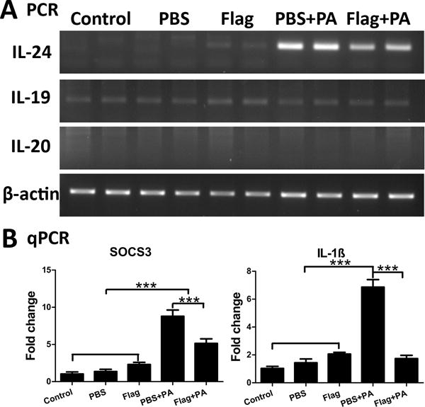
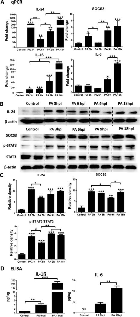
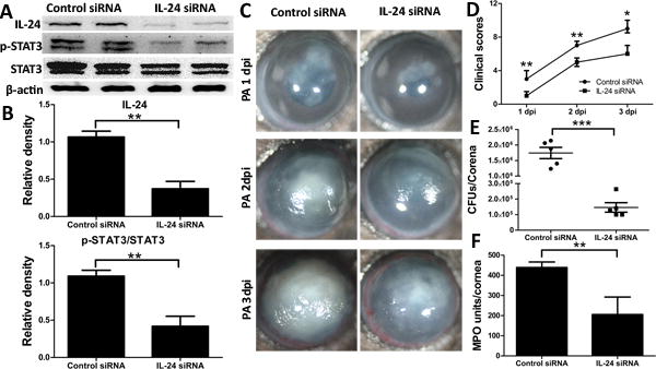
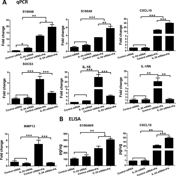
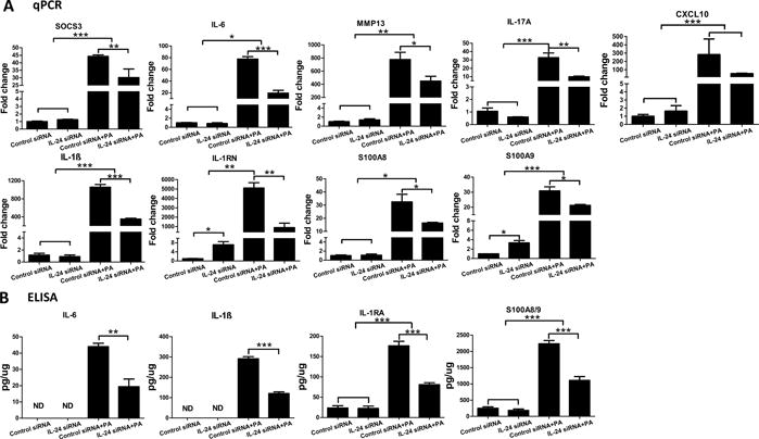
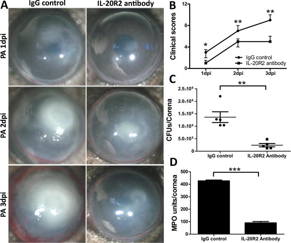
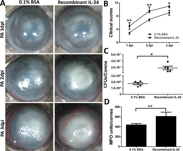
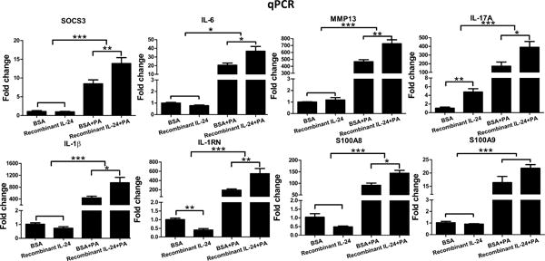
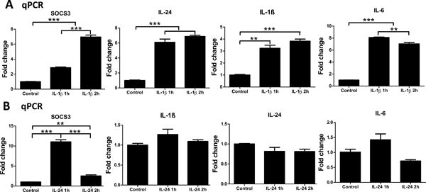
The aim of this study was to elucidate the expression and functions of IL-24 in C57BL/6 mouse corneas in response to Pseudomonas aeruginosa infection. Among IL-20R cytokines, only IL-24 was induced at both mRNA and protein levels by infection at early time points. The upregulation of IL-24 was dampened by flagellin pretreatment, which protects the corneas from microbial infection. Time course studies revealed bimodal early and later peaks of IL-24 expression, a pattern shared with suppressor of cytokine signaling (SOCS)3 but not IL-1β or IL-6. Silencing of IL-24 enhanced S100A8/A9 expression and suppressed SOCS3, IL-1β, IL-1RN, and matrix metalloproteinase 13 expression at 6 h postinfection. Downregulation of the IL-24 signaling pathway significantly reduced the severity of keratitis, whereas rIL-24 exacerbated P. aeruginosa-mediated tissue destruction. In vitro, rIL-1β induced the expression of SOCS3, IL-24, IL-1β, and IL-6 in primary cultured human corneal epithelial cells. rIL-24, alternatively, stimulated the expression of SOCS3, but not the others. In conclusion, IL-24 promotes P. aeruginosa keratitis through the suppression of early protective mucosal immunity, culminating in increased severity of P. aeruginosa keratitis.


Figures

Figure 1
Figure 1. P. aeruginosa
(PA) infection induces IL-24 and SOCS3 expression in B6 mouse corneal epithelial cells; Flagellin pretreatment attenuates their expression. Mouse corneas were scratched with a needle and topically pretreated with 500ng flagellin for 24 hours. PBS was used as control. Then the corneas were scratched again and inoculated with PA (ATCC 19660, CFUs: 1.0×10⁴). Cornea epithelia were collected at 6 hpi for semi-quantitative RT-PCR (A) or quantitative real-time PCR (B) analysis of IL-24, IL-19, and IL-20 (A), SOCS3 and IL-1β (B). Results are presented relative to those of naïve corneas, set as 1. P values were generated by one-way ANOVA. ***P < 0.001. Data are representative of three independent experiments (B: mean + s.e.m.). Flag: flagellin.
Figure 2
Figure 2
IL-24, SOCS3, and IL-1β are early responsive genes after PA infection in B6 mouse cornea. Mouse corneas were scratched with a needle and inoculated with PA. Whole corneas were collected at 3 hpi, 6 hpi, 9 hpi, and 18 hpi for qPCR analysis of IL-24, IL-1β, SOCS3, and IL-6 (A). Results are presented relative to those of naïve corneas, set as 1. (B) Immunoblot analysis of IL-24, SOCS3, p-STAT3, and STAT3 in cell lysates of whole corneas infected with PA for 3h, 6h, 9 h and 18 h. β-actin serves as the loading control. (C) Quantification of protein levels based on the densitometry of the Western blots in B. (D) ELISA assays of IL-1β and IL-6 in cell lysates of whole corneas infected with PA for 9 h or 18 h. Stars on top of columns are P value results comparing with the control. *P < 0.05, **P < 0.01, and ***P < 0.001 (ANOVA). Data are representative of three independent experiments (A, C and D: mean + s.e.m.). ND: not detected.
Figure 3
Figure 3
Silencing of IL-24 attenuates the severity of PA infection in mouse cornea. Mice were subconjunctivally injected with IL-24 siRNA (50 picomoles in 5ul RNase free water) twice in two days. Corneas were inoculated with PA 6h after the second injection. Non-specific siRNA serves as control. (A) Immunoblot analysis of IL-24, p-STAT3, and STAT3 in cell lysates of IL-24 siRNA- or control siRNA-treated corneas at 1 dpi. β-actin serves as the loading control. (B) Quantification of protein levels based on the densitometry of the Western blots in A. (C) Mice were monitored and photographed daily up to 3 dpi. (D) Clinical scores were assigned to each cornea daily and plotted as median + interquartile range. At 3 dpi, the corneas were excised and subjected to bacterial plate counting (E) and MPO assay (F). P values were generated by Man-Whitney test (D) or unpaired student t test (B, E and F). *P < 0.05, **P < 0.01 and ***P < 0.001. Data are representative of three independent experiments with five mice per group (B, E and F: mean + s.e.m.).
Figure 4
Figure 4
Silencing of IL-24 upregulates the expression of calprotectin and CXCL10 in mouse corneal epithelial cells in response to PA infection. Mouse corneas were treated with IL-24 siRNA or control siRNA and inoculated with PA as in figure 3. Corneal epithelial samples were collected at 6hpi for qPCR (A) or ELISA (B) analysis. For q-PCR, results are presented relative to those of control siRNA treated, uninfected corneas, set as 1. *P < 0.05, **P < 0.01, and ***P < 0.001 (ANOVA). Data are representative of three independent experiments with at least four corneas per group (mean + s.e.m.).
Figure 5
Figure 5
Silencing of IL-24 dampens the expression of cytokines and antimicrobial peptides in mouse cornea in response to PA infection. Mouse corneas were treated with IL-24 siRNA or control siRNA and inoculated with PA as in figure 3. (A) Whole cornea samples were collected at 1dpi for qPCR analysis. Results are presented relative to those of control siRNA treated, uninfected corneas, set as 1. (B) Cell lysates from whole corneal samples at 1dpi were subjected to ELISA analysis. *P < 0.05, **P < 0.01, and ***P < 0.001 (ANOVA or unpaired student T test). Data are representative of three independent experiments with at least four corneas per group (mean + s.e.m.). ND: not detected.
Figure 6
Figure 6
IL-20R2 neutralizing antibody decreases the severity of PA keratitis in mouse cornea. Mice were subconjunctivally injected with IL-20R2 neutralizing antibody (200ng/5ul) 4h before the inoculation with PA. Mouse IgG serves as the control. (A) Mice were monitored and photographed daily up to 3 dpi. (B) Clinical scores were assigned to each cornea daily and plotted as median + interquartile range. At 3 dpi, the corneas were excised and subjected to bacterial plate counting (C) and MPO assay (D). P values were generated by Man-Whitney test (B) or unpaired student t test (C and D). *P < 0.05, **P < 0.01 and ***P < 0.001. Data are representative of three independent experiments with five mice per group (C and D: mean + s.e.m.).
Figure 7
Figure 7
Recombinant IL-24 increases the susceptibility of the mouse cornea to PA infection. Mice were subconjunctivally injected with either recombinant mouse IL-24 (200ng in 5ul 0.1% BSA) or 0.1% BSA as control. Mouse corneas were inoculated with PA 4 h after the injection of recombinant IL-24. (A) The infected corneas were photographed daily. (B) The severity of keratitis was assessed with clinical scores. At 3 dpi, the corneas were excised and subjected to bacterial load (C) and MPO unit determination (D). P values were generated by Man-Whitney test (B) or unpaired student t test (C and D). *P < 0.05 and **P < 0.01. Data are representative of three independent experiments with five mice per group (B: median + interquartile range; C and D: mean + s.e.m.).
Figure 8
Figure 8
Recombinant IL-24 enhances cytokine expression in mouse cornea in response to PA infection. Mouse corneas were treated with recombinant mouse IL-24 or 0.1%BSA and inoculated with PA as in figure 7. Whole cornea samples were collected at 1dpi for qPCR analysis. Results are presented relative to those of 0.1% BSA treated, uninfected corneas, set as 1. *P < 0.05, **P < 0.01, and ***P < 0.001 (ANOVA). Data are representative of three independent experiments with at least four corneas per group (mean + s.e.m.).
Figure 9
Figure 9
Effects of recombinant human IL-1β or IL-24 on primary human corneal epithelial cells (HCECs). (A) HCECs were treated with recombinant human IL-1β (50ng/ml). Cells were collected at 1h and 2h for qPCR analysis of SOCS3, IL-24, IL-1β, and IL-6. (B) HCECs were treated with recombinant human IL-24 (100ng/ml). Cells were collected at 1h and 2h for qPCR analysis of SOCS3, IL-1β, IL-24, and IL-6. Results are presented relative to those of untreated control cells, set as 1 (mean+s.e.m.). **P < 0.01, and ***P < 0.001 (ANOVA). Data are representative of three independent experiments.

Similar articles

Cited by

References

    1. Whitcher JP, Srinivasan M, Upadhyay MP. Corneal blindness: a global perspective. Bull World Health Organ. 2001;79:214–221. - PMC - PubMed
    1. Stapleton F, Carnt N. Contact lens-related microbial keratitis: how have epidemiology and genetics helped us with pathogenesis and prophylaxis. Eye. 2012;26:185–193. - PMC - PubMed
    1. Molina DN, Colon M, Bermudez RH, Ramirez-Ronda CH. Unusual presentation of Pseudomonas aeruginosa infections: a review. Boletin de la Asociacion Medica de Puerto Rico. 1991;83:160–163. - PubMed
    1. Willcox MD. Pseudomonas aeruginosa infection and inflammation during contact lens wear: a review. Optometry and vision science: official publication of the American Academy of Optometry. 2007;84:273–278. - PubMed
    1. Hashimoto C, Hudson KL, Anderson KV. The Toll gene of Drosophila, required for dorsal-ventral embryonic polarity, appears to encode a transmembrane protein. Cell. 1988;52:269–279. - PubMed

MeSH terms

LinkOut - more resources