. 2017 Mar;5(6):e13146.
doi: 10.14814/phy2.13146.

Pressure overload leads to an increased accumulation and activity of mast cells in the right ventricle

Affiliations

Pressure overload leads to an increased accumulation and activity of mast cells in the right ventricle

Himal Luitel et al. Physiol Rep. 2017 Mar.

Abstract

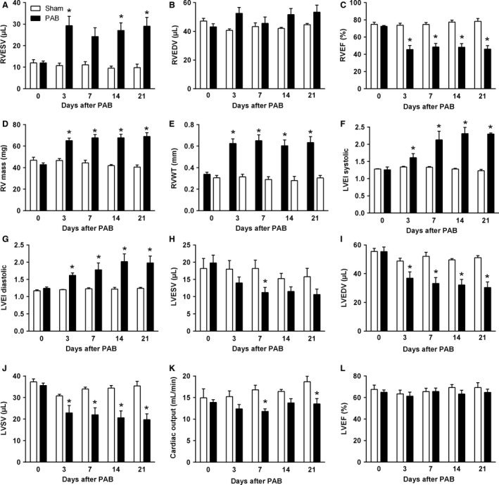
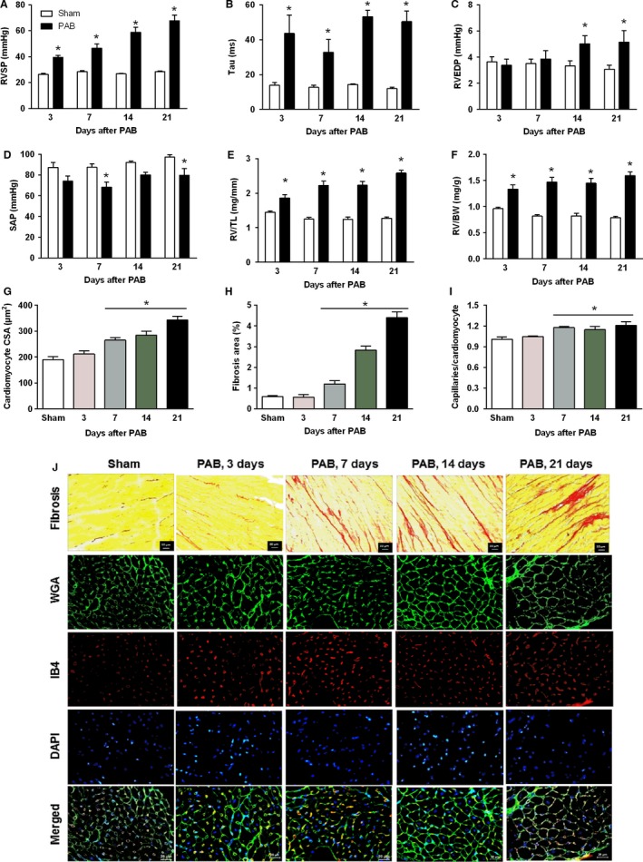
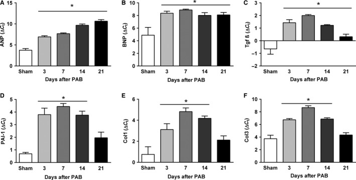
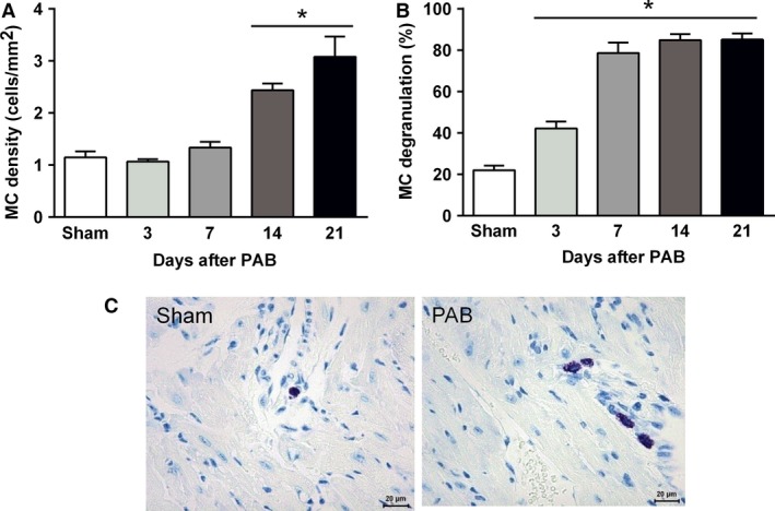
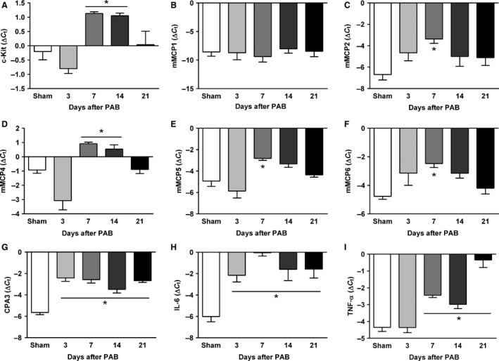
Right ventricular (RV) remodeling represents a complex set of functional and structural adaptations in response to chronic pressure or volume overload due to various inborn defects or acquired diseases and is an important determinant of patient outcome. However, the underlying molecular mechanisms remain elusive. We investigated the time course of structural and functional changes in the RV in the murine model of pressure overload-induced RV hypertrophy in C57Bl/6J mice. Using magnetic resonance imaging, we assessed the changes of RV structure and function at different time points for a period of 21 days. Pressure overload led to significant dilatation, cellular and chamber hypertrophy, myocardial fibrosis, and functional impairment of the RV Progressive remodeling of the RV after pulmonary artery banding (PAB) in mice was associated with upregulation of myocardial gene markers of hypertrophy and fibrosis. Furthermore, remodeling of the RV was associated with accumulation and activation of mast cells in the RV tissue of PAB mice. Our data suggest possible involvement of mast cells in the RV remodeling process in response to pressure overload. Mast cells may thus represent an interesting target for the development of new therapeutic approaches directed specifically at the RV.

Keywords: Mast cell; pulmonary artery banding; right ventricular hypertrophy.


Figures

Figure 1
Figure 1
Characterization of right and left ventricular structure and function and interventricular interaction. (A) Right ventricular end‐systolic volume. (B) Right ventricular end‐diastolic volume. (C) Right ventricular ejection fraction. (D) Right ventricular mass. (E) Right ventricular wall thickness. (F) Left ventricular eccentricity index during end‐systole. (G) Left ventricular eccentricity index during end‐diastole. (H) Left ventricular end‐systolic volume. (I) Left ventricular end‐diastolic volume. (J) Left ventricular stroke volume. (K) Cardiac output. (L) Left ventricular ejection fraction. Values are means ± SEM. *P < 0.05 versus sham at corresponding time points, n = 10 mice per group.
Figure 2
Figure 2
Characterization of hemodynamics and right ventricular remodeling. (A) Right ventricular systolic pressure. (B) Time constant of isovolumic relaxation of the right ventricle Tau. (C) Right ventricular end‐diastolic pressure. (D) Systemic arterial pressure. (E) Post‐mortem measured right ventricular weight values normalized to tibia length (RV/TL). (F) Post‐mortem measured right ventricular weight values normalized to body weight (RV/BW). (G) Bar graphs summarizing quantification of mean right ventricular cardiomyocyte cross‐sectional area. (H) Bar graphs summarizing quantification of interstitial fibrosis. (I) Bar graphs summarizing quantification of angiogenesis. (J) Representative images of picro‐sirius red staining of right ventricles cut in cross‐section and stained with wheat germ agglutinin‐FITC conjugate (WGA), isolectin B4‐TRITC conjugate (IB4), DAPI and merged. WGA marks cell boundaries (green), isolectin B4 (red) marks endothelial cells and DAPI marks nuclei (blue). Values are means ± SEM. *P < 0.05 vs. sham at corresponding time points, n = 10 mice per group for hemodynamic parameters and n = 5 mice per group for histological parameters.
Figure 3
Figure 3
Time course of the right ventricular mRNA gene expression of hypertrophic (A and B) and profibrotic markers (C–F) in sham and PAB mice. (A) Atrial natriuretic peptide. (B) B‐type natriuretic peptide. (C) Transforming growth factor 1. (D) Plasminogen activator inhibitor‐1. (E) Collagen 1 (Col1). (F) Collagen 3 (Col 3). Values are means ± SEM. *P < 0.05 versus sham at corresponding time points, n = 5 mice per group. SEM, standard error the mean.
Figure 4
Figure 4
Accumulation and activation of mast cells (MC) in the process of the RV remodeling. (A) Mast cell density. (B) Mast cell activity. (C) Representative images of mast cells in RV tissue from sham and PAB mice, n = 5 mice per group.
Figure 5
Figure 5
Time course of the right ventricular mRNA gene expression analysis of genes related to mast cells (A–G) and inflammation (H and I) in sham and PAB mice. (A) c‐Kit. (B) Murine mast cell protease (mMCP)‐1. (C) mMCP2. (D) mMCP4. (E) mMCP5. (F) mMCP6. (G) carboxypeptidase‐3 (CPA3). (H) IL‐6. (I) TNFα. Values are means ± SEM. *P < 0.05 versus sham at corresponding time points, n = 5 mice per group. PAB, pulmonary artery banding.

References

    1. Akers, I. A. , Parsons M., Hill M. R., Hollenberg M. D., Sanjar S., Laurent G. J., et al. 2000. Mast cell tryptase stimulates human lung fibroblast proliferation via protease‐activated receptor‐2. Am. J. Physiol. Lung Cell. Mol. Physiol. 278:L193–L201. - PubMed
    1. Ascah, K. J. , King M. E., Gillam L. D., and Weyman A. E.. 1990. The effects of right ventricular hemodynamics on left ventricular configuration. Can. J. Cardiol. 6:99–106. - PubMed
    1. Barnabei, M. S. , Palpant N. J., and Metzger J. M.. 2010. Influence of genetic background on ex vivo and in vivo cardiac function in several commonly used inbred mouse strains. Physiol. Genomics 42A:103–113. - PMC - PubMed
    1. Barrick, C. J. , Rojas M., Schoonhoven R., Smyth S. S., and Threadgill D. W.. 2007. Cardiac response to pressure overload in 129S1/SvImJ and C57BL/6J mice: temporal‐ and background‐dependent development of concentric left ventricular hypertrophy. Am. J. Physiol. Heart Circ. Physiol. 292:H2119–H2130. - PubMed
    1. Bartelds, B. , Borgdorff M. A., Smit‐van Oosten A., Takens J., Boersma B., Nederhoff M. G., et al. 2011. Differential responses of the right ventricle to abnormal loading conditions in mice: pressure vs. volume load. Eur. J. Heart Fail. 13:1275–1282. - PubMed

MeSH terms

LinkOut - more resources