. 2017 Jul 1;118(1):507-519.
doi: 10.1152/jn.00862.2016. Epub 2017 Mar 22.

Presynaptic and extrasynaptic regulation of posterior nucleus of thalamus

Affiliations

Presynaptic and extrasynaptic regulation of posterior nucleus of thalamus

Anthony Park et al. J Neurophysiol. .

Abstract

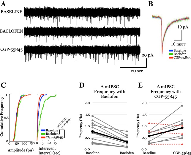
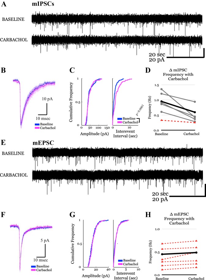
The posterior nucleus of thalamus (PO) is a higher-order nucleus involved in sensorimotor processing, including nociception. An important characteristic of PO is its wide range of activity profiles that vary across states of arousal, thought to underlie differences in somatosensory perception subject to attention and degree of consciousness. Furthermore, PO loses the ability to downregulate its activity level in some forms of chronic pain, suggesting that regulatory mechanisms underlying the normal modulation of PO activity may be pathologically altered. However, the mechanisms responsible for regulating such a wide dynamic range of activity are unknown. Here, we test a series of hypotheses regarding the function of several presynaptic receptors on both GABAergic and glutamatergic afferents targeting PO in mouse, using acute slice electrophysiology. We found that presynaptic GABAB receptors are present on both GABAergic and glutamatergic terminals in PO, but only those on GABAergic terminals are tonically active. We also found that release from GABAergic terminals, but not glutamatergic terminals, is suppressed by cholinergic activation and that a subpopulation of GABAergic terminals is regulated by cannabinoids. Finally, we discovered the presence of tonic currents mediated by extrasynaptic GABAA receptors in PO that are heterogeneously distributed across the nucleus. Thus we demonstrate that multiple regulatory mechanisms concurrently exist in PO, and we propose that regulation of inhibition, rather than excitation, is the more consequential mechanism by which PO activity can be regulated.NEW & NOTEWORTHY The posterior nucleus of thalamus (PO) is a key sensorimotor structure, whose activity is tightly regulated by inhibition from several nuclei. Maladaptive plasticity in this inhibition leads to severe pathologies, including chronic pain. We reveal here, for the first time in PO, multiple regulatory mechanisms that modulate synaptic transmission within PO. These findings may lead to targeted therapies for chronic pain and other disorders.

Keywords: GABA; glutamate; sensory processing; synaptic regulation.


Figures

Fig. 1.
Fig. 1.
A: schematic illustrating the major afferent and efferent pathways of posterior nucleus of thalamus (PO). PO receives excitatory drive from the periphery and from the cortex and projects excitatory projections to the cortex. It also receives inhibitory inputs from a number of GABAergic nuclei, including zona incerta (ZI), anterior pretectal nucleus (APT), and reticular nucleus of the thalamus (TRN). The reticular activating system, which regulates global levels of arousal, also acts on PO directly and indirectly through modulation of inhibition. B: schematic illustrating how modulation of presynaptic release from GABAergic afferents to PO can directly affect PO activity, a prediction of our previous computational work (Park et al. 2014).
Fig. 2.
Fig. 2.
GABAergic terminals synapsing onto PO neurons express GABAB receptors and are tonically active. A: representative whole cell voltage-clamp recordings of miniature inhibitory postsynaptic currents (mIPSCs) from a PO neuron [with high chloride intracellular solution, and in the presence of 0.5 µM tetrodotoxin (TTX) and 20.0 µM 6-cyano-7-nitroquinoxaline-2,3-dione (CNQX)] during baseline, baclofen-applied, and CGP-55845-applied conditions. Baclofen (10 µM) suppressed the frequency of mIPSCs, whereas CGP-55845 (2 μM) increased their frequency. B: overlaid averages of all individual events within each condition from the representative recording shown in A demonstrate that no postsynaptic changes occur with application of baclofen or CGP-55845. Shaded area around each curve depicts the 95% confidence interval. C: cumulative probability plots of the amplitude (left) and interevent interval (right) of the representative recording shown in A. Baclofen reduced mIPSC frequency by 65.3%. CGP-55845, not only reverses the effect of baclofen, but also further increases mIPSC frequency by 79.8% above baseline. No differences were observed in amplitude across conditions. D and E: group data plots depicting changes in frequency of mIPSCs in individual PO neurons between baseline and baclofen (D) or CGP-55845 (E). Solid black lines indicate average. Solid gray lines indicate statistically significant change from drug application by Kolmogorov-Smirnov test (K-S test), whereas dashed red lines indicate nonsignificant difference.
Fig. 3.
Fig. 3.
Glutamatergic terminals synapsing onto PO neurons express GABAB receptors but are not tonically active. A: representative whole cell voltage-clamp recordings of miniature excitatory postsynaptic currents (mEPSCs) from a PO neuron (in the presence of 0.5 µM TTX or 10.0 µM gabazine) during baseline, baclofen-applied, and CGP-55845-applied conditions. Baclofen (10 µM) suppressed the frequency of mEPSCs. CGP-55845 (2 µM) reversed these effects. B: overlaid averages of all individual events within each condition from the representative recording shown in A demonstrate that no postsynaptic changes occur with application of baclofen or CGP-55845. Shaded area around each curve depicts the 95% confidence interval. C: cumulative probability plots of the amplitude (left) and interevent interval (right) of the representative recording shown in A. No differences were observed in amplitude across conditions. Baclofen reduced mEPSC frequency by 25.2%. CGP-55845 reversed the effects of baclofen but did not produce further increases in frequency. D and E: group data plots depicting changes in mEPSC frequency in individual PO neurons between baseline and baclofen (D) or CGP-55845 (E). Solid black lines indicate average. Solid gray lines indicate statistically significant change from drug application by K-S test, whereas dashed red lines indicate nonsignificant difference.
Fig. 4.
Fig. 4.
GABAergic, but not glutamatergic, terminals synapsing onto PO neurons express functional cholinergic receptors. A and E: representative whole cell voltage-clamp recordings of mIPSCs (A) and mEPSCs (E) from a PO neuron [in the presence of 0.5 µM TTX, 20.0 µM CNQX (A), or 10.0 µM gabazine (E)] before and after application of carbachol (2.0 µM). B and F: overlaid averages of all individual events within each condition from the representative recording shown in A and E (respectively) demonstrate that no postsynaptic changes occur with application of carbachol. Shaded area around each curve depicts the 95% confidence interval. C and G: cumulative probability plots of the amplitude (left) and interevent interval (right) of the representative recordings shown in A and E, respectively. No differences were observed in amplitude across conditions in either mIPSC (C) or mEPSC (G) populations. Carbachol reduced mIPSC frequency by 47.7% (C), whereas mEPSC frequency remained unchanged (G). D and H: group data plots depicting changes in frequency of mIPSCs (D) and mEPSCs (H) in individual PO neurons before and after carbachol application. Solid black lines indicate average. Solid gray lines indicate statistically significant change from drug application by K-S test, whereas dashed red lines indicate nonsignificant difference.
Fig. 5.
Fig. 5.
Small subpopulation of GABAergic terminals in PO are influenced by cannabinoid signaling. A: representative whole cell voltage-clamp recordings of mIPSCs from a PO neuron (in the presence of 0.5 µM TTX or 20.0 µM CNQX) at baseline (top) and with application of 2.0 µM WIN 55,212-2 (middle) and 5.0 µM AM215 (bottom). WIN 55,212-2 produced a significant suppression of mIPSC frequency. AM251 application reversed these effects. B: cumulative probability plots of amplitude (left) and interevent interval (right) of the representative recording shown in A. WIN 55,212-2 reduced mEPSC frequency by 41.1%. AM251 reversed the effects of WIN 55,212-2, but did not produce further increases in frequency. No differences were observed in amplitude across conditions. C: overlaid averages of all individual events within each condition from the representative recording shown in A demonstrate that no postsynaptic changes occur with application of WIN 55,212-2 or AM251. Shaded area around each curve depicts the 95% confidence interval. D: representative whole cell voltage-clamp recording of mIPSCs, analogous to that in A, but of a PO neuron that does not respond to WIN 55,212-2 application. E: cumulative probability plots of amplitude (left) and interevent interval (right) of recording shown in D. No differences were observed in amplitude or frequency across conditions. F: overlaid averages of all individual events within each condition from the representative recording shown in D demonstrate that no postsynaptic changes occur with application of WIN 55,212-2 or AM251. Shaded area around each curve depicts the 95% confidence interval. G and H: group data plots depicting changes in frequency of mIPSCs in individual PO neurons between baseline and WIN 55,212-2 (G) or AM215 (H). No statistically significant difference was found between these groups despite 4 of 13 neurons exhibiting significant within-cell suppression of mIPSC frequency with WIN 55,212-2. I and J: group data plots depicting changes in frequency of mEPSCs in individual PO neurons between baseline and WIN 55,212-2 (I) or AM215 (J). No statistically significant difference was found between these groups. K: representative recording of mEPSCs from a PO neuron (in the presence of 0.5 µM TTX or 10.0 µM gabazine). L: cumulative probability plots of amplitude (left) and interevent interval (right) of the representative recording shown in K. No differences were observed in amplitude or interevent interval. M: overlaid averages of all individual events within each condition from the representative recording shown in K demonstrate that no postsynaptic changes occur with application of WIN 55,212-2 or AM251. Shaded area around each curve depicts the 95% confidence interval.
Fig. 6.
Fig. 6.
Some PO neurons exhibit tonic, GABAA-mediated currents. A: whole cell voltage-clamp recordings during bath application of 10 µM gabazine. Left: representative recording of a neuron responding to gabazine with an outward current (22.1 pA), indicating presence of extrasynaptic GABAA receptors that tonically mediate inward current (note, intracellular solution contains high [Cl]). Right: representative recording of a neuron lacking a response to gabazine application. B: histogram illustrating distribution of responses to GABAA receptor blockade (bin size = 3 pA).
Fig. 7.
Fig. 7.
Schematic showing PO presynaptic mechanisms revealed in the present study. PO neurons express presynaptic GABAB, cholinergic (m2) and CB1 receptors on GABAergic terminals, of which the GABAB receptor is tonically active. All 3 presynaptic mechanisms can suppress GABA release. On glutamatergic terminals, only presynaptic GABAB receptors are present but, in contrast to those found on GABAergic terminals, are not tonically active. These receptors can suppress release of glutamate. Extrasynaptic GABAA receptors can detect ambient GABA and/or GABA spillover to mediate tonic currents.

Similar articles

Cited by

References

    1. Ahissar E, Oram T. Thalamic relay or cortico-thalamic processing? Old question, new answers. Cereb Cortex 25: 845–848, 2015. doi:10.1093/cercor/bht296. - DOI - PubMed
    1. Alger BE. Endocannabinoids at the synapse a decade after the dies mirabilis (29 March 2001): what we still do not know. J Physiol 590: 2203–2212, 2012. doi:10.1113/jphysiol.2011.220855. - DOI - PMC - PubMed
    1. Andrade R. Blockade of neurotransmitter-activated K+ conductance by QX-314 in the rat hippocampus. Eur J Pharmacol 199: 259–262, 1991. doi:10.1016/0014-2999(91)90467-5. - DOI - PubMed
    1. Atwood HK, Malinow R, Manabe T, Poo M-m, Sakmann B, Silver PA. Central Synapses, Quantal Mechanisms and Plasticity (Human Frontier Workshop Reports, Workshop IV). Strasbourg, France: Human Frontier Science Program, 1998.
    1. Baksh MM, Kussrow AK, Mileni M, Finn MG, Bornhop DJ. Label-free quantification of membrane-ligand interactions using backscattering interferometry. Nat Biotechnol 29: 357–360, 2011. doi:10.1038/nbt.1790. - DOI - PMC - PubMed

Publication types

MeSH terms

LinkOut - more resources