

Case Reports
doi: 10.1136/bcr-2016-219048.
Pulmonary Kaposi sarcoma presenting as complete lung consolidation
Affiliations
- PMID: 28331023
- PMCID: PMC5372134
- DOI: 10.1136/bcr-2016-219048
Item in Clipboard
Case Reports
Pulmonary Kaposi sarcoma presenting as complete lung consolidation
BMJ Case Rep.
.
Abstract
The patient in our case presented with progressive dyspnoea and cough. Chest radiograph reveals complete opacification of the hemithorax. Complete lung consolidation was not seen on chest CT. The patient in this case had extensive pulmonary and endobronchial Kaposi sarcoma (KS) that led to complete consolidation of the right lung that was diagnosed via bronchoscopy. After diagnosis, he was restarted on antiretroviral therapy and single-agent chemotherapy for treatment of pulmonary KS.
2017 BMJ Publishing Group Ltd.
Conflict of interest statement
Figures
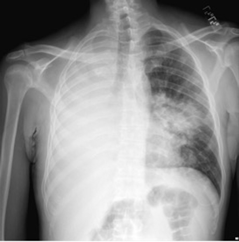
Chest radiograph with complete opacification of the right hemithorax, and multiple nodular opacities in the left lung field.
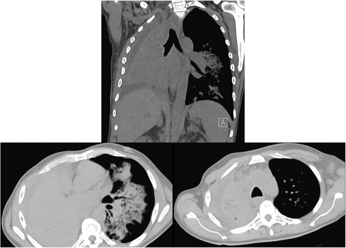
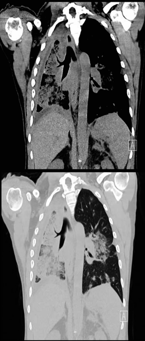
CT chest showing complete consolidation of the right lung with airbronchograms, right pleural effusion and multiple nodular opacities in the left lung centred around the bronchovascular bundles.
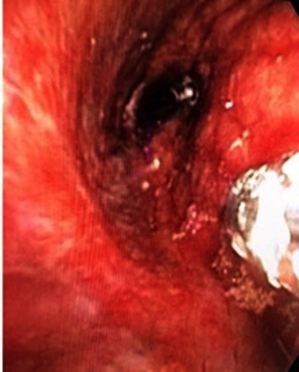
Bronchoscopic view showing narrowed right middle and lower lobe bronchi with diffuse red vascular lesions, biopsy forceps are in the right side of the image.
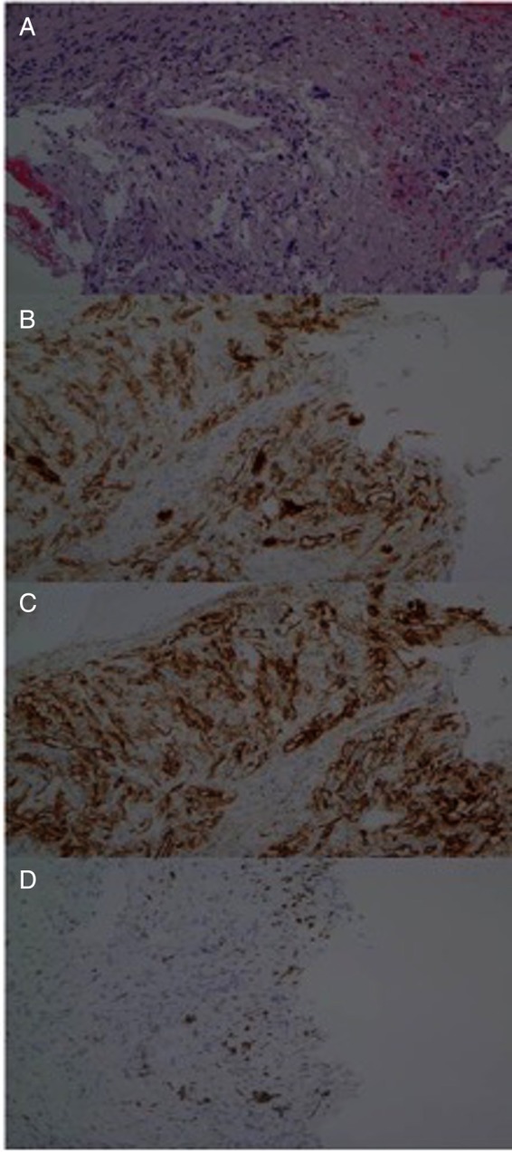
Histology of the endobronchial biopsy sample. (A) Spindle cells noted with ectatic blood vessels. (B) Spindle cells stain positive for CD31, CD34 in (C) and HHV-8 in (D).
CT chest 4 weeks after initiation of chemotherapy.
Similar articles
-
Pulmonary Kaposi's Sarcoma - Initial Presentation of HIV Infection.Folia Med (Plovdiv). 2019 Dec 31;61(4):643-649. doi: 10.3897/folmed.61.e47945. Folia Med (Plovdiv). 2019. PMID: 32337885
-
Chronic cough conundrum: a case report of a new diagnosis of HIV and pulmonary Kaposi's sarcoma.BMC Pulm Med. 2017 Mar 20;17(1):52. doi: 10.1186/s12890-017-0395-5. BMC Pulm Med. 2017. PMID: 28320359 Free PMC article.
-
A patient with HIV, dyspnea, and multiple pulmonary nodules: pulmonary Kaposi sarcoma.Chest. 2006 Dec;130(6):1924-8. doi: 10.1378/chest.130.6.1924. Chest. 2006. PMID: 17167017 No abstract available.
-
Pulmonary Kaposi's sarcoma revealed by a solitary nodule in a patient with acquired immunodeficiency syndrome.Am J Respir Crit Care Med. 1994 Apr;149(4 Pt 1):1041-3. doi: 10.1164/ajrccm.149.4.8143039. Am J Respir Crit Care Med. 1994. PMID: 8143039 Review.
-
Total resolution of ocular Kaposi sarcoma with different treatment approaches - a case series and review of literature.Int J STD AIDS. 2019 Jul;30(8):802-809. doi: 10.1177/0956462418825353. Epub 2019 May 2. Int J STD AIDS. 2019. PMID: 31046617 Review. No abstract available.
Cited by
-
Rare presentation of bronchopulmonary Kaposi sarcoma.BMJ Case Rep. 2019 Aug 26;12(8):e229436. doi: 10.1136/bcr-2019-229436. BMJ Case Rep. 2019. PMID: 31451457 Free PMC article.
-
Pulmonary involvement in acquired immunodeficiency syndrome-associated Kaposi's sarcoma: a descriptive analysis of thin-section manifestations in 29 patients.Quant Imaging Med Surg. 2021 Feb;11(2):714-724. doi: 10.21037/qims-20-284. Quant Imaging Med Surg. 2021. PMID: 33532271 Free PMC article.
-
Pulmonary Kaposi Sarcoma without Respiratory Symptoms and Skin Lesions in an HIV-Naïve Patient: A Case Report and Literature Review.Infect Dis Rep. 2022 Mar 25;14(2):228-242. doi: 10.3390/idr14020028. Infect Dis Rep. 2022. PMID: 35447880 Free PMC article.
-
Current and Future Tools for Diagnosis of Kaposi's Sarcoma.Cancers (Basel). 2021 Nov 25;13(23):5927. doi: 10.3390/cancers13235927. Cancers (Basel). 2021. PMID: 34885035 Free PMC article. Review.
References
Publication types
MeSH terms
Substances
LinkOut - more resources
Full Text Sources
Other Literature Sources
Medical