

Structural Characterization of Human Coronavirus NL63 N Protein
- PMID: 28331093
- PMCID: PMC5432860
- DOI: 10.1128/JVI.02503-16
Structural Characterization of Human Coronavirus NL63 N Protein
Abstract
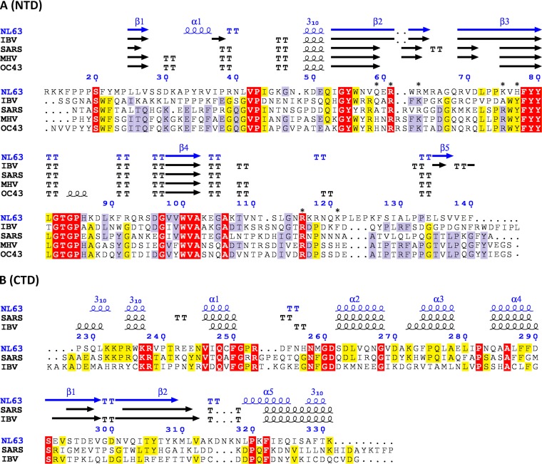
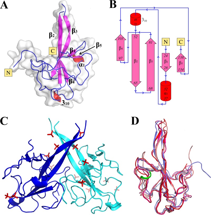
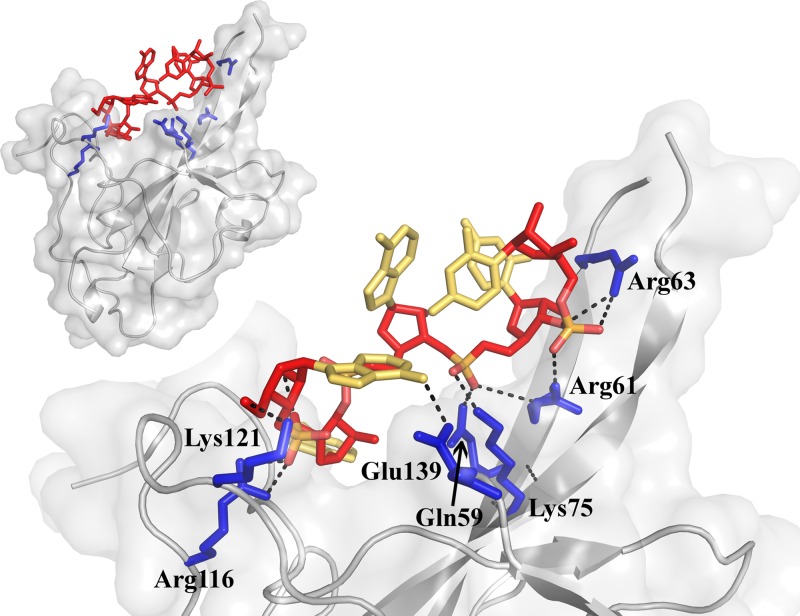
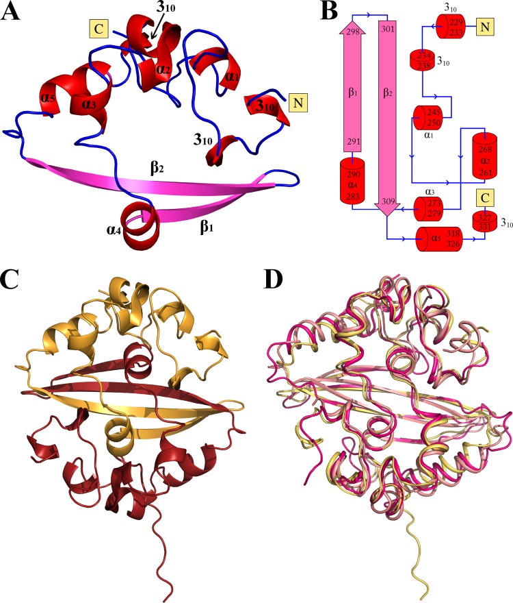
Coronaviruses are responsible for upper and lower respiratory tract infections in humans. It is estimated that 1 to 10% of the population suffers annually from cold-like symptoms related to infection with human coronavirus NL63 (HCoV-NL63), an alphacoronavirus. The nucleocapsid (N) protein, the major structural component of the capsid, facilitates RNA packing, links the capsid to the envelope, and is also involved in multiple other processes, including viral replication and evasion of the immune system. Although the role of N protein in viral replication is relatively well described, no structural data are currently available regarding the N proteins of alphacoronaviruses. Moreover, our understanding of the mechanisms of RNA binding and nucleocapsid formation remains incomplete. In this study, we solved the crystal structures of the N- and C-terminal domains (NTD, residues 10 to 140, and CTD, residues 221 to 340, respectively) of the N protein of HCoV-NL63, both at a 1.5-Å resolution. Based on our structure of NTD solved here, we proposed and experimentally evaluated a model of RNA binding. The structure of the CTD reveals the mode of N protein dimerization. Overall, this study expands our understanding of the initial steps of N protein-nucleic acid interaction and may facilitate future efforts to control the associated infections.IMPORTANCE Coronaviruses are responsible for the common cold and other respiratory tract infections in humans. According to multiple studies, 1 to 10% of the population is infected each year with HCoV-NL63. Viruses are relatively simple organisms composed of a few proteins and the nucleic acids that carry the information determining their composition. The nucleocapsid (N) protein studied in this work protects the nucleic acid from the environmental factors during virus transmission. This study investigated the structural arrangement of N protein, explaining the first steps of its interaction with nucleic acid at the initial stages of virus structure assembly. The results expand our understanding of coronavirus physiology and may facilitate future efforts to control the associated infections.
Keywords: CTD; N protein; NL63; NTD; coronavirus; nucleocapsid; structure.
Copyright © 2017 American Society for Microbiology.
Figures
References
-
- Drosten C, Gunther S, Preiser W, van der Werf S, Brodt HR, Becker S, Rabenau H, Panning M, Kolesnikova L, Fouchier RAM, Berger A, Burguiere AM, Cinatl J, Eickmann M, Escriou N, Grywna K, Kramme S, Manuguerra JC, Muller S, Rickerts V, Sturmer M, Vieth S, Klenk HD, Osterhaus ADME, Schmitz H, Doerr HW. 2003. Identification of a novel coronavirus in patients with severe acute respiratory syndrome. N Engl J Med 348:1967–1976. doi: 10.1056/NEJMoa030747. - DOI - PubMed
-
- Ksiazek TG, Erdman D, Goldsmith CS, Zaki SR, Peret T, Emery S, Tong S, Urbani C, Comer JA, Lim W, Rollin PE, Dowell SF, Ling AE, Humphrey CD, Shieh WJ, Guarner J, Paddock CD, Rota P, Fields B, DeRisi J, Yang JY, Cox N, Hughes JM, LeDuc JW, Bellini WJ, Anderson LJ, SARS Working Group. 2003. A novel coronavirus associated with severe acute respiratory syndrome. N Engl J Med 348:1953–1966. doi: 10.1056/NEJMoa030781. - DOI - PubMed
Publication types
MeSH terms
Substances
LinkOut - more resources
Full Text Sources
Other Literature Sources
Molecular Biology Databases
