. 2017 May 12;91(11):e00343-17.
doi: 10.1128/JVI.00343-17. Print 2017 Jun 1.

Recombinant Modified Vaccinia Virus Ankara Generating Ebola Virus-Like Particles

Affiliations

Recombinant Modified Vaccinia Virus Ankara Generating Ebola Virus-Like Particles

Marc Schweneker et al. J Virol. .

Abstract

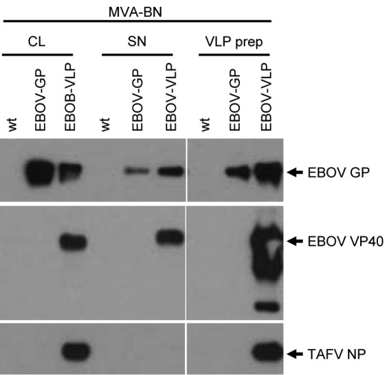
There are currently no approved therapeutics or vaccines to treat or protect against the severe hemorrhagic fever and death caused by Ebola virus (EBOV). Ebola virus-like particles (EBOV VLPs) consisting of the matrix protein VP40, the glycoprotein (GP), and the nucleoprotein (NP) are highly immunogenic and protective in nonhuman primates against Ebola virus disease (EVD). We have constructed a modified vaccinia virus Ankara-Bavarian Nordic (MVA-BN) recombinant coexpressing VP40 and GP of EBOV Mayinga and the NP of Taï Forest virus (TAFV) (MVA-BN-EBOV-VLP) to launch noninfectious EBOV VLPs as a second vaccine modality in the MVA-BN-EBOV-VLP-vaccinated organism. Human cells infected with either MVA-BN-EBOV-VLP or MVA-BN-EBOV-GP showed comparable GP expression levels and transport of complex N-glycosylated GP to the cell surface. Human cells infected with MVA-BN-EBOV-VLP produced large amounts of EBOV VLPs that were decorated with GP spikes but excluded the poxviral membrane protein B5, thus resembling authentic EBOV particles. The heterologous TAFV NP enhanced EBOV VP40-driven VLP formation with efficiency similar to that of the homologous EBOV NP in a transient-expression assay, and both NPs were incorporated into EBOV VLPs. EBOV GP-specific CD8 T cell responses were comparable between MVA-BN-EBOV-VLP- and MVA-BN-EBOV-GP-immunized mice. The levels of EBOV GP-specific neutralizing and binding antibodies, as well as GP-specific IgG1/IgG2a ratios induced by the two constructs, in mice were also similar, raising the question whether the quality rather than the quantity of the GP-specific antibody response might be altered by an EBOV VLP-generating MVA recombinant.IMPORTANCE The recent outbreak of Ebola virus (EBOV), claiming more than 11,000 lives, has underscored the need to advance the development of safe and effective filovirus vaccines. Virus-like particles (VLPs), as well as recombinant viral vectors, have proved to be promising vaccine candidates. Modified vaccinia virus Ankara-Bavarian Nordic (MVA-BN) is a safe and immunogenic vaccine vector with a large capacity to accommodate multiple foreign genes. In this study, we combined the advantages of VLPs and the MVA platform by generating a recombinant MVA-BN-EBOV-VLP that would produce noninfectious EBOV VLPs in the vaccinated individual. Our results show that human cells infected with MVA-BN-EBOV-VLP indeed formed and released EBOV VLPs, thus producing a highly authentic immunogen. MVA-BN-EBOV-VLP efficiently induced EBOV-specific humoral and cellular immune responses in vaccinated mice. These results are the basis for future advancements, e.g., by including antigens from various filoviral species to develop multivalent VLP-producing MVA-based filovirus vaccines.

Keywords: Ebola; MVA; VLP.


Figures

FIG 1
FIG 1
Cellular expression of Ebola virus GP, VP40, and NP driven by recombinant MVAs. (A) Schematic representation of wild-type MVA (MVA-BN) and MVA recombinants expressing Zaire ebolavirus glycoprotein (EBOV GP) either alone (MVA-EBOV-GP) or in combination with EBOV VP40 and TAFV NP (MVA-BN-EBOV-VLP). Expression cassettes were introduced into the MVA genome at IGRs (indicated by small arrows). The poxviral promoters PrS and PrS5E driving gene transcription of filoviral transgenes are indicated. (B to D) HeLa cells in 12-well plates were infected with the indicated viruses at an MOI of 10. (B) For Western blot analysis, cells were lysed at 6 h and 24 h p.i. in 150 μl of 1× Laemmli sample loading buffer. Cellular lysates prepared at 24 h p.i. were diluted 1:3, as indicated by the asterisks; separated by reducing SDS-PAGE; and transferred to polyvinylidene difluoride (PVDF) membranes. The blots were incubated with anti-GP antibody (MAb 6D8) and subsequently with anti-mouse IgG peroxidase conjugate. The immunoblots were developed with a standard chemiluminescent peroxidase substrate. (C) Analysis of N-glycosylation patterns of EBOV GP. HeLa cells were lysed in 150 μl of 1× RIPA buffer at 17 h p.i. Protein lysates were treated with Endo H or PNGase F or were left untreated (−). Following endoglycosidase treatment, the proteins were mixed with 3× Laemmli loading buffer, separated by reducing SDS-PAGE, and analyzed by immunoblotting using an anti-GP antibody (MAb 6D8). The immunoblot was developed using an ECL detection system with enhanced sensitivity. (D) Flow cytometric analysis of EBOV GP expression. At 4 h and 24 h p.i., HeLa cells were suspended in PBS-2% FCS and incubated with anti-EBOV GP antibody (MAb 6D8) and subsequently with anti-mouse IgG-allophycocyanin. The stained cells were analyzed by flow cytometry, and cell surface expression of EBOV GP is represented in the histogram plots. (E) HEK 293T/17 cells in 12-well plates were incubated for 2 h with the indicated viruses (MOI, 5). Thereafter, the inoculum was replaced with DMEM-2% FCS. One day after infection, adherent cells were lysed in 1× Laemmli loading buffer and analyzed by immunoblotting using antibodies directed to EBOV GP, EBOV VP40, or TAFV NP.
FIG 2
FIG 2
Intracellular distribution of EBOV GP, EBOV VP40, and TAFV NP expressed by recombinant MVAs. HeLa cells infected with MVA-BN-EBOV-VLP (expressing EBOV GP, EBOV VP40, and TAFV NP), MVA-BN-EBOV-GP (expressing EBOV GP alone), or wt MVA were fixed and permeabilized 6 h p.i. and incubated with either anti-EBOV GP MAb 6D8 (green), polyclonal rabbit anti-EBOV VP40 antibody (magenta), or polyclonal rabbit anti-TAFV NP antibody (cyan). Antigen-bound primary antibodies were detected with Alexa Fluor-conjugated secondary antibodies, and the cells were analyzed with an inverse confocal laser scanning microscope. MVA vector-infected cells were detected by either EGFP or RFP fluorescence (shown in gray).
FIG 3
FIG 3
Release of MVA-encoded EBOV VLPs and incorporation of TAFV NP. 293T/17 cells in T75 cell culture flasks were infected (MOI, 10) with MVA-BN-EBOV-VLP (expressing EBOV GP, EBOV VP40, and TAFV NP), MVA-BN-EBOV-GP (expressing EBOV GP alone), or MVA-BN wt. After 2 h, the inoculum was replaced with DMEM-5% FCS. One day after infection, supernatants from infected cells were precleared (400 × g for 5 min) and purified by centrifugation through a 20% sucrose cushion at 36,000 rpm in a Beckmann SW41 rotor for 2 h. The adherent cells were scraped in PBS and lysed in 1× Laemmli sample loading buffer. Cell lysates (CL), precleared supernatants (SN), and VLP preparations (VLP prep) were analyzed by immunoblotting using antibodies directed to either EBOV GP, EBOV VP40, or TAFV NP. Western blot images were acquired using Kodak BioMax Light films (Sigma-Aldrich, Munich, Germany).
FIG 4
FIG 4
TEM analysis of EBOV VLPs in MVA-BN-EBOV-VLP-infected cells and of concentrated VLP preparations. (A and B) TEM analysis of ultrathin sections. HeLa cells were infected with MVA-BN-EBOV-VLP (A) or MVA-BN (B) at an MOI of 10, fixed 1 day p.i. with glutaraldehyde and osmium tetroxide, and embedded in Epon. Ultrathin sections were stained with uranyl acetate and lead citrate and analyzed by TEM. (C to H) Immuno-EM analysis of concentrated VLP preparations from supernatants of HeLa cells infected for 24 h at an MOI of 10 with MVA-BN-EBOV-VLP (C and F), MVA-BN-EBOV-GP (D and G), and MVA-BN (E and H). (C, D, and E) Samples were adsorbed to EM grids and incubated with anti-EBOV GP MAb 6D8 and subsequently with secondary anti-mouse IgG antibody coupled with 18-nm colloidal gold. (F, G, and H) Samples were adsorbed to EM grids and incubated with anti-EBOV GP MAb 6D8 and polyclonal rabbit anti-vaccinia virus B5 antibody. The antigen-bound primary antibodies were detected with anti-mouse IgG conjugated with 12-nm colloidal gold and anti-rabbit IgG antibody conjugated with 18-nm colloidal gold.
FIG 5
FIG 5
Release of MVA-expressed EBOV VLPs and incorporation of NP. (A) HEK 293T/17 cells in 12 wells were infected with MVA-BN (MOI, 5) and subsequently transfected with plasmids encoding the indicated filoviral proteins under the control of a poxviral early/late promoter. After 17 h of infection/transfection, the cell supernatants were filtered and concentrated using Amicon Ultra 100K centrifugation columns according to the manufacturer's instructions. The adherent cells were lysed in 250 μl 1× Laemmli sample loading buffer. Cell lysates (CL) and Amicon column-purified supernatants (pur-SN) were analyzed by immunoblotting using antibodies directed to either TAFV NP, EBOV VP40, or EBOV NP. For detection of TAFV NP (bottom gel), a chemiluminescent substrate with enhanced sensitivity was used. (B) For analysis of B5 expression, cells were treated as described for panel A; an uninfected mock control was included as a control. A Western blot of CL and pur-SN was probed with a polyclonal rabbit anti-vaccinia virus B5 antibody. All the Western blot images were acquired using the ChemiDoc Touch System, and the images were analyzed and the signals quantified with Image Lab Software. The data shown are representative of the results of at least three independent experiments.
FIG 6
FIG 6
Transmission EM analysis of transfected VLP-producing cells. HeLa cells in 6 wells were infected with MVA-BN (MOI, 5) and subsequently transfected with plasmids encoding EBOV VP40 and EBOV NP (A) or EBOV VP40 and TAFV NP (B). At 12 h p.i., cells were harvested by scraping and fixed with 2.5% glutaraldehyde. Ultrathin sections of the fixed cells were prepared and analyzed by transmission EM. (A) The boxed area of the photomicrograph is shown at higher magnification at the lower right. The arrowheads indicate VLPs with an inner nucleocapsid-like structure appearing as a central ring (bull's eye). A cross-section through a cellular filopodium is marked with an arrow. (B) The arrowheads point to “empty” VLP cross sections. Note that only VLPs clearly showing a dark outer boundary were cut exactly in the perpendicular plane and therefore could reveal an inner ring.
FIG 7
FIG 7
EBOV GP-specific antibody responses in mice following immunization with MVA-BN-EBOV-VLP. CBA/J mice (group size, 5) were immunized intramuscularly in the hind leg on days 0 and 28 with either MVA-BN-EBOV-GP (GP) or MVA-BN-EBOV-VLP (VLP). Sera were sampled on days (d) 21, 42, and 56 and analyzed by PRNT50 using recombinant VSV-EBOV-GP as a surrogate virus (A) and for antigen-binding antibodies by EBOV GP-specific ELISA for total IgG (B), IgG1 (C), and IgG2a (D). Differences were not statistically significant. The PRNT and ELISA results shown are from one of two representative independent experiments. *, analysis of pooled sera from five mice. The error bars indicate standard errors of the means.
FIG 8
FIG 8
EBOV GP-specific CD8 T cell responses in mice following MVA-BN-EBOV-VLP immunization. CBA/J mice (group size, 5) were immunized on days 0 and 28 with either MVA-BN-EBOV-GP (GP) or MVA-BN-EBOV-VLP (VLP) by the i.m. or i.v. route. Mice were sacrificed at day 56, and spleen cell suspensions were restimulated in vitro with EBOV GP-derived peptide (TELRTFSI). The cells were stained for expression of CD4, CD8, CD44, and CD107a, and production of IFN-γ, TNF-α, and IL-2 was analyzed after intracellular cytokine staining by flow cytometry. The percentages of CD8 T cells expressing CD107a, IFN-γ, TNF-α, or IL-2 are shown on the left, and geometric mean fluorescence intensities (GMFI) of the signals for CD107a, IFN-γ, TNF-α, and IL-2 are shown on the right. *, P < 0.05 by unpaired two-tailed Student's t test. The error bars indicate standard errors of the means.

References

    1. Groseth A, Feldmann H, Strong JE. 2007. The ecology of Ebola virus. Trends Microbiol 15:408–416. doi: 10.1016/j.tim.2007.08.001. - DOI - PubMed
    1. Rougeron V, Feldmann H, Grard G, Becker S, Leroy EM. 2015. Ebola and Marburg haemorrhagic fever. J Clin Virol 64:111–119. doi: 10.1016/j.jcv.2015.01.014. - DOI - PMC - PubMed
    1. Kuhn JH, Becker S, Ebihara H, Geisbert TW, Johnson KM, Kawaoka Y, Lipkin WI, Negredo AI, Netesov SV, Nichol ST, Palacios G, Peters CJ, Tenorio A, Volchkov VE, Jahrling PB. 2010. Proposal for a revised taxonomy of the family Filoviridae: classification, names of taxa and viruses, and virus abbreviations. Arch Virol 155:2083–2103. doi: 10.1007/s00705-010-0814-x. - DOI - PMC - PubMed
    1. Geisbert TW, Geisbert JB, Leung A, Daddario-DiCaprio KM, Hensley LE, Grolla A, Feldmann H. 2009. Single-injection vaccine protects nonhuman primates against infection with marburg virus and three species of ebola virus. J Virol 83:7296–7304. doi: 10.1128/JVI.00561-09. - DOI - PMC - PubMed
    1. WHO Ebola Response Team. 2016. After Ebola in West Africa; unpredictable risks, preventable epidemics. N Engl J Med 375:587–596. doi: 10.1056/NEJMsr1513109. - DOI - PubMed

MeSH terms