

Galactose and its Metabolites Deteriorate Metaphase II Mouse Oocyte Quality and Subsequent Embryo Development by Disrupting the Spindle Structure
- PMID: 28331195
- PMCID: PMC5427935
- DOI: 10.1038/s41598-017-00159-y
Galactose and its Metabolites Deteriorate Metaphase II Mouse Oocyte Quality and Subsequent Embryo Development by Disrupting the Spindle Structure
Abstract
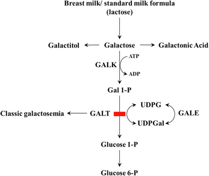
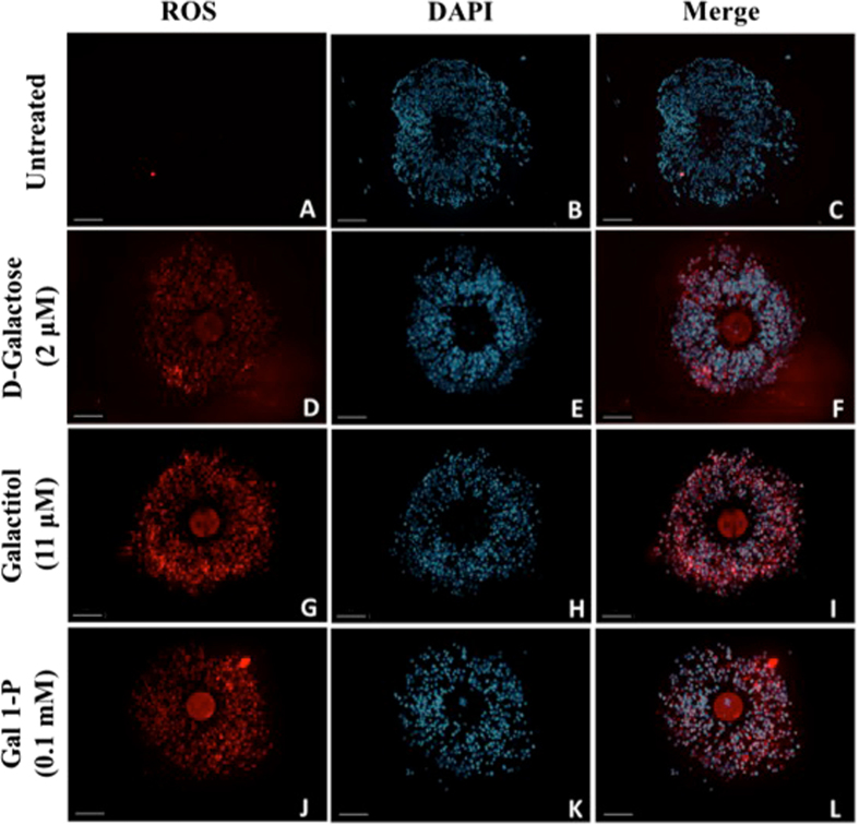
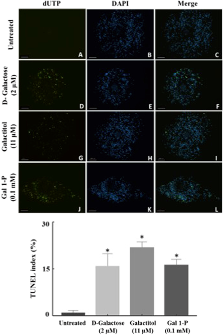
Premature ovarian insufficiency (POI) is a frequent long-term complication of classic galactosemia. The majority of women with this disorder develop POI, however rare spontaneous pregnancies have been reported. Here, we evaluate the effect of D-galactose and its metabolites, galactitol and galactose 1-phosphate, on oocyte quality as well as embryo development to elucidate the mechanism through which these compounds mediate oocyte deterioration. Metaphase II mouse oocytes (n = 240), with and without cumulus cells (CCs), were exposed for 4 hours to D-galactose (2 μM), galactitol (11 μM) and galactose 1-phosphate (0.1 mM), (corresponding to plasma concentrations in patients on galactose-restricted diet) and compared to controls. The treated oocytes showed decreased quality as a function of significant enhancement in production of reactive oxygen species (ROS) when compared to controls. The presence of CCs offered no protection, as elevated ROS was accompanied by increased apoptosis of CCs. Our results suggested that D-galactose and its metabolites disturbed the spindle structure and chromosomal alignment, which was associated with significant decline in oocyte cleavage and blastocyst development after in-vitro fertilization. The results provide insight into prevention and treatment strategies that may be used to extend the window of fertility in these patients.
Conflict of interest statement
The authors declare that they have no competing interests.
Figures

Similar articles
-
Peroxynitrite affects the cumulus cell defense of metaphase II mouse oocytes leading to disruption of the spindle structure in vitro.Fertil Steril. 2013 Aug;100(2):578-84.e1. doi: 10.1016/j.fertnstert.2013.04.030. Epub 2013 May 28. Fertil Steril. 2013. PMID: 23721714
-
The Defensive Role of Cumulus Cells Against Reactive Oxygen Species Insult in Metaphase II Mouse Oocytes.Reprod Sci. 2016 Apr;23(4):498-507. doi: 10.1177/1933719115607993. Epub 2015 Oct 14. Reprod Sci. 2016. PMID: 26468254 Free PMC article.
-
Cyclophosphamide and acrolein induced oxidative stress leading to deterioration of metaphase II mouse oocyte quality.Free Radic Biol Med. 2017 Sep;110:11-18. doi: 10.1016/j.freeradbiomed.2017.05.006. Epub 2017 May 9. Free Radic Biol Med. 2017. PMID: 28499912 Free PMC article.
-
Improved cryotolerance and developmental potential of in vitro and in vivo matured mouse oocytes by supplementing with a glutathione donor prior to vitrification.Mol Hum Reprod. 2016 Dec;22(12):867-881. doi: 10.1093/molehr/gaw059. Epub 2016 Sep 7. Mol Hum Reprod. 2016. PMID: 27604460
-
Glyphosate Induces Metaphase II Oocyte Deterioration and Embryo Damage by Zinc Depletion and Overproduction of Reactive Oxygen Species.Toxicology. 2020 Jun;439:152466. doi: 10.1016/j.tox.2020.152466. Epub 2020 Apr 19. Toxicology. 2020. PMID: 32315717
Cited by
-
Single-nucleus and spatial transcriptomics of paediatric ovary: Molecular insights into the dysregulated signalling pathways underlying premature ovarian insufficiency in classic galactosemia.Clin Transl Med. 2024 Oct;14(10):e70043. doi: 10.1002/ctm2.70043. Clin Transl Med. 2024. PMID: 39440457 Free PMC article.
-
Primary ovarian insufficiency in classic galactosemia: current understanding and future research opportunities.J Assist Reprod Genet. 2018 Jan;35(1):3-16. doi: 10.1007/s10815-017-1039-7. Epub 2017 Sep 20. J Assist Reprod Genet. 2018. PMID: 28932969 Free PMC article. Review.
-
The Implications of Insufficient Zinc on the Generation of Oxidative Stress Leading to Decreased Oocyte Quality.Reprod Sci. 2023 Jul;30(7):2069-2078. doi: 10.1007/s43032-023-01212-0. Epub 2023 Mar 15. Reprod Sci. 2023. PMID: 36920672 Free PMC article. Review.
-
Optimized platelet rich plasma releasate (O-rPRP) repairs galactosemia-induced ovarian follicular loss in rats by activating mTOR signaling and inhibiting apoptosis.Heliyon. 2020 Sep 21;6(9):e05006. doi: 10.1016/j.heliyon.2020.e05006. eCollection 2020 Sep. Heliyon. 2020. PMID: 33005806 Free PMC article.
-
Stem cell-based therapeutic potential in female ovarian aging and infertility.J Ovarian Res. 2024 Aug 24;17(1):171. doi: 10.1186/s13048-024-01492-3. J Ovarian Res. 2024. PMID: 39182123 Free PMC article. Review.
References
Publication types
MeSH terms
Substances
Grants and funding
LinkOut - more resources
Full Text Sources
Other Literature Sources
