. 2017 Mar 22;7(1):303.
doi: 10.1038/s41598-017-00412-4.

Immune evasion of Borrelia miyamotoi: CbiA, a novel outer surface protein exhibiting complement binding and inactivating properties

Affiliations

Immune evasion of Borrelia miyamotoi: CbiA, a novel outer surface protein exhibiting complement binding and inactivating properties

Florian Röttgerding et al. Sci Rep. .

Erratum in

Abstract

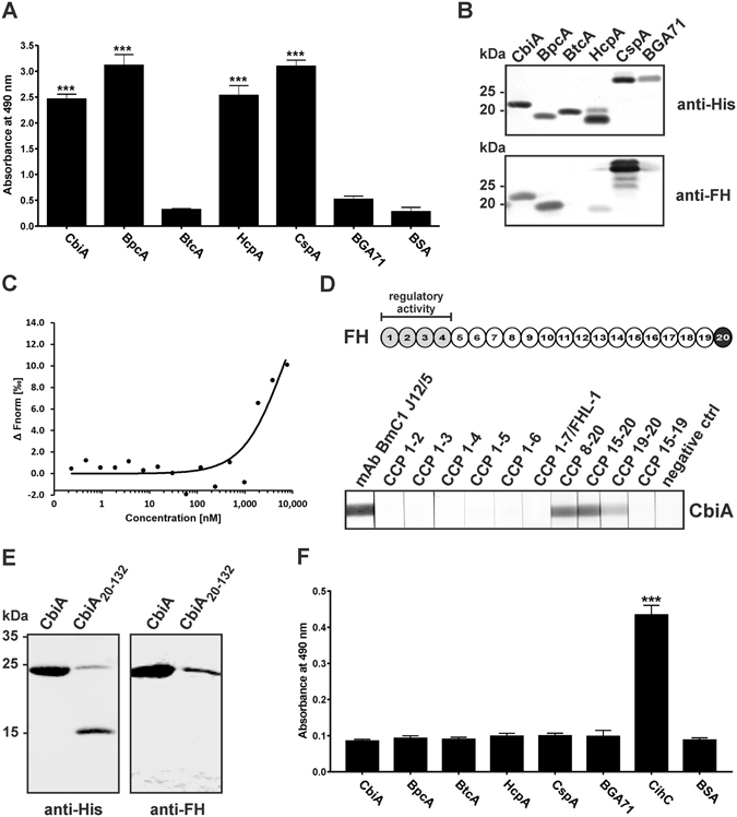
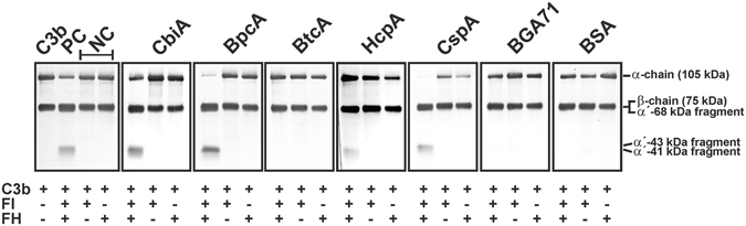
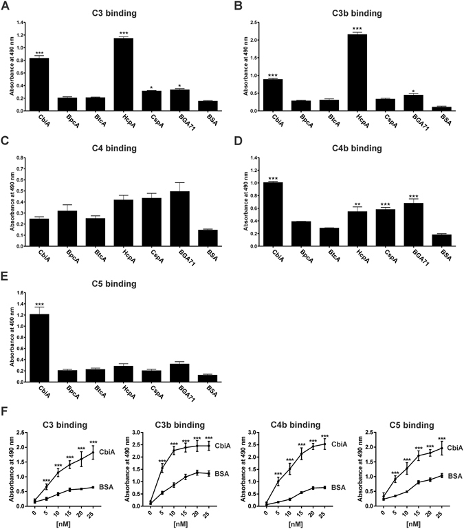
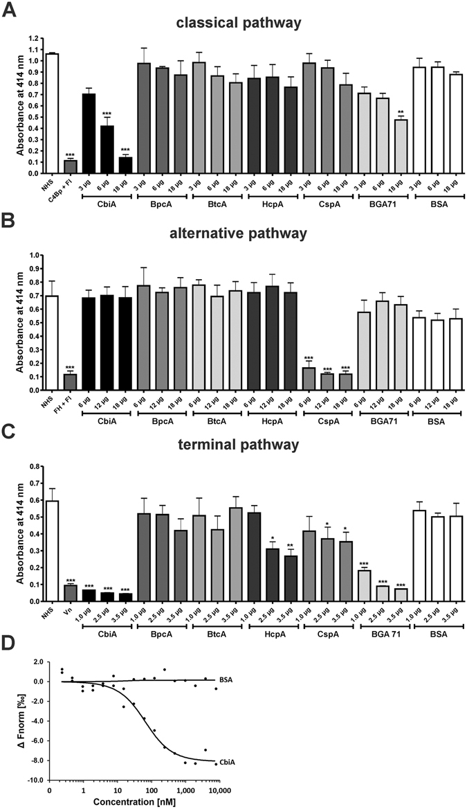
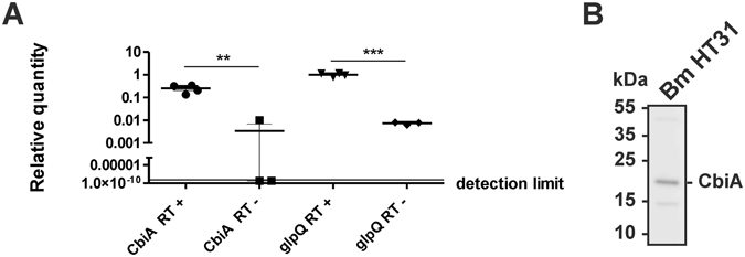
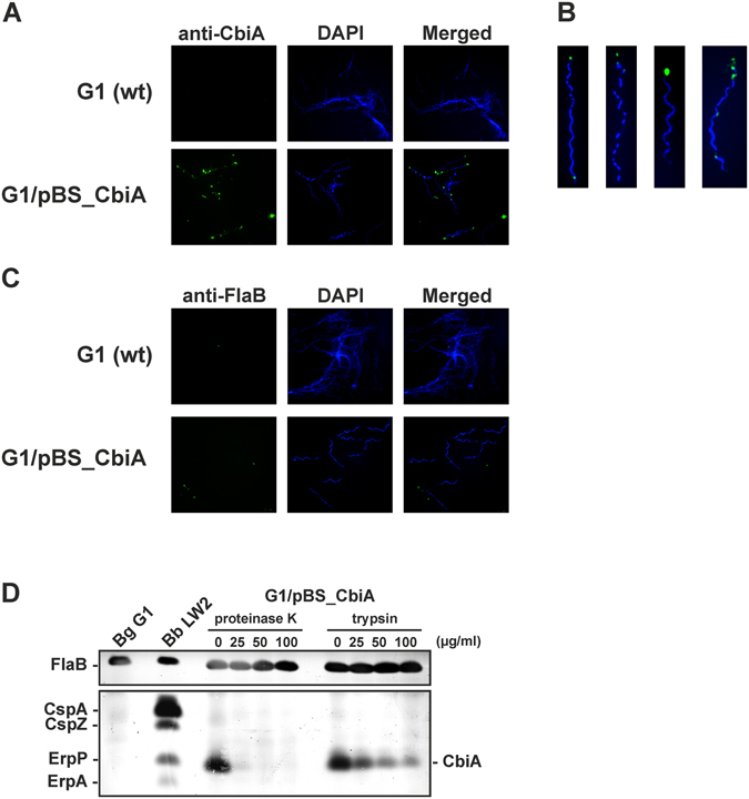
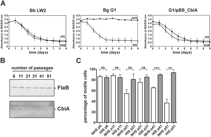
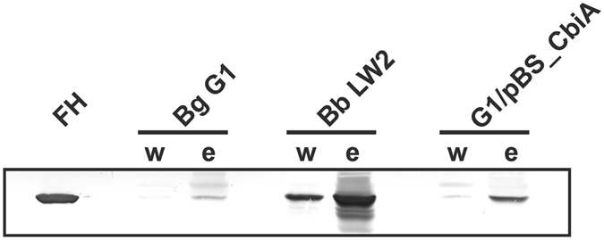
Borrelia (B.) miyamotoi, an emerging tick-borne relapsing fever spirochete, resists complement-mediated killing. To decipher the molecular principles of immune evasion, we sought to identify determinants contributing to complement resistance. Employing bioinformatics, we identified a gene encoding for a putative Factor H-binding protein, termed CbiA (complement binding and inhibitory protein A). Functional analyses revealed that CbiA interacted with complement regulator Factor H (FH), C3, C3b, C4b, C5, and C9. Upon binding to CbiA, FH retained its cofactor activity for Factor I-mediated inactivation of C3b. The Factor H-binding site within CbiA was mapped to domain 20 whereby the C-terminus of CbiA was involved in FH binding. Additionally, CbiA directly inhibited the activation of the classical pathway and the assembly of the terminal complement complex. Of importance, CbiA displayed inhibitory activity when ectopically produced in serum-sensitive B. garinii G1, rendering this surrogate strain resistant to human serum. In addition, long-term in vitro cultivation lead to an incremental loss of the cbiA gene accompanied by an increase in serum susceptibility. In conclusion, our data revealed a dual strategy of B. miyamotoi to efficiently evade complement via CbiA, which possesses complement binding and inhibitory activities.


Conflict of interest statement

The authors declare that they have no competing interests.

Figures

Figure 1
Figure 1
Binding of FH and C4BP to borrelial proteins and mapping of the interacting region in FH and CbiA. Binding of recombinant proteins to FH (A) and C4BP (B) was assessed by ELISA. Microtiter plates were coated with 500 ng His6-tagged proteins and incubated with FH or C4BP (5 µg/ml each). Bound FH and C4BP was detected using an anti-FH and anti-C4BP antiserum, respectively. All experiments were performed at least three times, with each individual test carried out in triplicate. **p ≤ 0.01, ***p ≤ 0.001, one-way ANOVA with Bonferroni post test. Far Western blot analysis of recombinant proteins (B). Proteins (500 ng each) were separated by SDS-PAGE and stained with silver or transferred to nitrocellulose. The membrane was incubated with NHS and subsequently probed with an anti-FH antiserum (lower panel). Binding of FH to CbiA (C). FH was labeled with NT-647 RED-NHS (NanoTemper technologies) and the interaction with CbiA was assessed in the fluid phase by microscale thermophoresis. The relative fluorescence in the thermophoresis phase has been plotted against the concentration of CbiA. The data shown are representative of three independent experiments. Localization of the binding domain in FH (D). Schematic representation of FH (upper panel). The CCP domains 1–4 are in light grey and the interacting domain is in black with white font. Mapping of the CbiA interacting domain in FH by Far Western blotting (lower panel). Purified recombinant CbiA was separated by SDS-PAGE, and transferred to nitrocellulose. The membrane strips were incubated with different constructs of FH (CCP1-2, CCP1-3, CCP1-4, CCP1-5, CCP1-6, CCP8-20, CCP15-20, CCP19-20, and CCP15-19), CCP1-7/FHL-1, mAb BmC1 J12/5, and with the secondary Ab (negative ctrl). Bound proteins were visualized using polyclonal anti-FH antibody. (E) Localization of the FH interacting domain in CbiA. His6-tagged CbiA and deletion mutant CbiA20–132 (500 ng each) were separated by SDS-PAGE and transferred to nitrocellulose. Membranes were probed with an anti-His6 antibody (upper panel) or incubated with NHS and subsequently probed with an anti-FH antiserum (lower panel). The full-length versions of (B,D and E) are presented in Supplementary Figure S4.
Figure 2
Figure 2
Analysis of functional activity of FH bound to borrelial proteins. Recombinant proteins immobilized to microtiter plates were used to capture FH. After extensive washing, C3b (100 ng/ml) and FI (200 ng/ml) were added and the mixture was incubated for 30 min at 37 °C. For control purposes, all reactions were also performed in the absence of FH and FI. Subsequently, samples were boiled for 5 min, subjected to 12.5% SDS-PAGE and transferred onto a nitrocellulose membrane. The various C3b degradation products were visualized by Western blotting using a polyclonal goat anti-human C3 antiserum. As additional controls, reaction mixtures containing purified C3b, C3b and FI were incubated with (+) or without (−) purified FH, respectively. The mobility of the α’- and the β-chain of C3 and the cleavage products of the α’-chain the α’68,’ α’46 and α’43 fragments are indicated. NC, negative controls; PC, positive control. A full-length version of the figures is presented in Supplementary Figure S5.
Figure 3
Figure 3
Binding of complement components to borrelial proteins. Binding of recombinant proteins to complement C3 (A), C3b (B), C4 (C), C4b (D), and C5 (E) was assessed by ELISA. Microtiter plates were coated with 500 ng His6-tagged proteins and incubated with the respective complement proteins (5 µg/ml each). Protein-protein complexes were detected using specific antibodies. Dose-dependent binding of C3, C3b, C4b, and C5 to CbiA (F). Microtiter plates were coated with 100 ng His6-tagged CbiA or 100 ng BSA and incubated with increasing concentrations with complement proteins and binding was analyzed with specific antibodies. Absorbance of each test was measured at 490 nm. Data represent means and SEM from three separate experiments, each performed at least in triplicate. Raw data were analyzed by one-way ANOVA with post hoc Bonferroni correction. ***P < 0.001, **P < 0.01, *P < 0.05.
Figure 4
Figure 4
Borrelial proteins inhibit TCC deposition of the CP and TP. In order to analyze the inhibitory effect of borrelial proteins on the AP (A), CP (B), and TP (C) a hemolytic assay was employed. Amboceptor-sensitized sheep erythrocytes (CP) and rabbit erythrocytes (AP) were incubated with NHS or with NHS pre-incubated with purified proteins or BSA in either Mg-EGTA buffer (AP) or GVB++ buffer (CP). Inhibition of the TP was investigated using sheep erythrocytes pre-incubated with the C5b-6 complex. A reaction mixture containing C7, C8, and C9 was pre-incubated with increasing concentrations of purified proteins or BSA. Following incubation, erythrocyte lysis was detected at 414 nm. Means from three separate experiments are shown and error bars correspond to SD. Raw data were analyzed using one-way ANOVA (A and B). ***P < 0.001, **P < 0.01, *P < 0.05. Binding of C9 to CbiA (D). C9 was labeled with NT-647 RED-NHS (NanoTemper technologies) and the interaction with CbiA was assessed in the fluid phase. The relative fluorescence in the thermophoresis phase has been plotted against the concentration of CbiA. The data shown are representative of three independent experiments.
Figure 5
Figure 5
CbiA is expressed in B. miyamotoi HT31. (A) Expression of cbiA and glpQ was determined by RT-PCR. B. miyamotoi cells were grown at 33 °C and harvested at mid logarithmic growth phase. After transcription of isolated RNA the relative expression levels of the cbiA and the glpQ gene were determined. Each experiment was performed at least three times in triplicate and error bars represent ±SD. (B) Western blot analysis of in vitro grown spirochetes. 250 ng cell lysates were subjected to 4–20% SDS-PAGE and proteins were transferred to nitrocellulose. The membrane was probed with polyclonal rabbit anti-CbiA antibody (1:500) and protein complexes were visualized by a HRP-conjugated anti-rabbit antibody (1:10,000) using a Lumilight LAS4000. A full-length version is presented in Supplementary Figure S6.
Figure 6
Figure 6
Surface exposure of CbiA in transformed B. garinii G1. (A) Surface localization of ectopically expressed CbiA was visualized by indirect immunofluorescence microscopy. Spirochetes (6 × 106) were incubated with rabbit anti-CbiA antiserum (1:50) for 1 h at RT with gentle agitation. After fixation, glass slides were incubated with an appropriate AlexaFluor 488-conjugated secondary antibody. For visualization of the spirochetes in a given microscopic field, the DNA-binding dye DAPI was used. The spirochetes were observed at a magnification of 100 × objective. The data were recorded with an Axio Imager M2 fluorescence microscope (Zeiss) equipped with a Spot RT3 camera (Visitron Systems). Each panel shown is representative of at least 20 microscope fields. (B) In situ protease accessibility assay. Native spirochetes were incubated with or without proteinases, then lysed by sonication and total proteins were separated by SDS-PAGE. CbiA was identified by Far Western blot analysis using NHS as source of FH. Flagellin (FlaB) was detected with mAb L41 1C11. FH-binding proteins of B. burgdorferi LW2 (CspA, CspZ, ErpP, ErpA) are indicated on the left and the band corresponding to CbiA on the right. A full-length version is presented in Supplementary Figure S7.
Figure 7
Figure 7
Serum susceptibility testing of spirochetes. (A) A growth inhibition assay was used to investigate susceptibility to human serum of B. burgdorferi (Bb) strain LW2, B. garinii (Bg) G1, and transformant G1/pBS_CbiA. Spirochetes were incubated in either 50% NHS (filled squares) or 50% HIS (filled triangles) over a cultivation period of 9 days at 33 °C, respectively. Color changes were monitored by measurement of the absorbance at 562/630 nm which is inversely proportional to the acidification of the culture medium. All experiments were performed at least three times, with each test conducted in triplicate with very similar results. For clarity, only data from one representative experiment is shown. Error bars represent ±SD. (B) Western blot analysis of spirochetes collected at different time points. Cell lysates (250 ng each) subjected to 4–20% SDS gels were analyzed by Western blotting to detect FlaB and CbiA using a monoclonal anti-FlaB antibody L41 1C11 (1:1000) and an anti-rabbit Ab (1:500), respectively. The full-length versions are presented in Supplementary Figure S8. (C) Serum susceptibility testing of spirochetes collected at different time points. Spirochetes were incubated in 50% NHS at 37 °C for 1 h. Dark-field microscopy were investigated for calculating the percentage of motile spirochetes. ns, not statistically significant. ***P < 0.001, **P < 0.01.
Figure 8
Figure 8
Binding of FH by B. garinii transformants. Serum adsorption assays were employed to detect binding of FH to viable spirochetes. Wild-type B. garinii (Bg) G1, B. burgdorferi (Bb) LW2, and transformant G1/pBS_CbiA (5 × 108 cells each) were incubated in NHS-EDTA to prevent complement activation, washed, and then bound proteins were eluted using 0.1 M glycine (pH 2.0). Both the last wash (w) and the eluate (e) fractions obtained from each strain were separated by 12.5% SDS-PAGE and transferred to nitrocellulose. A polyclonal anti-FH antibody was used to detect FH by Western blotting. The full-length version is presented in Supplementary Figure S9.

Similar articles

Cited by

References

    1. Fukunaga M, et al. Genetic and phenotypic analysis of Borrelia miyamotoi sp. nov., isolated from the ixodid tick Ixodes persulcatus, the vector for Lyme disease in Japan. Int. J. Syst. Bacteriol. 1995;45:804–810. doi: 10.1099/00207713-45-4-804. - DOI - PubMed
    1. Scoles GA, Papero M, Beati L, Fish D. A relapsing fever group spirochete transmitted by Ixodes scapularis ticks. Vector Borne Zoonotic Dis. 2001;1:21–34. doi: 10.1089/153036601750137624. - DOI - PubMed
    1. Richter D, Schlee DB, Matuschka FR. Relapsing fever-like spirochetes infecting European vector tick of Lyme disease agent. Emerg. Infect. Dis. 2003;9:697–701. doi: 10.3201/eid0906.020459. - DOI - PMC - PubMed
    1. Platonov AE, et al. Humans infected with relapsing fever spirochete Borrelia miyamotoi, Russia. Emerg. Infect. Dis. 2011;17:1816–1823. doi: 10.3201/eid1710.101474. - DOI - PMC - PubMed
    1. Hovius JW, et al. A case of meningoencephalitis by the relapsing fever spirochaete Borrelia miyamotoi in Europe. Lancet. 2013;382:658. doi: 10.1016/S0140-6736(13)61644-X. - DOI - PMC - PubMed

Publication types

MeSH terms