. 2017 Mar 22;7(1):300.
doi: 10.1038/s41598-017-00339-w.

Dentin sialoprotein facilitates dental mesenchymal cell differentiation and dentin formation

Affiliations

Dentin sialoprotein facilitates dental mesenchymal cell differentiation and dentin formation

Wentong Li et al. Sci Rep. .

Abstract

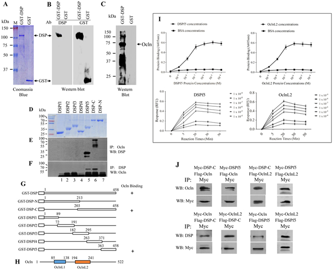
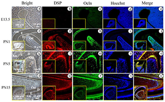
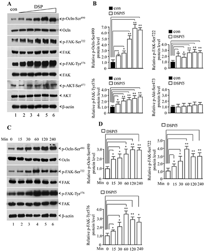
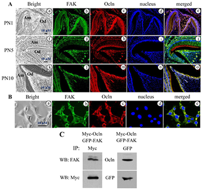
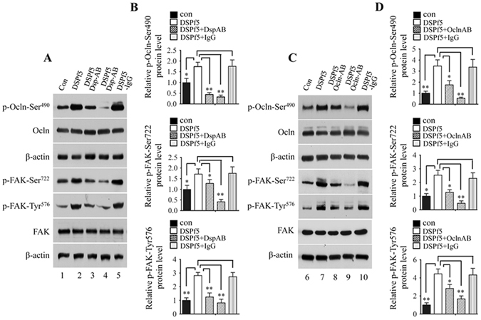
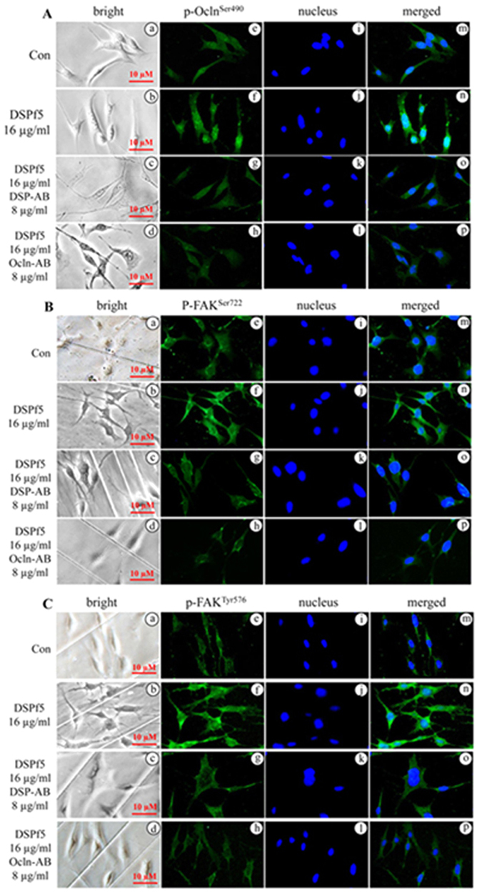
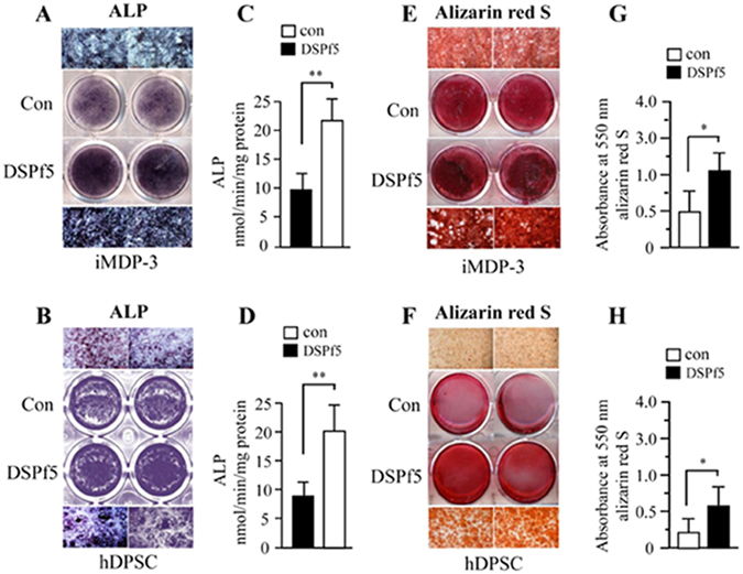
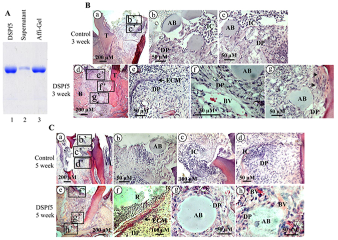
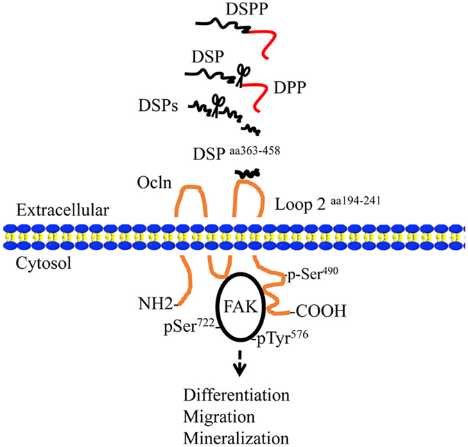
Dentin sialoprotein (DSP) is a dentin extracellular matrix protein. It is involved in dental mesenchymal cell lineages and dentin formation through regulation of its target gene expression. DSP mutations cause dentin genetic diseases. However, mechanisms of DSP in controlling dental mesenchymal cell differentiation are unknown. Using DSP as bait, we screened a protein library from mouse odontoblastic cells and found that DSP is a ligand and binds to cell surface receptor, occludin. Further study identified that the C-terminal DSP domainaa 363-458 interacts with the occludin extracellular loop 2aa 194-241. The C-terminal DSP domain induced phosphorylation of occludin Ser490 and focal adhesion kinase (FAK) Ser722 and Tyr576. Coexpression of DSP, occludin and FAK was detected in dental mesenchymal cells during tooth development. Occludin physically interacts with FAK, and occludin and FAK phosphorylation can be blocked by DSP and occludin antibodies. This DSP domain facilitates dental mesenchymal cell differentiation and mineralization. Furthermore, transplantation and pulp-capping procedures revealed that this DSP domain induces endogenous dental pulp mesenchymal cell proliferation, differentiation and migration, while stimulating blood vessel proliferation. This study elucidates the mechanism of DSP in dental mesenchymal lineages and implies that DSP may serve as a therapeutic agent for dentin-pulp complex regeneration in dental caries.


Conflict of interest statement

The authors declare that they have no competing interests.

Figures

Figure 1
Figure 1
DSP interacts with occludin. Recombinant DSP 1–458, 1–213, 203–458, 1–89, 72–191, 182–295, 263–371, 363–458 were expressed in Escherichia coli BL21 and purified according to the manufacturer's instruction as described by “Materials and methods”. The purified DSP fusion proteins were confirmed by Coomassie blue staining (A,D) and Western blot assays using either an anti-GST or anti-DSP antibody (B). Interaction between DSP polypeptides and Ocln by GST pull down was detected by Western blot using anti-DSP and anti-Ocln antibodies (C,E,F). Arrow shows Ocln band (C). Mixture of different fragments of DSP and Ocln fusion proteins was pulled down by Ocln antibody and interaction of DSP with Ocln was detected using DSP antibody (E) and vice versa (F). The GST-DSP fusion protein constructs and the localization of the DSP protein stretch that contains Ocln-binding domain are illustrated (G). The DSP number starts at the translational start site of DSPP (Met) as No. 1. (H) Schematic representation of mouse Ocln protein structure from the initiation of the translation start site 1 to the end 522. OclnL1 and OclnL2 indicate the extracellular loop 1 (aa 85–183) and loop 2 (aa 194–241). (I) Either recombinant OclnL2 protein or BSA as control was coated in 96-well plates. Serial diluted biotinylated DSPf5 was added to the wells and incubated with the unlabeled recombinant OclnL2 protein or BSA, respectively. Bound DSPf5 was detected using AP-conjugated streptavidin and 1 mg/ml PNPP as substrate at 405 nm using a microplate reader. Binding affinity of different concentrations of DSPf5 to its substrate at the given time periods were calculated. Using the same method, binding of the biotinylated OclnL2 to the unlabeled DSPf5 was examined. Data point represent the mean ± S.D. (n = 3). (J) For in vivo studies, different fragments of DSP and Ocln cDNAs were subcloned into a CMV mammalian expression plasmid tagged with Flag or Myc peptides, respectively. Myc-Ocln and Flag-DSP as well as Myc-DSP and Flag-Ocln expression vectors were transfected into HEK-293 cells, respectively. After 48 h transfection, the proteins were harvested and protein-protein interactions were immunoprecipitated using Myc antibody. Protein-protein interaction was detected by Western blotting using anti-Myc, anti-DSP or anti-Ocln antibody.
Figure 2
Figure 2
Expression of dentin sialoprotein and occludin in developing mouse teeth. At embryonic day (E) 13.5, DSP and Ocln expressions were not detected in tooth tissues (b,c) using double fluorescent immunohistochemistry, but at postnatal day (PN) 1, DSP expression (red) was observed in odontoblasts, ameloblasts and dental pulp cells (g), Ocln expression (green) was detected in these areas overlapped with DSP (h). At PN5 and PN15, expression of DSP and Ocln was apparently seen in odontoblasts and ameloblasts (l,m,q,r). However, Ocln expression was relatively wider than that of DSP. (a,f,k and p) show bright images. The cells were stained with Hoechst for nuclei (d,i,n,s). Images were merged (e,j,o,t). (at) show higher magnifications from the yellow boxes.
Figure 3
Figure 3
Effect of DSPf5 on occludin and FAK phosphorylation. (A) The mouse dental papilla mesenchymal cells were treated with or wither DSPf5 of 2 μg/ml (lane 2), 4 μg/ml (lane 3), 8 μg/ml (lane 4), 16 μg/ml (lane 5) and 24 μg/ml (lane 6) for 1 h at 37 °C. After cell harvest, protein expression was detected by Western blot assay using antibodies specific to p-Ocln-Ser490, Ocln, p-FAK-Ser722, p-FAK-Tyr576, FAK, p-AKT-Ser473, AKT and β-actin. (B) Protein expression levels were quantitated using imageJ software. Expression of p-Ocln-Ser490, p-FAK-Ser722, p-FAK-Tyr576 and p-AKT-Ser473 was normalized to Ocln, FAK and AKT, respectively. Protein on the control was considered as a one-fold increase. The expression level of proteins treated with DSPf5 was divided by protein level on the control group. The results showed the mean ± S.D. (n = 3). *p < 0.05; **p < 0.01. (C) The cells were treated with DSPf5 (8 μg/ml) in DMEM medium for 0. 15, 30, 60, 120 and 240 min. Protein expression was detected by Western blot analysis using antibodies specific to p-Ocln-Ser490, Ocln, p-FAK-Ser722, p-FAK-Tyr576, FAK, and β-actin, respectively. (D). Protein expression levels were quantitated using image J software. Expression of p-Ocln-Ser490, p-FAK-Ser722 and p-FAK-Tyr576 was normalized to Ocln and FAK, respectively. Protein treated with DSPf5 at 0 min as control group was considered as a one-fold increase. The expression level of proteins treated with DSPf5 at different time periods was divided by protein level on the control group.
Figure 4
Figure 4
Expression of occludin and FAK in odontoblasts during tooth development. (A) Fluorescent immunohistochemistry showed that at PN1, 5, 10 during mouse tooth development, Ocln (red) and FAK (green) were co-expressed in odontoblasts by the double immunofluorescent histochemistry using antibodies specific to Ocln and FAK (b,c,g,h,l,m). (a, f and k) show bright images. The cells were stained with Hoechst for nuclei (d,i,n). Images were merged (e,j,o). Am, Ameloblasts; Od, Odontoblasts. (B) Ocln (red) and FAK (green) were expressed in iMDP-3 cells using double immunofluorescent assay (b,c). a shows bright image. Hoechst was used for nuclear staining (d). Image was merged (e). (C) Ocln interacts with FAK in vivo. Myc-Ocln and GFP-FAK mammalian expression vectors were cotransfected into HEK-293 cells. After 48 h transfection, the cells were harvested. Ocln and FAK were immune-precipitated using Myc or GFP antibody. Interaction of Ocln with FAK was detected by Western blot assay using Ocln and FAK antibodies.
Figure 5
Figure 5
Effect of DSPf5 on occludin and FAK phosphorylation is blocked by DSP and occludin antibodies. (A,C) The mouse dental papilla mesenchymal cells were treated with or without DSPf5, or DSPf5 plus DSP antibody or DSPf5 plus Ocln antibody or DSPf5 with IgG as control for 1 h, respectively. Protein expression was detected by Western blotting using antibodies described above. Lanes, 1 and 6 as control without the DSPf5 induction; lanes 2 and 7 (8 μg/ml of DSPf5); lane 3 (8 μg/ml of DSPf5 plus 8 μg/ml of DSP antibody); lane 4 (8 μg/ml of DSPf5 plus 16 μg/ml of DSP antibody); lane 8 (8 μg/ml of the DSPf5 plus 8 μg/ml of Ocln antibody); lane 9 (8 μg/ml of DSPf5 plus 16 μg/ml of Ocln antibody); lanes 5 and 10 (8 μg/ml of DSPf5 plus 16 μg/ml of IgG). (B,D) Protein expression level was measured using imageJ software. Expression of p-Ocln-Ser490, p-FAK-Ser722 and p-FAK-Tyr576 was normalized to Ocln and FAK, respectively. Protein without treatment was considered as one-fold increase. The expression protein level of the treated groups was divided by protein level on the control group. The results are shown as the mean ± S.D. (n = 3). *p < 0.05; **p < 0.01.
Figure 6
Figure 6
DSP and occludin antibodies block phosphorylation of occludin and FAK proteins mediated by DSPf5 in mouse dental papilla mesenchymal cells. (A–C) The cells were treated with or without DSPf5 or DSPf5 (16 μg/ml) plus the DSP (8 μg/ml) or Ocln (8 μg/ml) antibody for 1 h at 37 °C. The cells were fixed and immune-stained using p-Ocln-Ser490 (A), p-FAK-Ser722 (B) and pFAK-Tyr576 (C) antibodies, respectively. Data showed that effect of DSPf5 on Ocln and FAK phosphorylation was blocked by the DSP or Ocln antibody (e–h). (a–d) are bright images. The cells were stained with Hoechst for nuclei (i–l). Images were merged (m–p).
Figure 7
Figure 7
Effect of DSP domain on dental cell differentiation and mineralization. Mouse dental papilla mesenchymal (iMDP-3) (A) and human dental pulp stem (hDPSC) cells (B) were treated with or without 10 μg/ml of DSPf5 in calcifying medium for 7 days. ALP activity was analyzed using in situ ALP staining (A, B). (C,D) Quantitative ALP activity of the cell lysates was assayed using ρ-nitrophenyl phosphate as a substrate. Protein concentration was determined using the BCA protein assay reagent as described in “Materials and methods”. There were significantly different between the DSPf5 treated groups and DSPf5 untreated groups. For cell biomineralization, iMDP-3 (E) and hDPSC cells (F) were maintained in the same condition for 14 days. The cells were fixed and stained for Alizarin red S dye. (G,H) The amount of calcium deposition was quantified by destaining with 10% cetylpyridinium chloride in 10 mM sodium phosphate at room temperature. Mineralization deposits were determined and data represent mean ± S.D. (n = 3). Con, control. *p < 0.05; **p < 0.01.
Figure 8
Figure 8
Effect of DSP domain on dental cell differentiation in vivo. (A) Lane 1, DSPf5 protein only; lane 2, DSPf5 was mixed with Affi-Gel Blue Gel Beads at room temperature for overnight. The mixture was centrifuged and supernatant was loaded onto a 7% SDS-PAGE gel; lane 3, the pellet was added to 1 x SDS-loading buffer and heated. The released DSPf5 from the compound was loaded onto a 7% SDS-PAGE gel. (B) The complex of DSPf5 coated to agarose beads was implanted into mouse dental pulp chambers. Compared to the control group after 3 week operation (Ba–c), in the DSPf5-treated group, DSPf5 was able to induce the dental pulp cell proliferation and differentiation. Dental pulp cells in the DSPf5-treated group secrete ECM (arrow) at the top “artificial hole” between resin and dental pulp. Blood vessels (BV) proliferate and migrate near agarose beads (AB). Arrowheads show that dental pulp cells differentiate and secrete ECM. b, c and e–g are enlarged from the boxes in the b’, c’ and e’–g’. In DSPf5-treated group after 5 week operation (C), ECM secreted by dental pulp cells forms a layer covering “the artificial hole” between resin and dental pup cells (Ce, f). Dental pulp cells surround the agarose beads (AB) and AB was resorbed and dental pulp cells and blood vessels invade into AB (Cg, h). A number of inflammatory cells were decreased compared to the control group. b–d and f–h are enlarged from the boxes in the b’–d’ and f’–h”. B, alveolar bone; BV, blood vessels; DP, dental pulp cells; ECM, extracellular matrix; IC, inflammatory cells; R, resin; T, teeth.
Figure 9
Figure 9
DSP domain regulates dental mesenchymal cell differentiation through occludin-FAK signaling. The hypothetical model depicts that the DSPaa 363–458 acts as a ligand and interacts with the extracellular loop 2 of Oclnaa 194–241, activating Ocln phosphorylation at Ser490. Furthermore, the DSP-Ocln complex activates FAK phosphorylation at Ser722 and Tyr576 and then induces dental mesenchymal cell differentiation and mineralization.

Similar articles

Cited by

References

    1. Erickson CA, Reedy MV. Neural crest development: the interplay between morphogenesis and cell differentiation. Curr Top Dev Biol. 1998;40:177–209. doi: 10.1016/S0070-2153(08)60367-1. - DOI - PubMed
    1. Discher DE, Mooney DJ, Zandstra PW. Growth factors, matrices, and forces combine and control stem cells. Science. 2009;324:1673–1677. doi: 10.1126/science.1171643. - DOI - PMC - PubMed
    1. Morrison SJ, Spradling AC. Stem cells and niches: mechanisms that promote stem cell maintenance throughout life. Cell. 2008;132:598–611. doi: 10.1016/j.cell.2008.01.038. - DOI - PMC - PubMed
    1. Bellahcene A, Castronovo V, Ogbureke KU, Fisher LW, Fedarko NS. Small integrin-binding ligand N-linked glycoproteins (SIBLINGs): multifunctional proteins in cancer. Nat Rev Cancer. 2008;8:212–226. doi: 10.1038/nrc2345. - DOI - PMC - PubMed
    1. D’Souza RN, et al. Gene expression patterns of murine dentin matrix protein 1 (Dmp1) and dentin sialophosphoprotein (DSPP) suggest distinct developmental functions in vivo. J Bone Miner Res. 1997;12:2040–2049. doi: 10.1359/jbmr.1997.12.12.2040. - DOI - PubMed

Publication types