. 2017:2017:5858315.
doi: 10.1155/2017/5858315. Epub 2017 Feb 26.

A Novel Role for Brain Natriuretic Peptide: Inhibition of IL-1β Secretion via Downregulation of NF-kB/Erk 1/2 and NALP3/ASC/Caspase-1 Activation in Human THP-1 Monocyte

Affiliations

A Novel Role for Brain Natriuretic Peptide: Inhibition of IL-1β Secretion via Downregulation of NF-kB/Erk 1/2 and NALP3/ASC/Caspase-1 Activation in Human THP-1 Monocyte

Letizia Mezzasoma et al. Mediators Inflamm. 2017.

Abstract

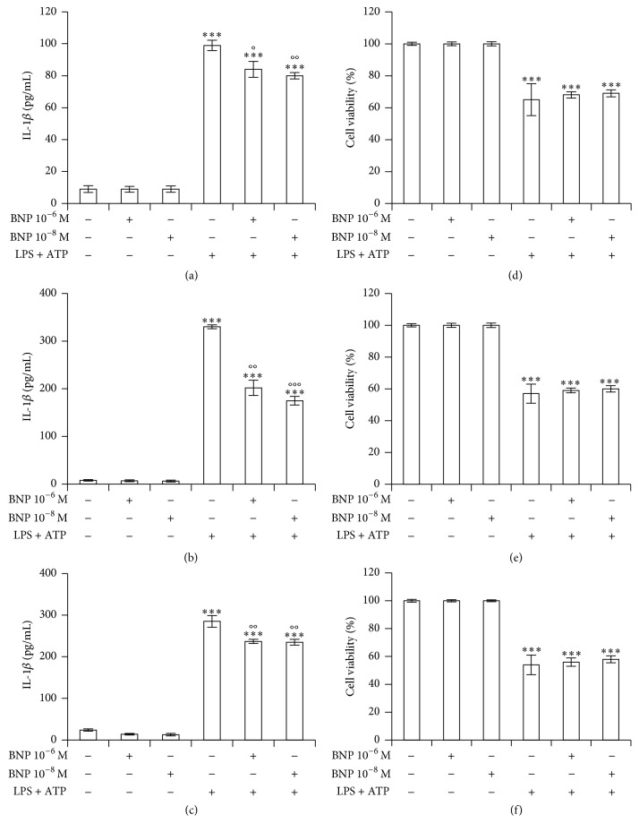
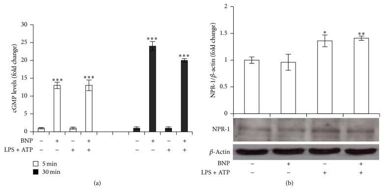
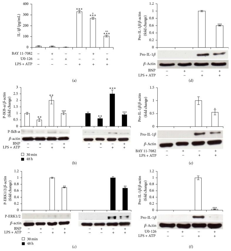
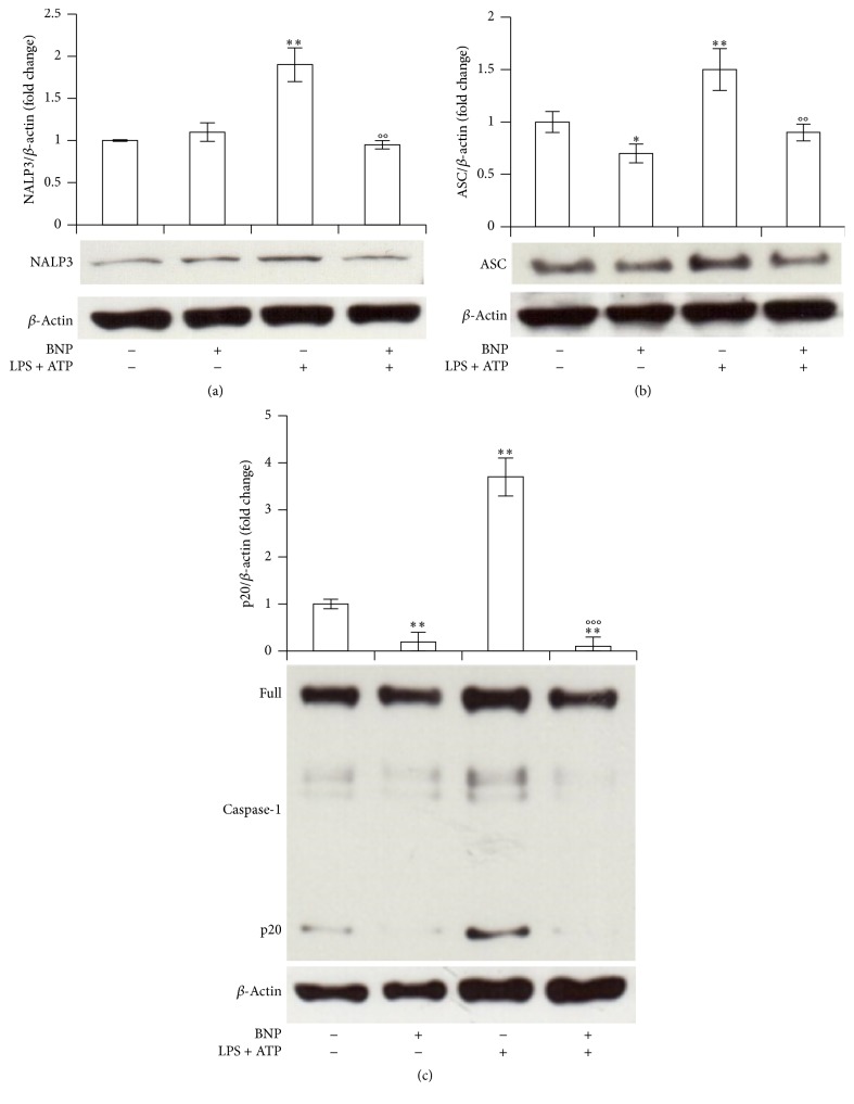
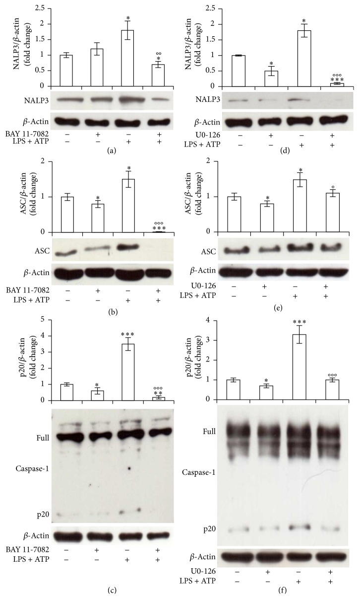
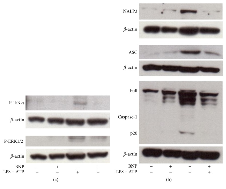
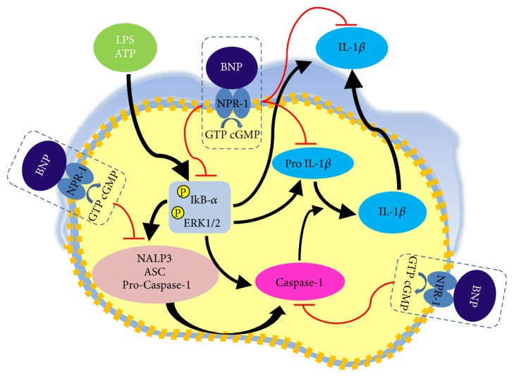
Interleukin-1β (IL-1β) is a pleiotropic cytokine and a crucial mediator of inflammatory and immune responses. IL-1β processing and release are tightly controlled by complex pathways such as NF-kB/ERK1/2, to produce pro-IL-1β, and NALP3/ASC/Caspase-1 inflammasome, to produce the active secreted protein. Dysregulation of both IL-1β and its related pathways is involved in inflammatory/autoimmune disorders and in a wide range of other diseases. Identifying molecules modulating their expression is a crucial need to develop new therapeutic agents. IL-1β is a strong regulator of Brain Natriuretic Peptide (BNP), a hormone involved in cardiovascular homeostasis by guanylyl cyclase Natriuretic Peptide Receptor (NPR-1). An emerging role of BNP in inflammation and immunity, although proposed, remains largely unexplored. Here, we newly demonstrated that, in human THP-1 monocytes, LPS/ATP-induced IL-1β secretion is strongly inhibited by BNP/NPR-1/cGMP axis at all the molecular mechanisms that tightly control its production and release, NF-kB, ERK 1/2, and all the elements of NALP3/ASC/Caspase-1 inflammasome cascade, and that NALP3 inflammasome inhibition is directly related to BNP deregulatory effect on NF-kB/ERK 1/2 activation. Our findings reveal a novel potent anti-inflammatory and immunomodulatory role for BNP and open new alleys of investigation for a possible employment of this endogenous agent in the treatment of inflammatory/immune-related and IL-1β/NF-kB/ERK1/2/NALP3/ASC/Caspase-1-associated diseases.


Conflict of interest statement

The authors declare that there is no conflict of interests regarding the publication of this study.

Figures

Figure 1
Figure 1
BNP decreases LPS/ATP-induced IL-1β release in THP-1 cells. THP-1 cells were treated with 10−6 and 10−8 M BNP in absence or presence of 10 μg/mL LPS/5 mM ATP for 24 (a, d), 48 (b, e), and 72 h (c, f). The supernatants were collected and IL-1β release was measured by ELISA (a, b, c). Cell viability was assessed by trypan blue uptake assay (d, e, f). Histograms indicate means ± SD of three separate experiments each one tested in triplicate. ∗∗∗P < 0.001 versus untreated cells and °P < 0.05, °°P < 0.01, and °°°P < 0.001 versus LPS/ATP treated cells.
Figure 2
Figure 2
BNP activates NPR-1 in THP-1 cells. (a) THP-1 cells were treated with 10−8 M BNP in absence or presence of 10 μg/mL LPS/5 mM ATP and cGMP intracellular levels were evaluated after 5 and 30 min treatment. (b) THP-1 cells were treated with 10−8 M BNP in absence or presence of 10 μg/mL LPS/5 mM ATP for 30 min. Cell lysates were immunoblotted for NPR-1. The blots were stripped of the bound Ab and reprobed with mouse anti-β-actin, to confirm equal loading. Western blots are representative of three separate experiments. Histograms indicate means ± SD of three separate experiments each one tested in triplicate. P < 0.05, ∗∗P < 0.01, and ∗∗∗P < 0.001 versus untreated cells.
Figure 3
Figure 3
BNP is involved in the priming mechanism controlling IL-1β production by inhibiting NF-kB and ERK 1/2 activation in THP-1 cells. (a) THP-1 cells were incubated with 10 μM BAY 11-7082 or U0-126 for 1 h and then treated with 10 μg/mL LPS/5 mM ATP for 48 h. The supernatants were collected and IL-1β release was measured by ELISA. (b, c, d) THP-1 cells were treated with 10−8 M BNP in absence or presence of 10 μg/mL LPS/5 mM ATP for 30 min (b, c) or 48 h (b, c, d). Cell lysates were immunoblotted for P-IkB-α (b), P-ERK 1/2 (c), or pro-IL-1β (d). (e, f) THP-1 cells were incubated with 10 μM BAY 11-7082 (e) or U0-126 (f) for 1 h and then treated with 10 μg/mL LPS/5 mM ATP for 48 h. Cell lysates were immunoblotted for pro-IL-1β. The blots were stripped of the bound Ab and reprobed with mouse anti-β-actin, to confirm equal loading. Western blots are representative of three separate experiments. All histograms indicate means ± SD of three separate experiments each one tested in triplicate. ∗∗P < 0.01 and ∗∗∗P < 0.001 versus untreated cells and °P < 0.05, °°P < 0.01, and °°°P < 0.001 versus LPS/ATP treated cells.
Figure 4
Figure 4
BNP is involved in the second mechanism controlling IL-1β production by inhibiting NALP3, ASC, and Caspase-1 cascade activation in THP-1 cells. THP-1 cells were treated with 10−8 M BNP in absence or presence of 10 μg/mL LPS/5 mM ATP for 30 min. Cell lysates were immunoblotted for NALP3 (a), ASC (b), or Caspase-1 (c). The blots were stripped of the bound Ab and reprobed with mouse anti-β-actin, to confirm equal loading. Western blots are representative of three separate experiments. Histograms indicate means ± SD of three separate experiments each one tested in triplicate. P < 0.05 and ∗∗P < 0.01 versus untreated cells and °°P < 0.01 and °°°P < 0.001 versus LPS/ATP treated cells.
Figure 5
Figure 5
NF-kB and ERK 1/2 are involved in NALP3/ASC/Caspase-1 cascade activation in THP-1 cells. THP-1 cells were incubated with 10 μM BAY 11-7082 (a, b, c) or U0-126 (d, e, f) for 1 h and then treated with 10 μg/mL LPS/5 mM ATP for 30 min. Cell lysates were immunoblotted for NALP3 (a, d), ASC (b, e), or Caspase-1 (c, f). The blots were stripped of the bound Ab and reprobed with mouse anti-β-actin, to confirm equal loading. Western blots are representative of three separate experiments. Histograms indicate means ± SD of three separate experiments each one tested in triplicate. P < 0.05, ∗∗P < 0.01, and ∗∗∗P < 0.001 versus untreated cells and °P < 0.05, °°P < 0.01, and °°°P < 0.001 versus LPS/ATP treated cells.
Figure 6
Figure 6
BNP is involved all the molecular mechanisms controlling IL-1β production and release by inhibiting the activation of NF-kB, ERK 1/2 and of all the elements of NALP3 inflammasome/ASC/Caspase-1 cascade in macrophage-like cells. PMA differentiated THP-1 cells were treated with 10−8 M BNP in absence or presence of 10 μg/mL LPS/5 mM ATP for 30 min. Cell lysates were immunoblotted for P-IkB-α and P-ERK1/2 (a) or NALP3, ASC, or Caspase-1 (b). The blots were stripped of the bound Ab and reprobed with mouse anti-β-actin, to confirm equal loading. Western blots are representative of three separate experiments.
Figure 7
Figure 7
Mechanism of action of BNP inhibitory effect on LPS/ATP-induced IL-1β secretion in human THP-1 monocyte. BNP/NPR1/cGMP axis strongly inhibits IL-1β secretion by acting both at transcriptional and at posttranslational level. Firstly, by inhibiting the activation of NF-kB and ERK 1/2 signaling pathway, BNP downregulates the production of the inactive cytoplasmic precursor pro-IL-1β. Subsequently, again by inhibiting NF-kB and ERK 1/2 activation, BNP strongly downregulates all the elements of NALP3 inflammasome/ASC/Caspase-1 cascade and Caspase -1 activation, essential steps for the formation of the active secreted IL-1β protein.

Similar articles

Cited by

References

    1. Dinarello C. A. Immunological and inflammatory functions of the interleukin-1 family. Annual Review of Immunology. 2009;27:519–550. doi: 10.1146/annurev.immunol.021908.132612. - DOI - PubMed
    1. Barker B. R., Taxman D. J., Ting J. P.-Y. Cross-regulation between the IL-1β/IL-18 processing inflammasome and other inflammatory cytokines. Current Opinion in Immunology. 2011;23(5):591–597. doi: 10.1016/j.coi.2011.07.005. - DOI - PMC - PubMed
    1. Martinon F., Mayor A., Tschopp J. The inflammasomes: guardians of the body. Annual Review of Immunology. 2009;27:229–265. doi: 10.1146/annurev.immunol.021908.132715. - DOI - PubMed
    1. Menu P., Vince J. E. The NLRP3 inflammasome in health and disease: the good, the bad and the ugly. Clinical and Experimental Immunology. 2011;166(1):1–15. doi: 10.1111/j.1365-2249.2011.04440.x. - DOI - PMC - PubMed
    1. Davis B. K., Wen H., Ting J. P.-Y. The inflammasome NLRs in immunity, inflammation, and associated diseases. Annual Review of Immunology. 2011;29:707–735. doi: 10.1146/annurev-immunol-031210-101405. - DOI - PMC - PubMed