

Preoperative subconjunctival combined injection of bevacizumab and mitomycin C before the surgical excision of primary pterygium: clinical and histological results
- PMID: 28331283
- PMCID: PMC5354544
- DOI: 10.2147/OPTH.S127700
Preoperative subconjunctival combined injection of bevacizumab and mitomycin C before the surgical excision of primary pterygium: clinical and histological results
Abstract
Purpose: The aim of this study was to detect the clinical and histological effects of preoperative subconjunctival injection of both bevacizumab and mitomycin C (MMC) 1 month before the surgical excision of primary pterygium using a bare sclera technique.
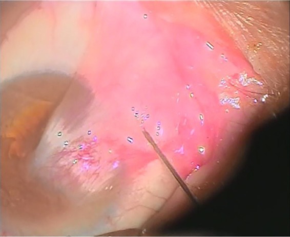
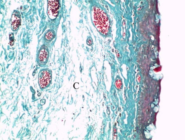
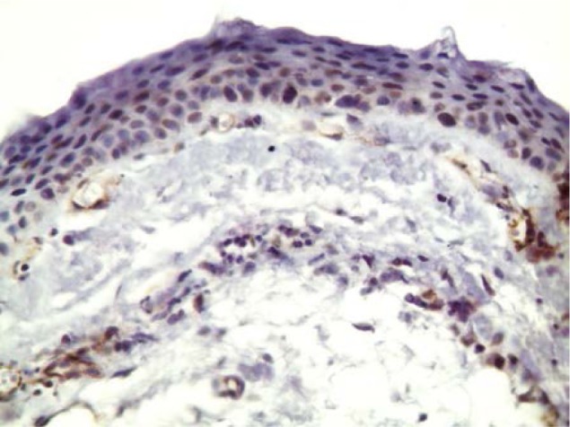
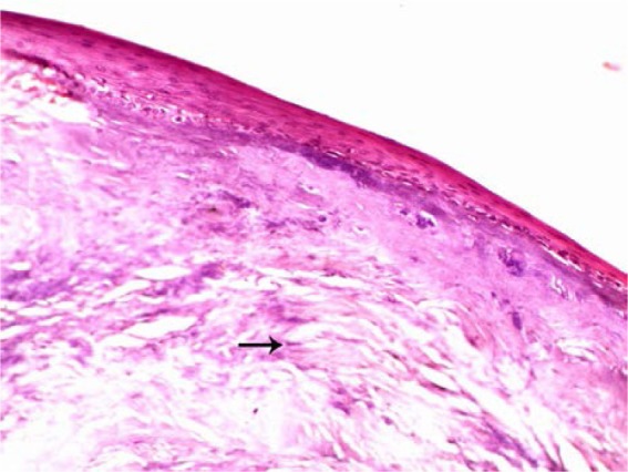
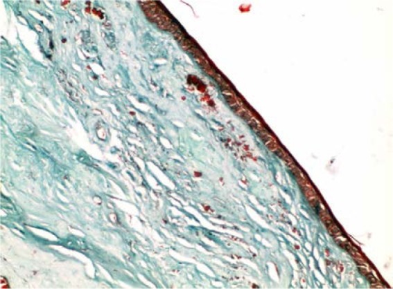
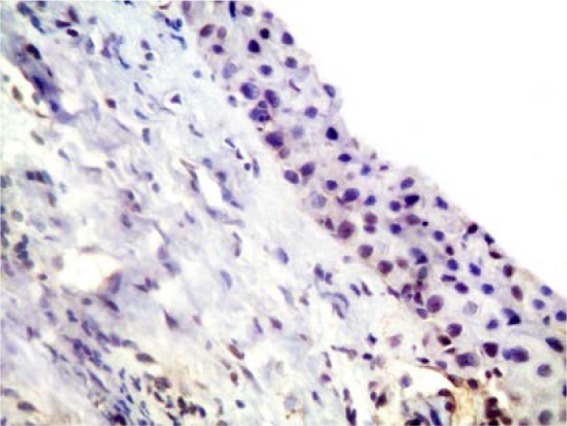
Patients and methods: A total of 20 patients with primary pterygium underwent subconjunctival combined injection of 0.1 mL of MMC (0.1 mg/mL) and 0.1 mL of bevacizumab (1.25 mg/0.1 mL) 1 month before bare sclera excision of the pterygium. The excised pterygium tissues were examined histologically and immunohistologically by CD31 staining, and the patients were followed up clinically for at least 2 years. The excised pterygia of two patients without preoperative injection were used for histological comparison.
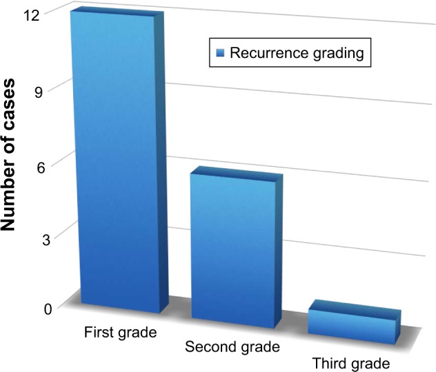
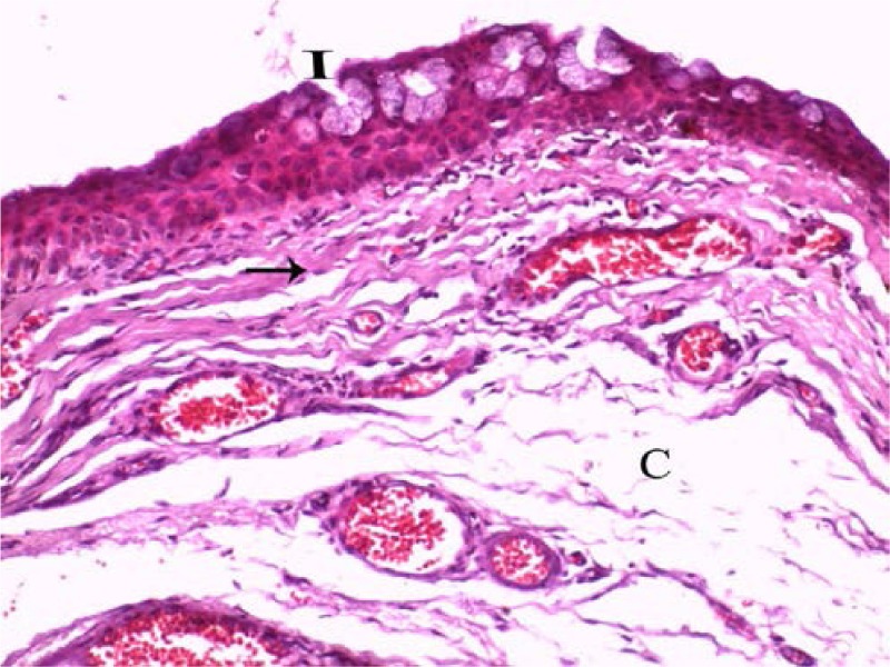
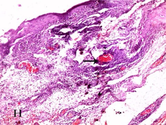
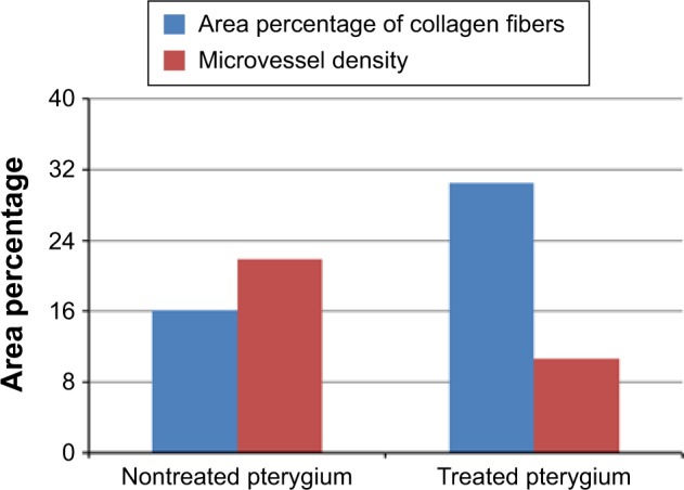
Results: Clinically, there were no intraoperative or postoperative complications. No recurrence was noted during the follow-up period. Histologically, the previously injected pterygia showed a decreased number of epithelial cells and stromal fibroblasts. The latter were rounded or oval and swollen rather than spindle shaped, and some were degenerating or apoptotic. Collagen and elastic fibers were degenerated, distorted, and decreased in density, while blood capillaries were obliterated. There was a significant decrease in CD31-positive cells in previously injected pterygia.
Conclusion: Preoperative subpterygium combined injection of bevacizumab and MMC is safe and effective in reducing the postoperative recurrence of primary pterygium. Histological and immunohistological changes in the form of decreased fibrovascular activity and degeneration of the extracellular matrix and nerve axons were noted.
Keywords: CD31; histological changes; primary pterygium; subconjunctival bevacizumab; subconjunctival mitomycin C.
Conflict of interest statement
Disclosure The authors report no conflicts of interest in this work.
Figures
References
-
- Hill JC, Maske R. Pathogenesis of pterygium. Eye (Lond) 1989;3(pt 2):218–226. - PubMed
-
- Kunimoto D, Kanitkar K, Makar M. The Wills Eye Manual: Office and Emergency Room Diagnosis and Treatment of Eye Disease. 4th ed. Philadelphia, PA: Lippincott Williams & Wilkins; 2004. pp. 50–51.
-
- Mauro J, Foster CS. Pterygia: pathogenesis and the role of subconjunctival bevacizumab in treatment. Semin Ophthalmol. 2009;24(3):130–134. - PubMed
-
- Kria L, Ohira A, Amemiya T. Immunohistochemical localization of basic fibroblast growth factor, platelet derived growth factor, transforming growth factor-beta and tumor necrosis factor-alpha in the pterygium. Acta Histochem. 1996;98(2):195–201. - PubMed
LinkOut - more resources
Full Text Sources
Other Literature Sources
