. 2017 Mar 8:10:1475-1485.
doi: 10.2147/OTT.S92389. eCollection 2017.

Investigating the expression, effect and tumorigenic pathway of PADI2 in tumors

Affiliations

Investigating the expression, effect and tumorigenic pathway of PADI2 in tumors

Wei Guo et al. Onco Targets Ther. .

Abstract

Background: Peptidylarginine deiminase (PAD) catalyzes the conversion of arginine residues to citrulline residues, termed citrullination. Recent studies have suggested that PAD isoform 2 (PADI2) plays an important role in tumors, although its tumorigenic effect and mechanism are largely unknown.

Materials and methods: Immunohistochemistry and enzyme-linked immunosorbent assay (ELISA) were used to investigate the expression level of PADI2 in various tumor tissues and patient blood samples, respectively. MNK-45 and Bel-7402 tumor cell lines originating from gastric and liver tumors, respectively, were treated with anti-PADI2 siRNA, and the subsequent cell proliferation, apoptosis and migration were observed. Polymerase chain reaction (PCR) arrays, including Cancer PathwayFinder, Oncogenes and Tumor Suppressor Genes, p53 Signaling Pathway, Signal Transduction Pathway and Tumor Metastasis PCR arrays, were used to investigate the tumorigenic pathway of PADI2 in the siRNA-treated tumor cells. This analysis was verified by real-time PCR.

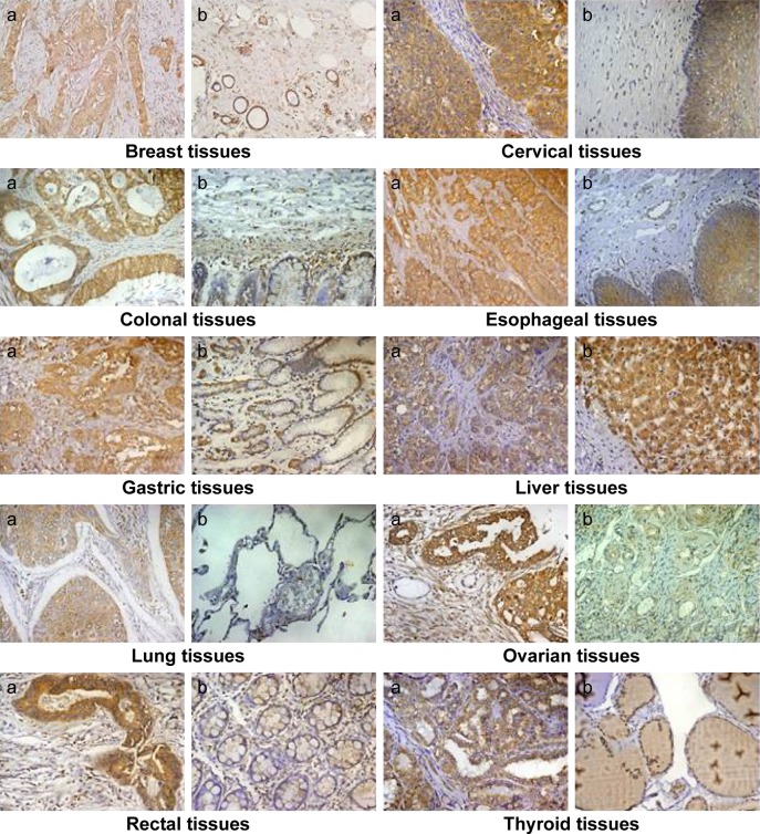
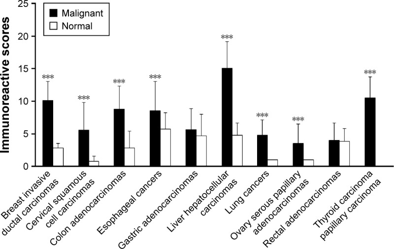
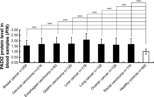
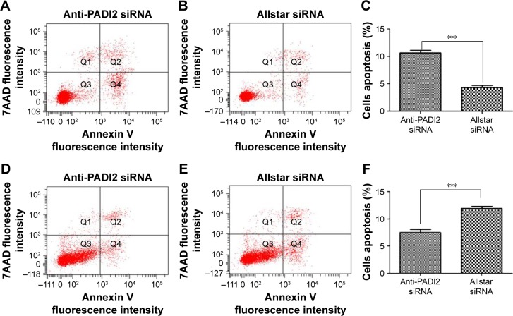
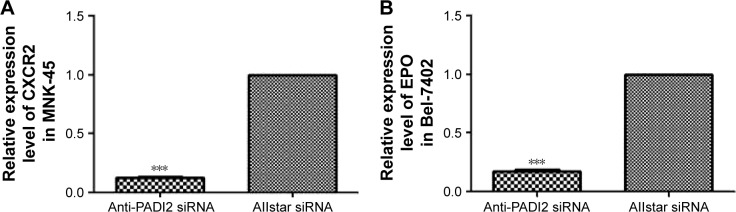
Results: Immunohistochemistry detected significantly increased expression of PADI2 in invasive breast ductal carcinoma, cervical squamous cell carcinoma, colon adenocarcinoma, liver hepatocellular carcinoma, lung cancer, ovarian serous papillary adenocarcinoma and papillary thyroid carcinoma samples. ELISA detected a twofold increase in PADI2 expression in the blood of 48.3% of patients with liver cancer, 38% of patients with cervical carcinoma and 32% of patients with gastric carcinoma. Increased apoptosis and decreased cell proliferation and migration were observed in the anti-PADI2 siRNA-treated MNK-45 cells, and increased cell proliferation and migration and decreased apoptosis were observed in the treated Bel-7402 cells with suppressed PADI2 expression. PCR arrays and real-time PCR detected significantly decreased CXCR2 and EPO expression in the MNK-45 cells and Bel-7402 cells, respectively, with the anti-PADI2 siRNA treatments.

Conclusion: PADI2 expression is increased in many types of tumor tissues and patient blood samples. PADI2 may advance abnormal cell behavior in gastric cancers by mediating CXCR2, a well-known gene that stimulates cell proliferation and invasion. However, PADI2 might have deleterious effects on tumor growth and metastasis in liver tumor cells by regulating the expression of EPO, a gene with controversial functions in tumor growth. The results suggest that the effect of PADI2 on tumorigenesis is multifactorial, depending on the tumor type.

Keywords: CXCR2; EPO; PADI2; pathway; tumorigenesis.


Conflict of interest statement

Disclosure The authors report no conflicts of interest in this work.

Figures

Figure 1
Figure 1
Immunodetection of PADI2 in various tumor tissues. Notes: PADI2 shows significantly increased expression in many tumor types. “a” represents tumor tissue, and “b” represents the parallel tumor adjacent tissues or normal tissues. Original magnification: 200×. Abbreviation: PADI2, peptidylarginine deiminase isoform 2.
Figure 2
Figure 2
Semiquantitation of the PADI2 expression level in various tumor tissues. Notes: The x-axis indicates tumor types and matched normal tissues, and the y-axis indicates the expression level of PADI2. ***P<0.001. Abbreviation: PADI2, peptidylarginine deiminase isoform 2.
Figure 3
Figure 3
Determination of PADI2 levels in the blood samples of patients with tumors using ELISA. Notes: The x-axis represents various tumor types, and the y-axis represents the PADI2 expression level with P/N values. The OD450 value of the positive serum (P-value) was divided by the OD405 value of the pooled negative sera (N value). The P/N ratio was calculated for each serum sample. ***P<0.001. Abbreviations: PADI2, peptidylarginine deiminase isoform 2; ELISA, enzyme-linked immunosorbent assay; OD, optical density.
Figure 4
Figure 4
Proliferation of MNK-45 cells and Bel-7402 cells treated with anti-PADI2 siRNA. Notes: (A) The CCK-8 assay detected MNK-45 viable cell numbers as represented by an OD value at 405 nm. (B) The CCK-8 assay detected Bel-7402 viable cell numbers as represented by an OD value at 405 nm. Cells treated with Allstar siRNA were used as negative controls. *P<0.05, **P<0.01 and ***P<0.001. Abbreviations: PADI2, peptidylarginine deiminase isoform 2; CCK-8, cell counting kit-8; OD, optical density.
Figure 5
Figure 5
Apoptosis in MNK-45 cells and Bel-7402 cells treated with anti-PADI2 siRNA, as measured using the annexin V cell apoptosis assay. Notes: (A) MNK-45 cells were treated with anti-PADI2 siRNA. (B) MNK-45 cells treated with Allstar siRNA were used as negative controls. (C) Result of the apoptosis assay for MNK-45 cells. (D) Bel-7402 cells were treated with anti-PADI2 siRNA. (E) Bel-7402 cells treated with Allstar siRNA were used as negative controls. (F) Result of the apoptosis assay for MNK-45 cells. ***P<0.05. Abbreviation: PADI2, peptidylarginine deiminase isoform 2.
Figure 6
Figure 6
Migration of MNK-45 and Bel-7402 cells treated with anti-PADI2 siRNA, as measured by the transwell migration assay. Notes: (A) MNK-45 cells were treated with anti-PADI2 siRNA. (B) MNK-45 cells treated with Allstar siRNA were used as negative controls. (C) Result of the migration measurement for MNK-45 cells. (D) Bel-7402 cells were treated with anti-PADI2 siRNA. (E) Bel-7402 cells treated with Allstar siRNA were used as negative controls. (F) Result of the migration measurement for Bel-7402 cells. Original magnification: 4.2×. ***P<0.001. Abbreviation: PADI2, peptidylarginine deiminase isoform 2.
Figure 7
Figure 7
Determination of significantly altered gene expression using PCR arrays. Notes: Tumor cell lines MNK-45 and Bel-7402 were treated with anti-PADI2 siRNA. Cells treated with Allstar siRNA were used as a negative control. Significantly altered expression of tumor-related genes was detected using (A) Cancer PathwayFinder PCR array, (B) Signal Transduction PCR array, (C) Tumor Metastasis PCR array, (D) Oncogenes and Tumor Suppressor Genes PCR array and (E) p53 Signaling PCR array. Abbreviations: PCR, polymerase chain reaction; PADI2, peptidylarginine deiminase isoform 2.
Figure 8
Figure 8
Determination of the mRNA expression levels of CXCR2 and EPO using real-time PCR. Notes: MNK-45 cells and Bel-7402 cells were treated with anti-PADI2 siRNA. The transcription levels of the target genes in Allstar siRNA-treated cells were set as 1. The target genes in the anti-PADI2 siRNA-treated cells were normalized to their expression in the controls. (A) CXCR2 expression level in MNK-45 cells and (B) EPO expression level in Bel-7402 cells. ***P<0.001. Abbreviations: PCR, polymerase chain reaction; PADI2, peptidylarginine deiminase isoform 2.

Similar articles

Cited by

References

    1. Pritzker LB, Nguyen TA, Moscarello MA. The developmental expression and activity of peptidylarginine deiminase in the mouse. Neurosci Lett. 1999;266(3):161–164. - PubMed
    1. Arita K, Hashimoto H, Shimizu T, Nakashima K, Yamada M, Sato M. Structural basis for Ca(2+)-induced activation of human PAD4. Nat Struct Mol Biol. 2004;11(8):777–783. - PubMed
    1. Chavanas S, Méchin MC, Nachat R, et al. Peptidylarginine deiminases and deimination in biology and pathology: relevance to skin homeostasis. J Dermatol Sci. 2006;44(2):63–72. - PubMed
    1. Mohanan S, Cherrington BD, Horibata S, McElwee JL, Thompson PR, Coonrod SA. Potential role of peptidylarginine deiminase enzymes and protein citrullination in cancer pathogenesis. Biochem Res Int. 2012;2012:895343. - PMC - PubMed
    1. Méchin MC, Coudane F, Adoue V, et al. Deimination is regulated at multiple levels including auto-deimination of peptidylarginine deiminases. Cell Mol Life Sci. 2010;67(9):1491–1503. - PMC - PubMed