

Silicone-induced granuloma of breast implant capsule (SIGBIC): similarities and differences with anaplastic large cell lymphoma (ALCL) and their differential diagnosis
- PMID: 28331364
- PMCID: PMC5354541
- DOI: 10.2147/BCTT.S126003
Silicone-induced granuloma of breast implant capsule (SIGBIC): similarities and differences with anaplastic large cell lymphoma (ALCL) and their differential diagnosis
Abstract
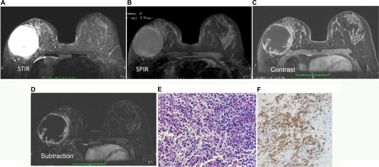
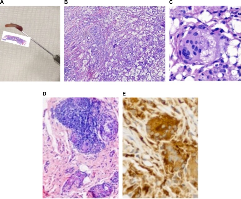
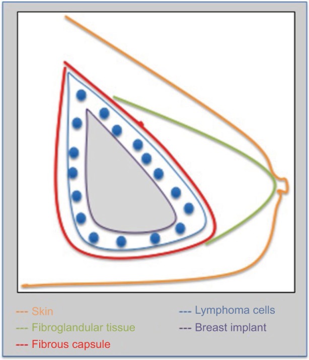
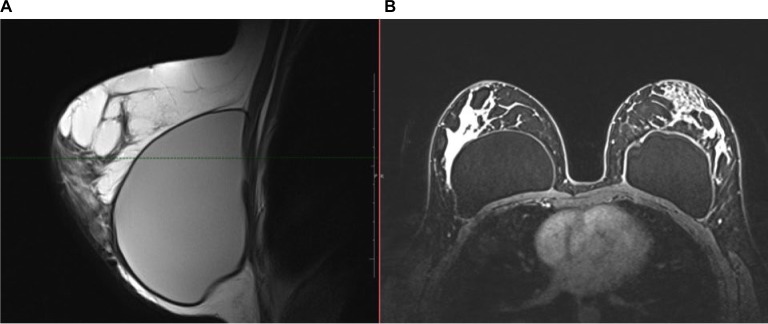
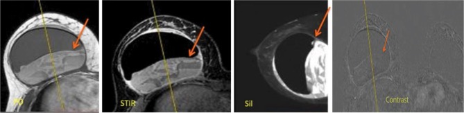
Primary breast lymphoma is a rare disease and accounts for 0.5% of cases of breast cancer. Most primary breast lymphomas develop from B cells, and the involvement of T cells is rare. Anaplastic large cell lymphoma (ALCL) is a recently discovered T-cell lymphoma associated with breast implants. Only a few cases have been reported to date. It is believed that the incidence of ALCL is increasing because of the increasing number of breast implants. The clinical presentation is variable and can manifest as a palpable mass in the breast or armpit, breast pain, or capsular contracture. Because of the rarity of the disease and the lack of knowledge to date, clinical diagnosis is often delayed, with consequent delays in treatment. The cause and pathogenesis have not been fully elucidated, and there are no evidence-based guidelines for diagnosis, treatment, or follow-up of this disease. We present a review of cases of patients with silicone breast implants, including ALCL, a rare type of breast cancer that is still under study, and silicone-induced granuloma of breast implant capsule and its differential diagnosis, and discuss if a silicone-induced granuloma of breast implant capsule could be the precursor of the disease.
Keywords: breast cancer; granuloma; implant; lymphoma.
Conflict of interest statement
Disclosure The authors report no conflicts of interest in this work.
Figures
Similar articles
-
Synchronous breast implant associated anaplastic large cell lymphoma (BIA-ALCL) and silicone induced granuloma of breast implant capsule (SIGBIC): What to learn.Radiol Case Rep. 2020 Aug 1;15(10):1736-1742. doi: 10.1016/j.radcr.2020.07.017. eCollection 2020 Oct. Radiol Case Rep. 2020. PMID: 32774571 Free PMC article.
-
Breast magnetic resonance imaging: tips for the diagnosis of silicone-induced granuloma of a breast implant capsule (SIGBIC).Insights Imaging. 2017 Aug;8(4):439-446. doi: 10.1007/s13244-017-0564-3. Epub 2017 Jul 14. Insights Imaging. 2017. PMID: 28710678 Free PMC article. Review.
-
Silicone-Induced Granuloma of Breast Implant Capsule (SIGBIC): Histopathology and Radiological Correlation.J Immunol Res. 2018 Sep 20;2018:6784971. doi: 10.1155/2018/6784971. eCollection 2018. J Immunol Res. 2018. PMID: 30327786 Free PMC article. Review.
-
The A, B and C's of Silicone Breast Implants: Anaplastic Large Cell Lymphoma, Biofilm and Capsular Contracture.Materials (Basel). 2018 Nov 28;11(12):2393. doi: 10.3390/ma11122393. Materials (Basel). 2018. PMID: 30486500 Free PMC article. Review.
-
Anaplastic large-cell lymphoma in women with breast implants.JAMA. 2008 Nov 5;300(17):2030-5. doi: 10.1001/jama.2008.585. JAMA. 2008. PMID: 18984890
Cited by
-
Not All That Shines on a PET Scan Is Cancer: A Silicone-Induced Granuloma Masquerading as Malignancy.Clin Pract. 2020 Dec 29;11(1):8-12. doi: 10.3390/clinpract11010003. Clin Pract. 2020. PMID: 33599216 Free PMC article.
-
BIA-ALCL-Horizon Scanning.JPRAS Open. 2022 Oct 21;34:245-251. doi: 10.1016/j.jpra.2022.09.007. eCollection 2022 Dec. JPRAS Open. 2022. PMID: 36483300 Free PMC article.
-
Fundamentals of Breast Implant Illness and Device Imaging.Int J Inflam. 2022 Aug 13;2022:4155530. doi: 10.1155/2022/4155530. eCollection 2022. Int J Inflam. 2022. PMID: 35996624 Free PMC article. Review.
-
Etiology of Breast Implant-Associated Anaplastic Large Cell Lymphoma (BIA-ALCL): Current Directions in Research.Cancers (Basel). 2020 Dec 21;12(12):3861. doi: 10.3390/cancers12123861. Cancers (Basel). 2020. PMID: 33371292 Free PMC article. Review.
-
Silicone Induced Granuloma of Breast Implant Capsule (SIGBIC) diagnosis: Breast Magnetic Resonance (BMR) sensitivity to detect silicone bleeding.PLoS One. 2020 Jun 26;15(6):e0235050. doi: 10.1371/journal.pone.0235050. eCollection 2020. PLoS One. 2020. PMID: 32589678 Free PMC article.
References
-
- inca.gov.br [homepage on the internet] Rio de Jajeiro: INCA- Ministério da Saúde; c1996-2016. [Accessed October 27, 2016]. Available from: http://www2.inca.gov.br/wps/wcm/connect/tiposdecancer/site/home/mama.
-
- Miranda RN, Lin L, Talwalkar SS, et al. Anaplastic large cell lymphoma involving the breast: a clinicopathologic study of 6 cases and review of the literature. Arch Pathol Lab Med. 2009;133:1383–1390. - PubMed
-
- Binmahfouz A, Steinke K. A case report of breast implantassociated anaplastic large cell lymphoma: The good, the bad, and the ugly. Int J Case Rep Images. 2016;7(8):537–541.
Publication types
LinkOut - more resources
Full Text Sources
Other Literature Sources
