

Long-term efficacy of ultrasound-guided high-intensity focused ultrasound treatment of breast fibroadenoma
- PMID: 28331611
- PMCID: PMC5353785
- DOI: 10.1186/s40349-017-0083-1
Long-term efficacy of ultrasound-guided high-intensity focused ultrasound treatment of breast fibroadenoma
Abstract
Background: To assess the long term efficacy and tolerability of one or two ultrasound (US)-guided high-intensity focused ultrasound (HIFU) treatment in patients with breast fibroadenoma (FA).
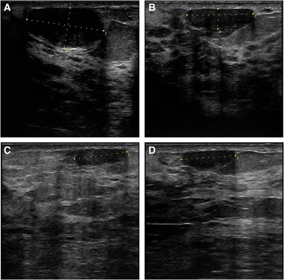
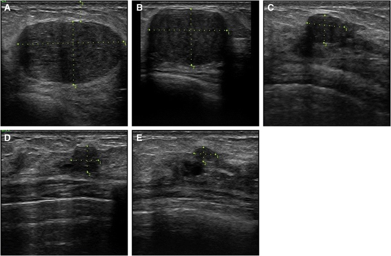
Methods: Twenty patients with 26 FA were selected for US-guided HIFU. The therapy was performed in one or two sessions. FA volume was assessed before and followed up to 24 months after the last HIFU. After each treatment, adverse events were evaluated.
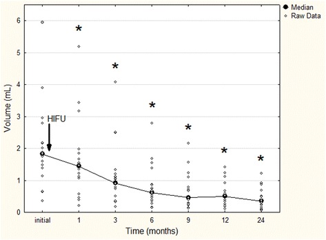
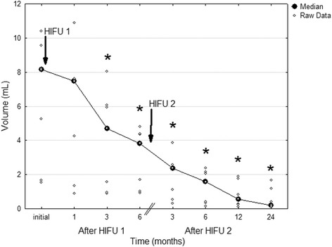
Results: In 19/26 FA (73.1%) one HIFU was performed (group 1), whereas 7/26 FA (26.9%) received second HIFU (group 2) 6-9 months (median, 7 months) after the first session. In group 1 and 2, FA volume decreased significantly at 1-month (p < 0.001) and 3-month follow-up (p = 0.005), respectively, and continued to reduce until 24-month follow-up (p < 0.001 and p = 0.003, respectively). At 24 months, mean volume reduction was 77.32% in group 1 and 90.47% in group 2 (p = 0.025). Mild subcutaneous edema was observed in 4 patients and skin erythema in 3 patients.
Conclusions: US-guided HIFU represents a promising non-invasive method with sustainable FA volume reduction and patient's tolerability. Although one treatment is highly efficient, the volume reduction can be increased with second treatment.
Trial registration: NCT01331954. Registered 07 April 2011.
Keywords: Ablation techniques; Breast fibroadenoma; High-intensity focused ultrasound (HIFU); Interventional ultrasonography; Ultrasound (US) guidance.
Figures
Similar articles
-
Five-year follow-up after a single US-guided high intensity focused ultrasound treatment of breast fibroadenoma.Sci Rep. 2024 Aug 7;14(1):18370. doi: 10.1038/s41598-024-68827-4. Sci Rep. 2024. PMID: 39112604 Free PMC article.
-
Ultrasound-guided high-intensity focused ultrasound treatment of breast fibroadenoma-a multicenter experience.J Ther Ultrasound. 2015 Jan 22;3(1):1. doi: 10.1186/s40349-014-0022-3. eCollection 2015. J Ther Ultrasound. 2015. PMID: 25635224 Free PMC article.
-
High intensity focused ultrasound (HIFU) for the treatment of symptomatic breast fibroadenoma.Int J Hyperthermia. 2018;35(1):463-470. doi: 10.1080/02656736.2018.1508757. Epub 2018 Sep 11. Int J Hyperthermia. 2018. PMID: 30204024
-
Clinical efficacy and safety of ultrasound-guided high-intensity focused ultrasound for breast fibroadenoma: a systematic review and meta-analysis.Int J Hyperthermia. 2024;41(1):2374874. doi: 10.1080/02656736.2024.2374874. Epub 2024 Jul 25. Int J Hyperthermia. 2024. PMID: 39053900
-
[Treatment of fibroadenomas by high-intensity focused ultrasound: What results? Review].Gynecol Obstet Fertil Senol. 2018 Jun;46(6):524-529. doi: 10.1016/j.gofs.2018.05.001. Gynecol Obstet Fertil Senol. 2018. PMID: 29773521 Review. French.
Cited by
-
Portable ultrasound-guided high-intensity focused ultrasound with functions for safe and rapid ablation: prospective clinical trial for uterine fibroids-short-term and long-term results.Eur Radiol. 2020 Mar;30(3):1554-1563. doi: 10.1007/s00330-019-06468-2. Epub 2019 Nov 8. Eur Radiol. 2020. PMID: 31705252 Clinical Trial.
-
Ultrasound radiomics features predicting the dosimetry for focused ultrasound surgery of benign breast tumor: A retrospective study.Front Genet. 2022 Sep 6;13:969409. doi: 10.3389/fgene.2022.969409. eCollection 2022. Front Genet. 2022. PMID: 36118892 Free PMC article.
-
Five-year follow-up after a single US-guided high intensity focused ultrasound treatment of breast fibroadenoma.Sci Rep. 2024 Aug 7;14(1):18370. doi: 10.1038/s41598-024-68827-4. Sci Rep. 2024. PMID: 39112604 Free PMC article.
-
Clinical Efficacy of Ultrasound-guided High-intensity Focused Ultrasound Ablation for Treating Breast Fibroadenoma of Different Sizes: A Retrospective Study.Gynecol Minim Invasive Ther. 2025 Feb 27;14(1):72-80. doi: 10.4103/gmit.GMIT-D-24-00035. eCollection 2025 Jan-Mar. Gynecol Minim Invasive Ther. 2025. PMID: 40143974 Free PMC article.
-
The Role of High Intensity Focused Ultrasound in the Treatment of Fibroadenomas: A Systematic Review.Breast Care (Basel). 2023 Aug;18(4):279-288. doi: 10.1159/000524738. Epub 2022 May 2. Breast Care (Basel). 2023. PMID: 37900548 Free PMC article. Review.
References
Associated data
LinkOut - more resources
Full Text Sources
Other Literature Sources
Medical
Miscellaneous
