Review
. 2017 Mar 11:3:6.
doi: 10.1186/s40748-017-0045-1. eCollection 2017.

Congenital Diaphragmatic hernia - a review

Affiliations
Review

Congenital Diaphragmatic hernia - a review

Praveen Kumar Chandrasekharan et al. Matern Health Neonatol Perinatol. .

Abstract

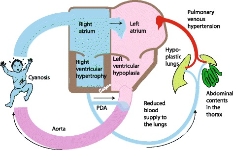
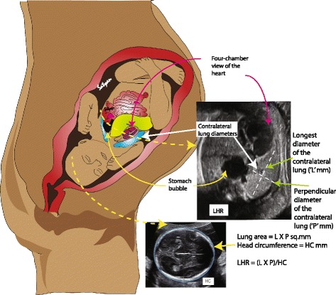
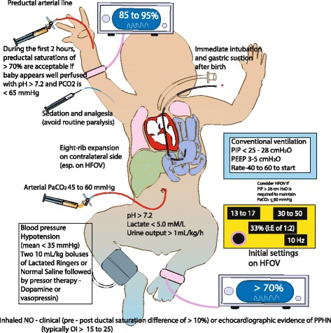
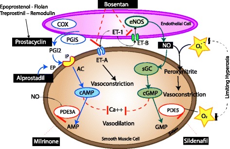
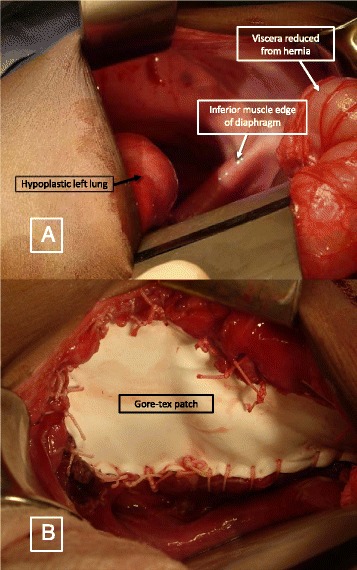
Congenital Diaphragmatic hernia (CDH) is a condition characterized by a defect in the diaphragm leading to protrusion of abdominal contents into the thoracic cavity interfering with normal development of the lungs. The defect may range from a small aperture in the posterior muscle rim to complete absence of diaphragm. The pathophysiology of CDH is a combination of lung hypoplasia and immaturity associated with persistent pulmonary hypertension of newborn (PPHN) and cardiac dysfunction. Prenatal assessment of lung to head ratio (LHR) and position of the liver by ultrasound are used to diagnose and predict outcomes. Delivery of infants with CDH is recommended close to term gestation. Immediate management at birth includes bowel decompression, avoidance of mask ventilation and endotracheal tube placement if required. The main focus of management includes gentle ventilation, hemodynamic monitoring and treatment of pulmonary hypertension followed by surgery. Although inhaled nitric oxide is not approved by FDA for the treatment of PPHN induced by CDH, it is commonly used. Extracorporeal membrane oxygenation (ECMO) is typically considered after failure of conventional medical management for infants ≥ 34 weeks' gestation or with weight >2 kg with CDH and no associated major lethal anomalies. Multiple factors such as prematurity, associated abnormalities, severity of PPHN, type of repair and need for ECMO can affect the survival of an infant with CDH. With advances in the management of CDH, the overall survival has improved and has been reported to be 70-90% in non-ECMO infants and up to 50% in infants who undergo ECMO.

Keywords: Extracorporeal membrane oxygenation; Lung Hypoplasia; Pulmonary Hypertension.


Figures

Fig. 1
Fig. 1
Classification of CDH based on location of the diaphragmatic hernias: Most common type of hernias are the posterior lateral hernias (70–75%) also known as Bochdalek hernias, with majority occurring on the left side (85%) and less frequently on the right side (13%) or bilateral (2%). Other types of hernias are the anterior defects or Morgagni hernias (23–28%) followed by the rare central hernias (2–7%). (Copyright Satyan Lakshminrusimha)
Fig. 2
Fig. 2
Size of the defect – The size of the defect may vary between small (A) to diaphragmatic agenesis (D). Defects B & C are considered moderate to large (Tsao et al. 2008). (Copyright Satyan Lakshminrusimha)
Fig. 3
Fig. 3
Anatomical and radiological features of CDH – A defect in the diaphragm causes the abdominal viscera to herniate into the thoracic cavity. Left sided hernias are common (85%) which results in herniation of both small and large intestines along with solid intra-abdominal organs. Herniation of viscera into the thoracic cavity results in abnormal lung development on the ipsilateral side with variable effect on the contralateral side. The effect of abnormal lung development on the contralateral side depends on the extent of herniation and the effect on mediastinal shift. Pulmonary hypoplasia results in abnormal pulmonary vasculature resulting in persistent pulmonary hypertension leading to right ventricular dysfunction. This is more pronounced after transitioning from fetal circulation. Left ventricular dysfunction can be secondary to direct compression in left sided hernia and secondary to low ventricular volumes in right sided hernias. Pre-operative chest and abdomen x-ray shows the air and fluid filled loops of bowels on the left side of the thorax with the endotracheal tube above the thoracic vertebra level 4 pushed towards the right side signifying mediastinal shift. (Copyright Satyan Lakshminrusimha)
Fig. 4
Fig. 4
Two-hit hypothesis for CDH (Copyright Satyan Lakshminrusimha)
Fig. 5
Fig. 5
Cardiovascular effects of CDH – Hypoplastic lungs secondary to herniation of abdominal viscera leads to concomitant hypoplasia of the pulmonary vessels. This results in reduced blood supply to the hypoplastic alveolar-capillary unit. Once the infant transitions from fetal circulation, this effect is more pronounced resulting in pulmonary hypertension which leads to right ventricular dysfunction. Secondary to pulmonary hypertension, there is shunting of blood from right to left across the patent foramen ovale and the patent ductus arteriosus. Left ventricular dysfunction along with left atrial dysfunction results in pulmonary venous hypertension and worsening of pulmonary arterial hypertension. This presents clinically in a wide spectrum of labile pre & postductal saturations to profound cyanosis. (Copyright Satyan Lakshminrusimha)
Fig. 6
Fig. 6
Lung to head ratio (LHR) measurement – Obstetric ultrasound technique is used to measure the lung to head ratio known as LHR to assess the severity of CDH. The head circumference is measured as shown. The contralateral lung area is calculated as a product of the longest and perpendicular diameter of the contralateral lung. The ratio of this area to the head circumference gives the LHR. A LHR of <0.6 has been associated with poor outcome while a ratio of >1.35 has been associated with survival. Alternatively, an observed to expected LHR measurement is used in order to overcome bias secondary to gestational age. (Copyright Satyan Lakshminrusimha)
Fig. 7
Fig. 7
Management of CDH – At birth infants with CDH or suspected CDH should have an orogastric/nasogastric tube with suction to attain bowel decompression. Bag-mask ventilation should be avoided. The majority of these infants (especially with prenatal diagnosis of CDH) require intubation in the delivery room. A pre-ductal pulse oximeter is placed on the right upper extremity as soon as possible. Oxygen saturation targets are based on NRP guidelines. Ventilation using a T-piece resuscitator is preferred to avoid high airway pressures. Ventilator parameters are as shown in the figure. Preductal blood gases and invasive blood pressure monitoring are preferred. Inhaled nitric oxide is often used for the management of PPHN. For blood pressure management, fluid boluses and vasopressor agents are used based on the parameters in the figure. (Copyright Satyan Lakshminrusimha)
Fig. 8
Fig. 8
Management of pulmonary hypertension in CDH: pulmonary vasodilators and nitric oxide – prostacyclin – endothelin pathways. AC – acetylcholine, Ca- calcium, cAMP - cyclic adenosine monophosphate, cGMP – cyclic guanosine monophosphate, COX – cycloxygenase, eNOS – endothelial nitric oxide synthase, ET – endothelin, EP – prostaglandin E receptor, IP – prostacyclin I receptor, NO – nitric oxide, PGI – prostaglandin I, sGC - soluble guanylyl cyclase, PDE – phosphodiesterase inhibitor (Copyright Satyan Lakshminrusimha)
Fig. 9
Fig. 9
a Left sided diaphragmatic hernia showing the hypoplastic left lung, inferior muscle edge of the diaphragm and reduced viscera. b Prosthetic patch – Gore-Tex patch used to close the defect

References

    1. Colvin J, Bower C, Dickinson JE, Sokol J. Outcomes of congenital diaphragmatic hernia: a population-based study in Western Australia. Pediatrics. 2005;116(3):e356–363. doi: 10.1542/peds.2004-2845. - DOI - PubMed
    1. Gallot D, Boda C, Ughetto S, Perthus I, Robert-Gnansia E, Francannet C, Laurichesse-Delmas H, Jani J, Coste K, Deprest J, et al. Prenatal detection and outcome of congenital diaphragmatic hernia: a French registry-based study. Ultrasound Obstet Gynecol. 2007;29(3):276–283. doi: 10.1002/uog.3863. - DOI - PubMed
    1. Yang W, Carmichael SL, Harris JA, Shaw GM. Epidemiologic characteristics of congenital diaphragmatic hernia among 2.5 million California births, 1989–1997. Birth Defects Res A Clin Mol Teratol. 2006;76(3):170–174. doi: 10.1002/bdra.20230. - DOI - PubMed
    1. McGivern MR, Best KE, Rankin J, Wellesley D, Greenlees R, Addor MC, Arriola L, de Walle H, Barisic I, Beres J, et al. Epidemiology of congenital diaphragmatic hernia in Europe: a register-based study. Arch Dis Child Fetal Neonatal Ed. 2015;100(2):F137–144. doi: 10.1136/archdischild-2014-306174. - DOI - PubMed
    1. Tennant PW, Samarasekera SD, Pless-Mulloli T, Rankin J. Sex differences in the prevalence of congenital anomalies: a population-based study. Birth Defects Res A Clin Mol Teratol. 2011;91(10):894–901. doi: 10.1002/bdra.22846. - DOI - PubMed