Review
. 2017:2017:8081758.
doi: 10.1155/2017/8081758. Epub 2017 Feb 26.

Emerging Synaptic Molecules as Candidates in the Etiology of Neurological Disorders

Affiliations
Review

Emerging Synaptic Molecules as Candidates in the Etiology of Neurological Disorders

Viviana I Torres et al. Neural Plast. 2017.

Abstract

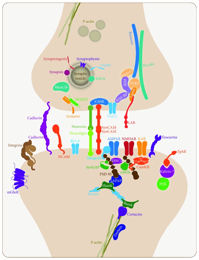
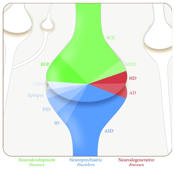
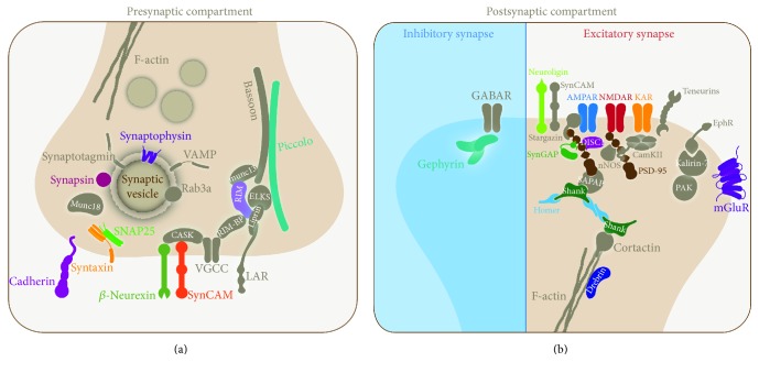
Synapses are complex structures that allow communication between neurons in the central nervous system. Studies conducted in vertebrate and invertebrate models have contributed to the knowledge of the function of synaptic proteins. The functional synapse requires numerous protein complexes with specialized functions that are regulated in space and time to allow synaptic plasticity. However, their interplay during neuronal development, learning, and memory is poorly understood. Accumulating evidence links synapse proteins to neurodevelopmental, neuropsychiatric, and neurodegenerative diseases. In this review, we describe the way in which several proteins that participate in cell adhesion, scaffolding, exocytosis, and neurotransmitter reception from presynaptic and postsynaptic compartments, mainly from excitatory synapses, have been associated with several synaptopathies, and we relate their functions to the disease phenotype.


Conflict of interest statement

All authors declare no conflict of interests.

Figures

Figure 1
Figure 1
Molecular composition of a central chemical synapse. The image shows a typical excitatory synapse in the CNS. Pre- and postsynaptic proteins are organized in macromolecular functional complexes playing different roles in scaffolding, exocytosis, endocytosis, and signaling in their respective compartments. In addition, the most relevant adhesion molecules are represented.
Figure 2
Figure 2
Schematic representation of neurological disorders associated with synaptic protein dysfunction. The image summarizes the neurological diseases described in this review represented by color code: neurodevelopmental (green spectrum), neuropsychiatric (blue spectrum), and neurodegenerative (red spectrum). The number of synaptic proteins involved in each category is proportionally illustrated. AD, Alzheimer's disease; ADHD, attention deficit hyperactivity disorder; ASD, autism spectrum disorder; BPD, bipolar spectrum disorder; FXS, Fragile X syndrome; HD, Huntington's Disease; ID, intellectual disability; MDD, major depressive disorder; SCZ, schizophrenia.
Figure 3
Figure 3
Schematic representation of synaptic proteins associated with synaptopathies. (a) Presynaptic and (b) postsynaptic proteins involved in human synaptopathies described in this review are color highlighted. Mutations in a gene or gene combination for a synaptic protein may lead to neurodevelopment, neuropsychiatric, and neurodegenerative diseases.
Figure 4
Figure 4
Domain structure of two active zone proteins associated with synaptopathies. The diagrams show the multimodular organization of (a) Piccolo and (b) RIM, and their interaction with other proteins. Arrows indicate binding reactions. Domains are shown in colored boxes and designations are indicated by standard abbreviations.

Similar articles

Cited by

References

    1. Ackermann F., Waites C. L., Garner C. C. Presynaptic active zones in invertebrates and vertebrates. EMBO Reports. 2015;16(8):923–938. doi: 10.15252/embr.201540434. - DOI - PMC - PubMed
    1. Südhof T. C. The presynaptic active zone. Neuron. 2012;75(1):11–25. doi: 10.1016/j.neuron.2012.06.012. - DOI - PMC - PubMed
    1. Okabe S. Molecular anatomy of the postsynaptic density. Molecular and Cellular Neuroscience. 2007;34(4):503–518. doi: 10.1016/j.mcn.2007.01.006. - DOI - PubMed
    1. Dani A., Huang B., Bergan J., Dulac C., Zhuang X. Superresolution imaging of chemical synapses in the brain. Neuron. 2010;68(5):843–856. doi: 10.1016/j.neuron.2010.11.021. - DOI - PMC - PubMed
    1. Sigrist S. J., Sabatini B. L. Optical super-resolution microscopy in neurobiology. Current Opinion in Neurobiology. 2012;22(1):86–93. doi: 10.1016/j.conb.2011.10.014. - DOI - PubMed

Publication types