. 2016 Aug;1(2):eaah3539.
doi: 10.1126/sciimmunol.aah3539. Epub 2016 Aug 19.

IL-1β is an innate immune sensor of microbial proteolysis

Affiliations

IL-1β is an innate immune sensor of microbial proteolysis

Christopher N LaRock et al. Sci Immunol. 2016 Aug.

Abstract

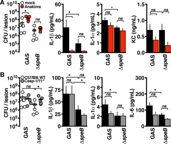
Interleukin-1β (IL-1β) is a key proinflammatory cytokine that drives antimicrobial immune responses. IL-1β is aberrantly activated in autoimmune diseases, and IL-1β inhibitors are used as therapeutic agents to treat patients with certain autoimmune disorders. Review of postmarketing surveillance of patients receiving IL-1β inhibitors found a disproportionate reporting of invasive infections by group A Streptococcus (GAS). IL-1β inhibition increased mouse susceptibility to GAS infection, but IL-1β was produced independent of canonical inflammasomes. Newly synthesized IL-1β has an amino-terminal prodomain that blocks signaling activity, which is usually proteolytically removed by caspase-1, a protease activated within the inflammasome structure. In place of host caspases, the secreted GAS cysteine protease SpeB generated mature IL-1β. During invasive infection, GAS isolates may acquire pathoadaptive mutations eliminating SpeB expression to evade detection by IL-1β. Pharmacological IL-1β inhibition alleviates this selective pressure, allowing invasive infection by nonpathoadapted GAS. Thus, IL-1β is a sensor that directly detects pathogen-associated proteolysis through an independent pathway operating in parallel with host inflammasomes. Because IL-1β function is maintained across species, yet cleavage by caspases does not appear to be, detection of microbial proteases may represent an ancestral system of innate immune regulation.


Conflict of interest statement

Competing interests: The authors declare that they have no competing interests.

Figures

Fig. 1
Fig. 1. IL-1 inhibition is associated with severe invasive GAS infection
(A) Proportional reporting risk of bacterial species overrepresented in infections associated with the IL-1 signaling inhibitor anakinra (M. tb., Mycobacterium tuberculosis) and the types of GAS infection associated with IL-1 inhibition (APSGN, acute poststreptococcal glomerulonephritis). Proportional reporting of streptococcal NF infection in individuals taking IL-1 inhibitors compared with incidents associated with other classes of anti-inflammatories prescribed for the same conditions, including tumor necrosis factor (TNF), IL-6, CD20 (rituximab), disease- modifying antirheumatic drugs (DMARDs) such as methotrexate, and nonsteroidal anti-inflammatory drugs (NSAIDs). Several of these drugs have been described in single case reports to be associated with streptococcal NF (–44) except for anakinra, although it is the most overreported in the FAERS. (B) Reported outcomes of streptococcal NF infections associated with anakinra compared to all other drugs.
Fig. 2
Fig. 2. IL-1 restricts invasive GAS independent of inflammasome regulation
(A) Quantification of GAS in the lesions or the blood of anakinra-treated or control mice (n = 5) 72 hours after intradermal infection (P = 0.016 and P = 0.026), measurement of lesion sizes (n = 5, P = 0.0004), and quantification of lesion (n = 5) cytokines by ELISA (P = 0.0264, P = 0.0281, and P = 0.1631). ND, none detected; ns, not significant. (B) Total and active IL-1β production by GAS-infected BMM (n = 3) 2 hours after infection via immunoblot or IL-1R reporter assay (P < 0.0001). RU, relative unit. (C) GAS survival in mock- or anakinra-treated C57Bl/6, IL-1R−/− or Casp-1/11−/− BMMs (n = 4; P = 0.012, P = 0.006, and P = 0.262). (D) Seventy-two hours after intradermal GAS infection into anakinra- or mock-treated C57BL/6, IL-1R−/−, or Casp1/11−/− mice (n = 5), lesions were excised and CFU were quantified (P = 0.0004, P = 0.081, P = 0.046, and P = 0.029). (E) BMMs treated with NLRP3 inhibitor MCC950, caspase-1 inhibitor VX-765, or the IL-1 inhibitor anakinra for 1 hour and then treated for 2 hours with ATP (NLRP3 inflammasome inducer), MSU (monosodium urate crystals; NLRP3 inflammasome inducer), and dA:dT [poly(dA:dT); AIM2 inflammasome inducer) or infected with GAS were examined for IL-1 signaling via reporter assay (n = 4). Data are means ± SEM and are representative of at least three experiments; statistical significance is determined by Student’s t test.
Fig. 3
Fig. 3. GAS protease SpeB cleaves and activates IL-1β
(A) IL-1 signaling from infected BMM detected after 2 hours of incubation with GAS using IL-1R reporter cells. Where indicated, bacteria (GAS) or macrophages (BMM) were pretreated for 1 hour with protease inhibitor cocktail (PI) (n = 3; P = 0.002 and P = 0.040). BMMs infected with the indicated GAS strains (n = 3) were examined by IL-1R reporter assay for IL-1 signaling (P ≤ 0.0001, P = 0.427, and P = 0.937) or ELISA specific for each cytokine (P = 0.043 and P = 0.038). (B) Immunoblot of total and active IL-1β production by GAS-infected C57BL/6 or Casp1/11−/− BMM 2 hours after infection. WT, wild type. (C) Casp1/11−/− macrophages (n = 3) mock or pretreated with YVAD-fmk (caspase-1 inhibitor), IETD-fmk (caspase-8 inhibitor), DEVD-fmk (caspase-3/6/7 inhibitor), E-64 (broad-spectrum cysteine protease inhibitor), or BCAN [N-(benzyloxycarbonyl)-2-aminoacetonitrile; SpeB inhibitor] were infected with GAS for 2 hours, and then, supernatants were examined by IL-1R reporter assay (P = 0.266, P = 0.232, P = 0.380, P = 0.0005, and P = 0.0005). (D) Examination of IL-1 signaling activity by IL-1R reporter assay from HEK293T cells 8 hours after transfection with p-proil1b (n = 3) or cotransfection with pSpeB (P = 0.002) compared to cotransfected pSpeBΔ (C192S catalytic mutation), pCasp1, or pCasp7. (E) Diagram of reporter assay for normalized IL-1–converting enzyme analysis. Two hours after transfection with the indicated PAMPs, DAMPs, and proteases, proteolysis was measured by FRET, and supernatants were analyzed for conversion of pro–IL-1β into a signaling-competent form. Am, Ametrine; To, Tomato; Ex, excitation; Em, emission. (F) Silver-stained gel of recombinant human pro–IL-1β incubated with SpeB and biological activation of pro–IL-1β (n = 4) at the indicated time points (P = 0.61 and P = 0.07; <0.0001 remainder). The asterisk indicates the band of mature IL-1β sequenced for cleavage site identification. Rx, reaction. (G) Cleavage of internally quenched fluorescent peptides with the indicated sequences was monitored after incubation with SpeB, caspase-1, or neutrophil elastase (NE) (n = 3; P = 0.0001, P = 0.002, P = 0.012, P = 0.0001, and P = 0.0005). Data are means ± SEM and are representative of at least three experiments. Statistical significance was determined by Student’s t test. RFU, relative fluorescence unit.
Fig. 4
Fig. 4. IL-1β activation by SpeB limits GAS invasive potential
(A) Seventy-two hours after intradermal infection by the GAS strains indicated into anakinra-treated or control C57BL/6 mice (n = 5), lesions were excised, CFU were quantified (P = 0.03 and P = 0.57), and cytokines were quantified by ELISA (*P < 0.05). KC, keratinocyte chemoattractant. (B) Seventy-two hours after intradermal infection by the GAS strains indicated into C57BL/6 or caspase-1/11−/− mice (n = 5), lesions were excised, CFU were quantified (P = 0.42 and P = 0.03), and cytokines were quantified by ELISA (*P < 0.05). Data are means ± SEM (n = 5) and are representative of at least three experiments. Statistical significance was determined by Student’s t test.
Fig. 5
Fig. 5. IL-1β selects for bacterial protease inactivation
(A) GAS isolates recovered 72 hours after intradermal infection (n = 5 samples of 8 isolates each) examined for switching by testing for expression of SpeB by casein hydrolysis assay (P < 0.0001 and P = 0.034). (B) GAS (n = 5 samples of 8 isolates each) recovered from infected macrophages (Mac), keratinocytes (Ker), and neutrophils (PMN) 4 hours after infection examined for SpeB expression by casein hydrolysis assay (P < 0.0001, P = 0.25, and P = 0.67). (C) BMM treated 1 hour with inflammasome-targeting drugs or mutant for the indicated inflammasome components infected with GAS 4 hours and the recovered CFU (n = 5 samples of 8 isolates each) examined for SpeB expression by casein hydrolysis assay (*P < 0.005). Data are means ± SEM and are representative of at least three experiments. Statistical significance was determined by Student’s t test.
Fig. 6
Fig. 6. CovRS/SpeB GAS hypoactivate IL-1β
(A and B) IL-1 signaling in BMM infected with AP GAS deficient in SpeB determined by ELISA (P = 0.034 and P = 0.003) and bioactive IL-1 reporter assay (*P < 0.05). (C) GAS replication with the cells was concurrently monitored by dilution plating (*P < 0.05). Data are means ± SEM (n = 4) and are representative of at least three experiments. Statistical significance was determined by Student’s t test.
Fig. 7
Fig. 7. Models of IL-1β activation
(A) Diagram of IL-1β signaling during GAS infection. IL-1β is processed by both caspase-1 through canonical inflammasome pathways and directly by SpeB via an alternative pathway to result in pathogen-restrictive inflammation. The covR/S pathoadaptation that occurs during invasive infection represses SpeB to prevent alternative activation of IL-1β and allow evasion of IL-1β-induced immune mechanisms. mIL-1b, mature IL-1β. (B) Alignment of the activation region of IL-1β in vertebrates with potentially caspase-targeted aspartic acids highlighted, and compared to the conservation of related signaling and inflammasome proteins. PRR, pattern recognition receptor; D. melanogaster, Drosophila melanogaster; NLRC, nuclear oligomerization domain-like protein subfamily C; PHYIN, pyrin and HIN domain; ASC, apoptotic speck-like protein containing a caspase recruitment domain.

Comment in

Similar articles

Cited by

References

    1. Saavedra PH, Demon D, Van Gorp H, Lamkanfi M. Protective and detrimental roles of inflammasomes in disease. Semin Immunopathol. 2015;37:313–322. - PubMed
    1. Broderick L, De Nardo D, Franklin BS, Hoffman HM, Latz E. The inflammasome and autoinflammatory syndromes. Ann Rev Pathol. 2015;10:395–424. - PubMed
    1. LaRock CN, Cookson BT. Burning down the house: cellular actions during pyroptosis. PLoS pathogens. 2013;9:e1003793. - PMC - PubMed
    1. Walker MJ, Barnett TC, McArthur JD, Cole JN, Gillen CM, Henningham A, Sriprakash KS, Sanderson-Smith ML, Nizet V. Disease manifestations and pathogenic mechanisms of group A Streptococcus. Clin Microbiol Rev. 2014;27:264–301. - PMC - PubMed
    1. Hsu LC, Enzler T, Seita J, Timmer AM, Lee CY, Lai TY, Yu GY, Lai LC, Temkin V, Sinzig U, Aung T, Nizet V, Weissman I, Karin M. IL-1β-driven neutrophilia preserves antibacterial defense in the absence of the kinase IKKβ. Nat Immunol. 2011;12:144–150. - PMC - PubMed