

General features of inhibition in the inner retina
- PMID: 28332227
- PMCID: PMC5556161
- DOI: 10.1113/JP273648
General features of inhibition in the inner retina
Abstract
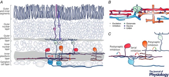
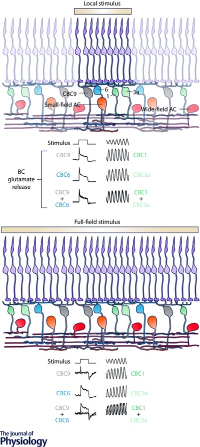
Visual processing starts in the retina. Within only two synaptic layers, a large number of parallel information channels emerge, each encoding a highly processed feature like edges or the direction of motion. Much of this functional diversity arises in the inner plexiform layer, where inhibitory amacrine cells modulate the excitatory signal of bipolar and ganglion cells. Studies investigating individual amacrine cell circuits like the starburst or A17 circuit have demonstrated that single types can possess specific morphological and functional adaptations to convey a particular function in one or a small number of inner retinal circuits. However, the interconnected and often stereotypical network formed by different types of amacrine cells across the inner plexiform layer prompts that they should be also involved in more general computations. In line with this notion, different recent studies systematically analysing inner retinal signalling at a population level provide evidence that general functions of the ensemble of amacrine cells across types are critical for establishing universal principles of retinal computation like parallel processing or motion anticipation. Combining recent advances in the development of indicators for imaging inhibition with large-scale morphological and genetic classifications will help to further our understanding of how single amacrine cell circuits act together to help decompose the visual scene into parallel information channels. In this review, we aim to summarise the current state-of-the-art in our understanding of how general features of amacrine cell inhibition lead to general features of computation.
Keywords: GABA; amacrine cell; computation; glycine; inhibition; neuronal network; retina; vision.
© 2017 The Authors. The Journal of Physiology © 2017 The Physiological Society.
Figures
Similar articles
-
The Synaptic and Morphological Basis of Orientation Selectivity in a Polyaxonal Amacrine Cell of the Rabbit Retina.J Neurosci. 2015 Sep 30;35(39):13336-50. doi: 10.1523/JNEUROSCI.1712-15.2015. J Neurosci. 2015. PMID: 26424882 Free PMC article.
-
A structural basis for omnidirectional connections between starburst amacrine cells and directionally selective ganglion cells in rabbit retina, with associated bipolar cells.Vis Neurosci. 2002 Mar-Apr;19(2):145-62. doi: 10.1017/s0952523802191139. Vis Neurosci. 2002. PMID: 12385627
-
Timing and computation in inner retinal circuitry.Annu Rev Physiol. 2007;69:271-90. doi: 10.1146/annurev.physiol.69.120205.124451. Annu Rev Physiol. 2007. PMID: 17059359 Review.
-
Cholinergic excitation complements glutamate in coding visual information in retinal ganglion cells.J Physiol. 2018 Aug;596(16):3709-3724. doi: 10.1113/JP275073. Epub 2018 Jun 21. J Physiol. 2018. PMID: 29758086 Free PMC article.
-
Receptor targets of amacrine cells.Vis Neurosci. 2012 Jan;29(1):11-29. doi: 10.1017/S0952523812000028. Vis Neurosci. 2012. PMID: 22310370 Free PMC article. Review.
Cited by
-
Rod and cone interactions in the retina.F1000Res. 2018 May 23;7:F1000 Faculty Rev-657. doi: 10.12688/f1000research.14412.1. eCollection 2018. F1000Res. 2018. PMID: 29899971 Free PMC article. Review.
-
Retinal primary cilia and their dysfunction in retinal neurodegenerative diseases: beyond ciliopathies.Mol Med. 2024 Jul 26;30(1):109. doi: 10.1186/s10020-024-00875-y. Mol Med. 2024. PMID: 39060957 Free PMC article. Review.
-
Understanding the retinal basis of vision across species.Nat Rev Neurosci. 2020 Jan;21(1):5-20. doi: 10.1038/s41583-019-0242-1. Epub 2019 Nov 28. Nat Rev Neurosci. 2020. PMID: 31780820 Review.
-
Shining new light into the workings of photoreceptors and visual interneurons.J Physiol. 2017 Aug 15;595(16):5425-5426. doi: 10.1113/JP274464. J Physiol. 2017. PMID: 28809044 Free PMC article. No abstract available.
-
Spike desensitisation as a mechanism for high-contrast selectivity in retinal ganglion cells.Front Cell Neurosci. 2024 Jan 10;17:1337768. doi: 10.3389/fncel.2023.1337768. eCollection 2023. Front Cell Neurosci. 2024. PMID: 38269116 Free PMC article.
References
Publication types
MeSH terms
Grants and funding
LinkOut - more resources
Full Text Sources
Other Literature Sources