

doi: 10.1038/ncomms14862.
Correspondence: Oncogenic MYC persistently upregulates the molecular clock component REV-ERBα
Affiliations
- PMID: 28332504
- PMCID: PMC5376640
- DOI: 10.1038/ncomms14862
Item in Clipboard
Correspondence: Oncogenic MYC persistently upregulates the molecular clock component REV-ERBα
Nat Commun.
.
No abstract available
Conflict of interest statement
The authors declare no competing financial interests.
Figures

(a,b) U2OS BMAL1::Luc cells expressing MYC-ER were cultured±4OHT (with ethanol serving as a loading control for all experiments) for 24 h, then changed to ‘lumicycle' media (see Methods)±4OHT and+0.1 μM dexamethasone. (a) mRNA was collected at the indicated time points after synchronization, and endogenous REV-ERBα (NR1D1), BMAL1 (ARNTL), and PER2 were determined by RT-PCR, normalized to β2M. mRNA (FC)=fold change. (b) Lysates were collected at the indicated time points after synchronization and processed for protein expression of REV-ERBα, BMAL1, and PER2 (indicated by <). Tubulin serves as a loading control. Portions of a and b were previously published, but instead labelled as 0–48 h. (c) REV-ERBα protein from (b) was quantitated using Image Studio software (Licor, Lincoln, NE, USA), normalized to the relevant Tubulin and plotted over time. (d,e) SHEP N-MYC-ER expressing cells were cultured±4OHT for 24 h, then 0.1 μM dexamethasone was added. (d) mRNA was collected at the indicated time points after synchronization, and endogenous REV-ERBα (NR1D1), BMAL1 (ARNTL) and PER2, were determined by RT-PCR, normalized to β2M. (e) Lysates were collected at the indicated time points after synchronization and processed for protein expression of REV-ERBα (‘short' and ‘long' indicate exposure time), BMAL1 and PER2. Tubulin serves as a loading control. For all panels, CT (cell time) indicate time of collection after synchronization. For immunoblots, molecular weights are noted in kDa. Portions of a–c were previously published and are reprinted with permission from Elsevier.
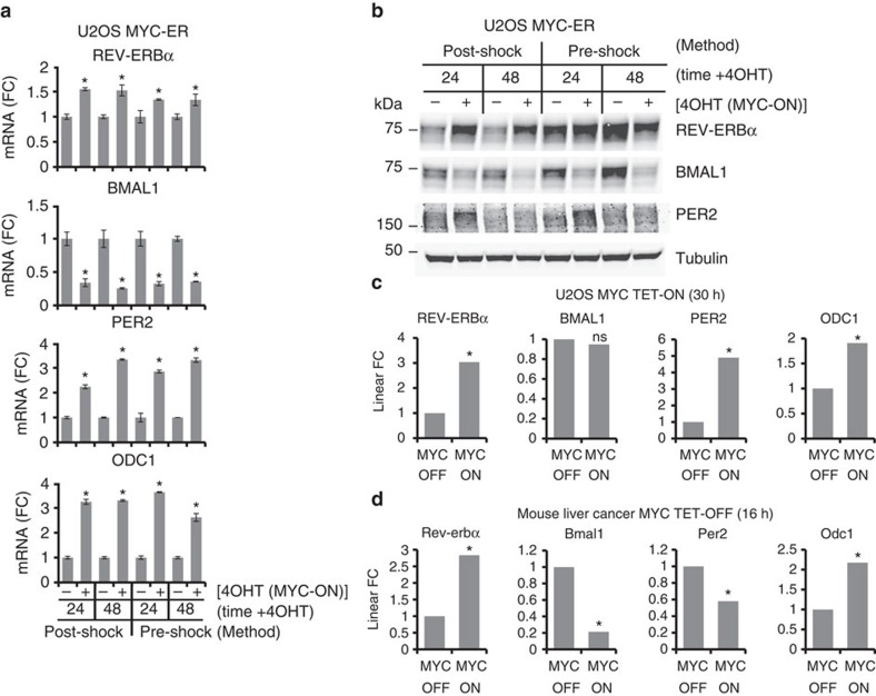
(a,b) MYC was induced in U2OS MYC-ER cells that were synchronized with dexamethasone in two different schemes. ‘Post-Shock': as in Fig. 1, cells were treated±4 OHT (with ethanol serving as a loading control for all experiments) for 24 h. The ‘24' h sample was collected, then 0.1 μM dexamethasone was added to media and the ‘48' h sample was collected 24 h later. ‘Pre-Shock': 1 μM dexamethasone was added for 20 min, then cells were washed once in PBS and fresh media was added±4OHT. Cells were collected at the indicated times after media change. (a) mRNA was collected at the indicated times, and endogenous REV-ERBα (NR1D1), BMAL1 (ARNTL), PER2, and ODC1 were determined by RT-PCR, normalized to β2M. mRNA (FC)=fold change. Data are averages of biological triplicates with error bars representing s.d., and *P<0.05 by Student's t-test of 4 OHT (MYC-ON) samples relative to ethanol (MYC-OFF) samples at each time point. (b) Samples from (a) were processed for protein expression of REV-ERBα, BMAL1 and PER2. Tubulin serves as a loading control. Molecular weights are noted in kDa. (c) Previously published RNA-Seq data from U2OS cells expressing exogenous MYC under the control of a TET-ON system and treated±1 μg ml−1 doxycycline for 30 h. REV-ERBα (NR1D1), BMAL1 (ARNTL), PER2 and ODC1 were determined. Data are presented as linear fold change (FC) and represent biological triplicates, and *P<0.05 of MYC-ON samples relative to MYC-OFF samples as previously described; NS, not significant. (d) Previously published RNA-seq data from liver tumors driven by a MYC-TET-OFF system. Mice were fed water or doxycycline for 16 h to turn off MYC. Rev-erbα (Nr1d1), Bmal1 (Arntl), Per2 and Odc1 were determined. Data are presented as linear FC and represent biological duplicates, and *P<0.05 of MYC-ON samples relative to MYC-OFF samples as previously described.
References
Publication types
MeSH terms
Substances
Grants and funding
LinkOut - more resources
Full Text Sources
Other Literature Sources
